


Granulomatous lobular mastitis (GLM) is a non-infectious, nonspecific inflammatory breast disease. In recent years, its rising incidence and increasingly complex clinical manifestations, coupled with a lack of robust evidence-based medical data, have rendered GLM a formidable diagnostic and therapeutic challenge [1]. Accurate and timely diagnosis is essential for initiating effective treatment and improving clinical outcomes. Owing to its speed, convenience, non-invasiveness, and absence of ionizing radiation, breast ultrasonography has become the preferred imaging modality for the diagnosis of GLM [2]. However, in the past, ultrasound primarily focused on detecting breast masses and their advanced stages. In particular, during the non-mass stage, limitations in probe frequency and resolution hinder the identification of subtle ductal changes and periductal abnormalities, contributing to delayed or missed diagnoses. Nonetheless, early detection remains critical, as it offers the greatest potential for effective intervention and screening.
Our institution is recognized as one of the earliest and most prominent centers for the diagnosis and treatment of GLM in China [3-5]. Over the past decade, our center has accumulated extensive clinical experience through a high volume of cases. This has enabled us to build a substantial institutional repository of ultrasound images and clinical data, forming a solid foundation for research. Through a systematic analysis of this repository, we previously identified and described a set of stage-specific ultrasonographic features of GLM. Building upon this foundation, the present study aims to further evaluate and validate the reliability of these ultrasound features, providing objective statistical evidence to support their diagnostic value. The primary aim is to comprehensively delineate the ultrasonographic manifestations of GLM across its various clinical stages—with a particular focus on the diagnostically challenging non-mass stage—to provide a valuable reference for clinical practice.
Method
Case data
All cases in the validation cohort were derived from female patients aged 18 years and older, who were diagnosed with GLM either by initial assessment at external hospitals or through outpatient consultation at Longhua Hospital, Shanghai University of Traditional Chinese Medicine, between June 2022 and December 2023. A total of 273 patients were enrolled. The study, which built upon prior retrospective analysis of historical ultrasound images for feature summarization, was conducted under the approval of the Ethics Committee of Longhua Hospital, Shanghai University of Traditional Chinese Medicine (approval number: 2021LCSY047), and all patients in the validation cohort provided informed consent.
Assessment strategy
Based on the clinical and imaging characteristics of GLM, disease progression was classified into 6 stages: Non-Mass Stage, Mass Stage, Abscess Stage, Late Ulceration Stage, Fistula Stage, and Complex Type (defined as the simultaneous presence of ≥ 2 ultrasonographic features corresponding to different GLM stages within the same breast, characterized by the coexistence of Mass, Abscess, Late Ulceration, and Fistula stages). In our preliminary research, we summarized a set of stage-specific ultrasonographic features of GLM, including five distinct types of the “track sign,” the “donut sign,” the “false burr sign,” features indicative of abscess formation, post-ulceration manifestations, and duct-like hypoechoic patterns, among others.
Clinical assessment of each case was determined through multidisciplinary evaluation involving two senior breast specialists, one radiologist, and one pathologist. This assessment was based on findings from specialized clinical examinations, MRI, and pathological biopsy. Ultrasound assessment was independently performed by two senior sonographers, who evaluated each case using the predefined ultrasonographic criteria developed during our preliminary studies. In cases where the assessments differed, a consensus meeting was held to reach a final diagnosis. The sonographers were blinded to the patients’ clinical and pathological results during image interpretation to minimize bias.
Ultrasonic examination
Ultrasonic examinations were performed using the Hitachi ALOKA ARIETTA 850 color Doppler ultrasound system, equipped with the L64 probe operating within a probe frequency range of 5-18 MHz. Patients were positioned in the supine position with both arms relaxed and placed at their sides. The patient's position could be adjusted during the examination as needed. Scanning was performed in a radial pattern from the nipple, utilizing fan-shaped and circular multi-sectional scanning techniques. The following parameters were recorded: lesion location, size, shape, echogenicity, blood flow signals, duct diameter, presence of thickening around the duct, secretions within the duct lumen, and calcification.
Examination flow and operational details:
① A routine ultrasound (US) examination of the lesion was conducted, documenting the lesion's location, size, shape, echogenicity, blood flow signals, duct diameter, and calcification.
② The acoustic beam was focused on the region of interest.
③ The “ZOOM” function was applied to magnify and visualize the entire duct in cases of ductal dilation, observing for lipid secretions, whether the duct extends toward the nipple, and assessing the smoothness or roughness of the duct wall, as well as any thickening around the duct and reduction in interstitial echogenicity.
④ A coupling agent was applied generously to the areola, and gentle pressure was applied to the probe without excessive force, preventing ductal closure.
⑤ The color scale was adjusted to the lowest feasible setting, ideally below 2 cm/s, and blood flow signals within the hypoechoic area of the duct were carefully examined.
⑥ For suspected lipid secretions, light probe compression was applied, allowing the visualization of low echogenicity or fine, punctate high echogenicity patterns within the duct, with a floating or flowing sensation.
Results
The clinical and ultrasound assessment results are summarized in table 1. A total of 261 cases were clinically assessed as GLM, while 12 cases were not GLM, including 4 cases of breast cancer, 2 cases of intraductal papilloma, and 4 cases of breast hyperplasia. Ultrasound assessment of GLM demonstrated a sensitivity of 98.85%, with high specificity of 83.33% and positive predictive value of 99.23%. To further evaluate diagnostic reliability, we calculated the positive likelihood ratio (LR+ = 5.93; 95% CI: 1.82–19.29), which—together with the high sensitivity—supports the reliability and stability of the ultrasonographic diagnostic performance for GLM.
Table 1 Clinical and ultrasound assessment results
| Clinical assessment | Frequency | Percentage | Ultrasound assessment | |||
| Sensitivity | Specificity | PPV | NPV | |||
| GLM, granulomatous lobular mastitis; PPV, positive predictive value; NPV, negative predictive value | ||||||
| GLM | 261 | 95.60% | 98.85% | 83.33% | 99.23% | 76.92% |
| Not GLM | 12 | 4.40% | ||||
The distribution of ultrasonographic features across different stages of GLM in 261 patients is presented in table 2. Cases categorized as Complex Type were excluded from the main statistical analysis because their overlapping ultrasonographic features across multiple stages could confound comparisons among mutually exclusive groups. Therefore, the Chi-square tests in table 2 were conducted only for cases assigned to a single, clearly defined stage to avoid confounding the interpretation of stage-specific ultrasonographic findings. We observed a significant association between specific ultrasonographic features features and the clinical stage of GLM. Various types of track signs, with the exception of the track sign with periductal abscess formation, were significantly more prevalent in the Non-Mass Stage compared to other stages. Notably, the track sign with periductal abscess formation also demonstrated a higher tendency to appear in the Non-Mass Stage. The donut sign was significantly more frequent in both the Non-Mass Stage and Abscess Stage than in other stages. The false burr sign was predominantly observed during the Mass Stage, showing a significantly higher distribution compared to other stages.
Table 2 Distribution of ultrasonographic features in different stages of GLM
| Ultrasonographic features | Non-mass stage (n = 42) | Mass stage (n = 61) | Abscess stage (n = 33) | Late ulceration stage (n = 27) | Fistula stage (n = 41) | Complex type# (n = 57) | χ2 | P value |
| # Statistical analysis was not included for the Complex Type as this group may exhibit characteristics from the preceding stages, leading to potential confounding and dilution of effects. * Different types of track signs may occur concurrently within the same case; therefore, the sum of the frequencies of each track sign subtype exceeds the total frequency of the track sign (All Types). a Fisher’s exact test; b The imaging features marked in this study are strictly stage-dependent (appearing only in specific pathological stages or later stages), and theoretically, they cannot appear prior to the specified stages (with observed values being 0 in this study). Given their 100% specificity (no false positives observed), we conducted the analysis only on subgroups where these features were actually present. | ||||||||
| Track Ssign * (All types) | 39 (92.86%) | 15 (24.59%) | 4 (12.12%) | 5 (18.52%) | 3 (7.32%) | 31 (54.39%) | 92.207 | < 0.001 |
| Track sign with high echogenicity on the inner wall* | 24 (57.14%) | 7 (11.48%) | 2 (6.06%) | 3 (11.11%) | 2 (4.88%) | 16 (28.07%) | 52.721 | < 0.001 |
| Track sign with low echogenicity on the inner wall* | 17 (40.48%) | 4 (6.56%) | 1 (3.03%) | 1 (3.7%) | 1 (2.44%) | 7 (12.28%) | 31.689a | < 0.001a |
| Track sign with isoechogenicity on the inner wall* | 11 (26.19%) | 2 (3.28%) | 1 (3.03%) | 1 (3.7%) | 1 (2.44%) | 5 (8.77%) | 17.256a | < 0.001a |
| Track sign with periductal abscess formation* | 7 (16.67%) | 2 (3.28%) | 1 (3.03%) | 1 (3.7%) | 1 (2.44%) | 3 (5.26%) | 7.870a | 0.057a |
| Track sign coexisting with ductal inflammation, periductal inflammation, and nodular structures* | 10 (23.81%) | 1 (1.64%) | 1 (3.03%) | 1 (3.7%) | 1 (2.44%) | 6 (10.53%) | 16.773a | <0.001a |
| Donut sign | 29 (69.05%) | 4 (6.56%) | 22 (66.67%) | 2 (7.41%) | 2 (4.88%) | 7 (12.28%) | 88.196 | < 0.001 |
| False burr sign | 0 (0%) | 48 (78.69%) | 2 (6.06%) | 8 (29.63%) | 7 (17.07%) | 17 (29.82%) | 64.005b | < 0.001b |
| Large areas of mixed echogenicity with irregularly thickened and rough abscess walls; a flowing sensation within the abscess cavity was observed upon probe compression. | 0 (0%) | 0 (0%) | 33 (100%) | 8 (29.63%) | 4 (9.76%) | 11 (19.30%) | 63.599b | <0.001b |
| A relatively well-defined and smooth-edged hypoechoic area was observed in the areolar region, communicating with the ductal system. | 0 (0%) | 0 (0%) | 0 (0%) | 19 (70.37%) | 19 (46.34%) | 23 (40.35%) | 3.813b | 0.043b |
| A duct-like hypoechoic structure extending toward the skin was observed, with thickening of the inner wall of the duct. | 0 (0%) | 0 (0%) | 0 (0%) | 3 (7.32%) | 38 (92.68%) | 26 (45.61%) | 21.815b | < 0.001b |
Another characteristic ultrasonographic finding of the Abscess Stage was the presence of large areas of mixed echogenicity with irregularly thickened and rough abscess walls; a flowing sensation within the abscess cavity was elicited upon probe compression. In the Late Ulceration Stage, a specific ultrasonographic feature was the appearance of a relatively well-defined and smooth-edged hypoechoic area in the areolar region, which communicated with the ductal system. In the Fistula Stage, the characteristic feature was a duct-like hypoechoic structure extending toward the skin, with evident thickening of the inner ductal wall.
Building on these validated ultrasonographic features, we provide a comprehensive overview of the characteristic and stage-specific imaging manifestations of GLM, supplemented with representative figures to enhance clinical recognition and interpretation.
Non-mass stage: solely pain as the clinical manifestation
In the early stages of GLM, the disease may present with pain as the only clinical manifestation, often leading to misdiagnosis as mammary gland hyperplasia (a disorder of breast structure), corresponding to the Non-Mass Stage. The underlying pathological basis at this stage involves irregular hyperplasia of ductal epithelium, ductal dilation, and periductal tissue fibrosis. As a result, the ultrasonographic appearance exhibits similar characteristics, with dilated and thickened ductal cross-sections resembling a "train track" structure, known as the track sign. As the extent of inflammatory cell infiltration increases and the severity of the disease progresses, the sonographic features evolve. Five distinct types of ultrasonographic changes have been summarized, although these five types of track signs do not necessarily appear in isolation; different track signs may coexist in the same patient.
Type 1: track sign with high echogenicity on the inner wall
The ultrasonographic appearance shows thickening of the ductal wall, with the inner wall displaying a strip-like or linear high echogenicity. The ductal wall is smooth, and lipid-containing secretions are seen floating within the duct. A thin, uniformly hypoechoic halo is observed around the duct (Fig. 1).
Figure 1
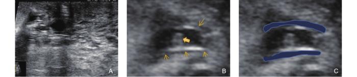
Figure 1
Ultrasonographic features of the Non-Mass Stage in GLM (type 1: Track Sign with High Echogenicity on the Inner Wall). (A) shows the appearance of several ducts behind the areola, exhibiting irregular dilation and thickening of the ductal walls; (B) magnified image, reveals the thickened ductal walls with a strip-like or linear high echogenicity on the inner wall. The duct wall is smooth, and lipid-containing secretions (bold arrow) are visible inside the duct. A thin, uniformly hypoechoic halo (thin arrow) surrounds the duct; (C) highlights the blue region, which represents the hypoechoic track sign due to the thickening around the duct.
Type 2: track sign with low echogenicity on the inner wall
The ultrasonographic appearance shows a single or several ducts behind the areola with irregular thickening of the ductal walls. The surrounding ductal wall is hypoechoic, and the duct is notably dilated. A large amount of hyperechoic secretion is accumulated within the lumen. Upon probe compression, a floating sign is observed (Fig. 2).
Figure 2
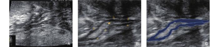
Figure 2
Ultrasonographic features of the Non-Mass Stage in GLM (type 2: Track Sign with Low Echogenicity on the Inner Wall). (A) shows the appearance of several ducts behind the areola with irregular dilation and thickening of the ductal walls; (B) a magnified image, reveals the thickened ductal walls with roughened inner walls. Dense lipid-containing secretions (bold arrow) are visible within the lumen, and a floating sign is observed upon probe compression. A thick, uneven hypoechoic area (thin arrow) surrounds the duct; (C) highlights the blue region, representing the hypoechoic track sign with uneven thickening around the duct.
Type 3: track sign with isoechogenicity on the inner wall
The ultrasonographic appearance shows significant thickening of the inner wall of some ducts, presenting as isoechoic. The surrounding ductal wall is further thickened and hypoechoic. A small amount of anechoic secretion is accumulated within the duct (Fig. 3).
Figure 3
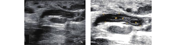
Figure 3
Ultrasonographic features of the Non-Mass Stage in GLM (type 3: Track Sign with isoechogenicity on the Inner Wall). (A) shows the ultrasonographic appearance of several ducts behind the areola with irregular dilation and thickening of the ductal walls; (B) a magnified image, reveals significant thickening of the walls of some ducts, with the inner walls becoming thickened and rough, presenting as isoechoic (triangle arrow). No anechoic secretion is observed within the lumen (bold arrow). The surrounding ductal wall is notably thickened, presenting as an uneven hypoechoic area (thin arrow).
Type 4: track sign with periductal abscess formation
As the disease progresses, the ultrasonographic appearance shows high echogenicity on the inner wall of the duct, with further thickening of the surrounding ductal wall, presenting as a hypoechoic area that merges with the surrounding stroma, forming a large hypoechoic region. A small amount of pus is observed accumulating around the duct. Clinically, some cases present with palpable localized thickening and flaky nodules, signaling the transition from the non-mass stage to the mass stage (Fig. 4).
Figure 4
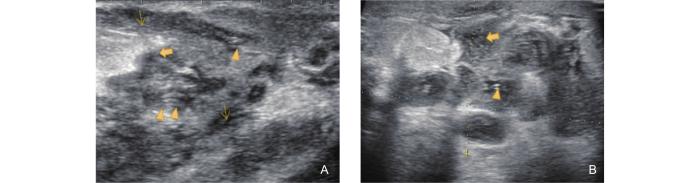
Figure 4
Ultrasonographic features of the Non-Mass Stage in GLM (type 4: Track Sign with Periductal Abscess Formation). (A) shows the ultrasonographic appearance of several ducts behind the areola with irregular dilation. The inner wall of the duct displays linear high echogenicity (triangle arrow), while the surrounding ductal wall is markedly thickened and presents as an extremely hypoechoic area (thin arrow). The stroma around the duct shows decreased echogenicity, slightly higher than that of the surrounding ductal wall (area marked with asterisks); (B) shows the same patient's ipsilateral breast, with a local quadrant demonstrating an irregular mixed echogenic area around the duct, which corresponds to fat necrosis and abscess formation (bold arrow).
Type 5: Track sign coexisting with ductal inflammation, periductal inflammation, and nodular structures
This represents the most severe lesion manifestation in the non-mass stage, marking the transition from the non-mass stage to the mass stage. At this point, the inflammatory response within the duct is at its peak, and the duct diameter is widest. Contrast-enhanced ultrasound at this stage reveals ring-like enhancement of the dilated ductal walls, which provides valuable information about the location and extent of the lesion (Fig. 5).
Figure 5
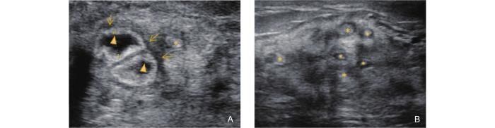
Figure 5
Ultrasonographic features of the Non-Mass Stage in GLM (type 5: coexistence of ductal inflammation, periductal Inflammation, and nodular Structures). (A) shows the sonographic appearance of several ducts behind the areola with irregular dilation. The ductal lumen is further expanded, with the inner walls thickened and rough, presenting as a ring-like high echogenicity pattern, consistent with the donut sign (triangle arrow). Two ducts merge with each other, and the surrounding ductal wall is markedly thickened, presenting as an extremely hypoechoic area (thin arrow). Another duct shows a relatively smaller diameter, with slight thickening of the ductal wall (area marked with an asterisk); (B) shows the same patient's ipsilateral breast, with a local quadrant revealing a nodular structure formed by the coexistence of dilated ducts and periductal fibrosis. Several ductal cross-sections with thickened walls are visible inside, showing high echogenicity, while the surrounding ductal area presents as hypoechoic (area marked with an asterisk).
Mass stage
The ultrasonographic features observed at this stage warrant particular attention, as the mass-like lesions must be differentiated from breast carcinoma. Ultrasound typically reveals an ill-defined hypoechoic mass with angular margins and the appearance of a false burr sign, which may be associated with compressive expansion of the lesion into adjacent tissue planes and its expansive growth pattern [6]. The pathological basis of the false burr sign lies in the adhesion of mammary trabeculae or the formation of fibrous strands. Color Doppler imaging often shows relatively abundant internal vascularity within the mass. In addition, localized skin thickening and edema may be present, and in some cases, thickening of Cooper’s ligaments can also be observed (Fig. 6).
Figure 6
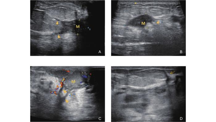
Figure 6
Ultrasonographic features of the Mass Stage in GLM. (A-B) show hypoechoic masses (M) with irregular, ill-defined margins. The masses exhibit angular extensions and a false burr sign, characterized by relatively uniform, slender hypoechoic projections radiating from the lesion edges (triangle arrows); (C) demonstrates abundant blood flow within and surrounding the mass, accompanied by skin thickening and edema in the affected area (asterisk); (D) shows thickening of Cooper’s ligaments in some cases (thin arrow).
Abscess stage
Patients with GLM may develop abscesses. On ultrasonography, this is manifested as large areas of mixed echogenicity in the areolar or peripheral regions. The inner wall of the abscess cavity appears irregularly thickened, with overlying skin edema and thickening. A substantial amount of purulent fluid is present within the cavity, and a flowing sensation can be elicited upon probe compression. Dilated ducts extending toward the nipple can also be observed [7] (Fig. 7).
Figure 7
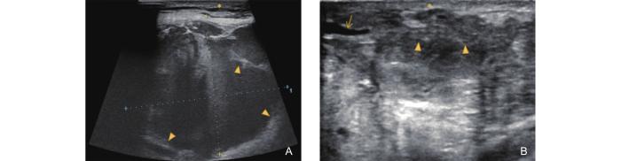
Figure 7
Ultrasonographic features of the Abscess Stage in GLM. (A-B) shows a large area of mixed echogenicity in the areolar region. The inner wall of the abscess is irregularly thickened with a rough surface (triangle arrow). Overlying skin edema and thickening are observed (asterisk). (B) shows a dilated duct extending toward the nipple is also visible (thin arrow).
Late ulceration stage
Following the abscess stage, the disease progresses to the late ulceration stage, during which the extent of the lesion is significantly reduced, and the inflammatory response associated with ductitis and periductal inflammation is markedly alleviated. On ultrasound, a hypoechoic or mixed echogenic area with relatively smooth and well-defined margins can be observed in the periareolar region. This area communicates with the ductal system and extends toward the nipple (Fig. 8).
Figure 8
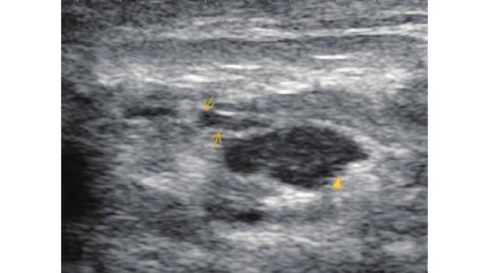
Figure 8
Ultrasonographic features of the Late Ulceration Stage in GLM.
Fistula stage
In some patients with GLM, the disease may progress to the fistula stage [8]. Ultrasonographically, this stage is characterized by the presence of one or more quadrants showing irregular sheet-like or duct-like hypoechoic structures traversing from the glandular layer through the adipose tissue and extending into the subcutaneous region. These structures often interconnect with one another and, in some cases, extend as deep as the retromammary space. The inner walls of the ducts are thickened, with or without associated pus accumulation. Skin ulceration may also be observed (Fig. 9).
Figure 9
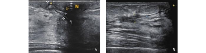
Figure 9
Ultrasonographic features of the Fistula Stage in GLM. (A) shows a duct-like hypoechoic structure beneath the nipple (N) extending toward the areolar skin. The inner wall of the duct is thickened (thin arrow), and a skin ulceration is visible at the areola (asterisk); (B) demonstrates an irregular sheet-like hypoechoic area extending from the glandular layer through the adipose tissue into the subcutaneous region, with a visible skin ulceration at the surface (asterisk).
Complex type (coexistence of non-mass, mass, abscess, late ulceration, and fistula stage)
In patients with GLM whose disease duration exceeds one month, it is common to observe a combination of clinical features such as masses, abscesses, ulcers, and fistulas. In other words, the progression of GLM in some patients does not follow a linear sequence from the non-mass stage to the mass stage, abscess stage, late ulceration stage, and fistula stage. Instead, the disease may follow a fluctuating course, with some lesions stagnating at a particular stage while new lesions emerge or progress into subsequent stages. This results in a complex pattern of overlapping old and new lesions (Fig. 10).
Figure 10
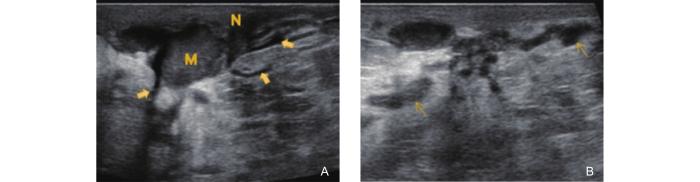
Figure 10
Ultrasonographic features of the Complex Type in GLM (coexistence of multiple stages). (A) shows a hypoechoic nodule located posterior to the nipple (N), with indistinct margins and angular contours. Several dilated ducts are visible surrounding the lesion (bold arrows); (B) demonstrates multiple hypoechoic ducts around the mass with luminal obliteration and thickening of both the ductal walls and surrounding tissues (thin arrows).
Discussion
Ultrasound is the first-line imaging modality for GLM, aiding in the detection of inflammatory lesions, abscess formation, and fistulous tracts. It also facilitates ultrasound-guided biopsy and serves as a valuable tool for monitoring disease remission or progression during follow-up [9]. Although the imaging features of GLM are generally considered nonspecific, complex, and variable, a combination of clinical context and stage-specific ultrasonographic characteristics can significantly improve diagnostic accuracy. Common ultrasonographic findings include a hypoechoic or heterogeneous mass, an irregular hypoechoic lesion with tubular extension [10], and abscess formation. The diagnostic accuracy of ultrasound is closely related to the ultrasonographic subtype of GLM [11], with higher accuracy observed in patchy heterogeneous echo types and nodular or mass-forming types, while diffuse-type GLM remains more challenging to diagnose.
Early-stage GLM is the most easily overlooked phase in clinical practice and is frequently misdiagnosed as mammary ductal hyperplasia. In both domestic and international literature, the ultrasonographic features of GLM in the non-mass stage have been briefly summarized as parenchymal heterogeneity and distortion with or without acoustic shadowing [12], without further detailed characterization. Based on case review and validation at our center, we highlight that the presence of dilated and enlarged ductal cross-sections—collectively referred to as the track sign—is a key diagnostic indicator during this stage. Furthermore, we propose a five-type classification system for the non-mass stage based on disease severity. Among them, the latter two types reflect the most extensive inflammatory cell infiltration, characterized by significantly widened ductal lumens and more pronounced periductal thickening. These may appear as large, irregular or nodular hypoechoic structures within the gland. Importantly, these nodular structures are not true solid granulomas but are formed by the confluence of dilated ducts and surrounding fibrotic tissue [13,14].
Histologically, these findings are accompanied by focal fat necrosis, periductal fibrosis, and lymphocytic infiltration. On physical examination, broad regions of indurated nodularity can often be palpated. It is noteworthy that these five ultrasonographic patterns do not occur in isolation, nor do they necessarily follow a sequential progression. Multiple ultrasonographic features may coexist across different stages of the disease, in various regions of the same breast, or even bilaterally. Therefore, it is essential to recognize the characteristic track sign and distinguish it from other types of intraductal lesions. We believe that early detection of GLM should be a primary focus in clinical management. Enhancing awareness and understanding of early-stage GLM may help improve diagnostic accuracy.
In addition to detailing the ultrasonographic features of the non-mass stage, this study also summarizes the imaging characteristics of GLM across its full spectrum of clinical stages—including the mass stage, abscess stage, late ulceration stage, fistula stage, and complex type. Notably, complex-type GLM presents with diverse and overlapping ultrasonographic findings. During ultrasound examination, the sonographer must carefully document the condition of each lesion, including the location and extent of masses and abscesses, whether pus cavities are interconnected, the depth of any ulcerated tract from the skin surface, and the number, position, and orientation of fistulas. These ultrasound findings can guide clinicians in determining the appropriate timing and method of surgical intervention.
Moreover, we have standardized a systematic ultrasound examination protocol and operational procedure specific to GLM. Considering the common overlap, inconsistency, and ambiguity in ultrasonographic terminology for breast imaging, this study provides clearly defined sonographic terms and representative images to improve diagnostic clarity. This effort not only enhances clinicians’ understanding of GLM-specific ultrasound features but also lays the groundwork for more standardized reporting and examination practices in breast ultrasonography.
One limitation of this study is the lack of quantitative interobserver agreement analysis (e.g., Kappa statistics). The ultrasonographic assessments were conducted independently by two senior sonographers, and discrepancies were resolved through consensus discussion. As this study primarily focused on the qualitative characterization and validation of previously undefined imaging features, quantitative analysis such as interobserver Kappa testing was not included at this stage. Future research will incorporate Kappa consistency analysis and quantitative imaging evaluation methods to further enhance the objectivity and reproducibility of ultrasonographic assessment. Furthermore, this study did not include a comparative analysis with magnetic resonance imaging (MRI). With the growing use of MRI in GLM assessment, we acknowledge its valuable complementary role in delineating disease extent and aiding in staging [15]. Nonetheless, ultrasound remains the first-line and most accessible imaging modality for GLM, offering real-time, dynamic, and radiation-free assessment that cannot be replaced in routine clinical practice. In future research, we plan to perform a systematic comparison between ultrasonographic and MRI features across various GLM stages to better clarify their respective diagnostic strengths and complementary value in disease staging.
Conclusion
In conclusion, the findings of this study demonstrate that specific ultrasonographic features of GLM can be reliably used to identify the disease and determine its clinical stage. We established a standardized operating procedure for ultrasonographic evaluation of GLM, systematically characterized the ultrasonographic features across different stages, and provided representative imaging examples. These efforts aim to improve the accuracy of ultrasound-based diagnosis and staging of GLM, thereby enhancing the reproducibility and applicability of this diagnostic approach in clinical practice.
Acknowledgment
This research was based on data collected at Longhua Hospital. The authors sincerely thank all the clinicians, ultrasonographers, and staff who contributed to patient recruitment, imaging acquisition, and data management for this study.
Authors’ Contributions
MNY and HQY contributed to the study conception and design. Data collection and image analysis were performed by YJR, DWG and LMZ. Statistical analysis was performed by YYZ, BW and JJW. The study was supervised by HFC. The first draft of the manuscript was written by YLY and YFC, and all authors commented on previous versions of the manuscript. All authors read and approved the final manuscript. YLY and YFC contributed equally to this work. MNY and HQY are co-corresponding authors.
Funding
This study was supported by the Major Difficult and Complicated Disease Clinical Collaboration Project of Integrated Traditional Chinese and Western Medicine from the National Administration of Traditional Chinese Medicine (No. 19), and the National Natural Science Foundation of China (No. 82104854). 2024 Annual Science and Technology Development Project of Shanghai University of Traditional Chinese Medicine (No. 24BZH11).
Data Availability
The datasets used and analyzed during the current study available from the corresponding author on reasonable request.
Declarations
Ethics approval and consent to participate
The study was approved by the Ethics Committee of Longhua Hospital, Shanghai University of Traditional Chinese Medicine (approval number: 2021LCSY047), and all patients in the validation cohort provided informed consent.
Consent for publication
Not applicable.
Conflict of interest
The authors declare no conflicts of interest related to this work.
Reference
Etiology of granulomatous lobular mastitis based on metagenomic next-generation sequencing
Management of granulomatous lobular mastitis: an international multidisciplinary consensus (2021 edition)
A cohort study of prolactin and non-puerperal mastitis using real world data
DOI:10.1038/s41598-025-92504-9 [Cited within: 1]
Clinical study on the treatment of 120 cases of acne mastoid carbuncle with GUs’ surgery comprehensive external therapy
Granulomatous lobular mastitis: a clinicopathological analysis of 300 cases
Imaging features of granulomatous mastitis in 36 patients with new sonographic signs
Multimodality imaging features of idiopathic granulomatous mastitis: outcome of 12 years of experience
Granulomatous lobular mastitis: imaging, diagnosis, and treatment
Analysis of the clinical character-istics and ultrasonographic features in 141 cases of cystic neutrophilic granulomatous mastitis
Idiopathic granulomatous mastitis: manifestations at multimodality imaging and pitfalls
Diagnostic value of ultrasonography in different types of granulomatous lobular mastitis: a multicenter retrospective analysis of 120 cases
Idiopathic granulomatous mastitis: a diagnostic dilemma for the breast radiologist
Cystic neutrophilic granulomatous mastitis: Imaging features with histopathologic correlation
Differentiation of malignancy and idiopathic granulomatous mastitis presenting as nonmass lesions on MRI: Radiological, clinical, radiomics, and clinical-radiomics models


