From Non-Mass Stage to Complex Type: Ultrasonographic Assessment of Granulomatous Lobular Mastitis Across Six Clinical Stages
Yin Yuliana,1, Cheng Yifana,1, Zhou Liangmeib, Zhong Yuanyuana, Wang Binga, Wu Jingjinga, Ren Yajuanb, Gao Dongwenb, Chen Hongfenga, Ye Meinaa,*( ), Yin Haoqiangb,*()
), Yin Haoqiangb,*()
.
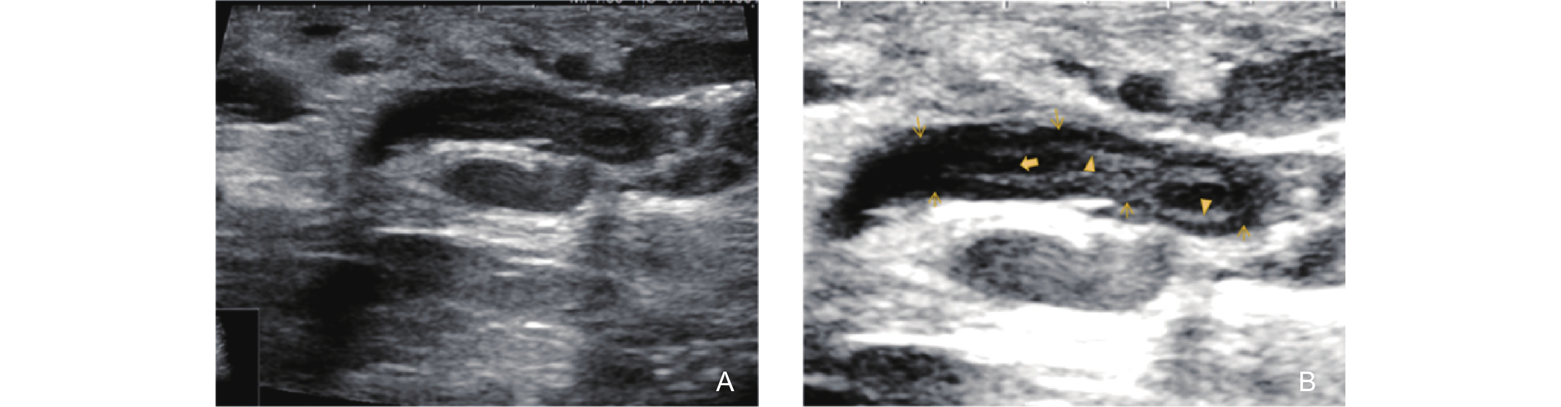
Figure 3. Ultrasonographic features of the Non-Mass Stage in GLM (type 3: Track Sign with isoechogenicity on the Inner Wall). (A) shows the ultrasonographic appearance of several ducts behind the areola with irregular dilation and thickening of the ductal walls; (B) a magnified image, reveals significant thickening of the walls of some ducts, with the inner walls becoming thickened and rough, presenting as isoechoic (triangle arrow). No anechoic secretion is observed within the lumen (bold arrow). The surrounding ductal wall is notably thickened, presenting as an uneven hypoechoic area (thin arrow).