), Yin Haoqiangb,*()
.
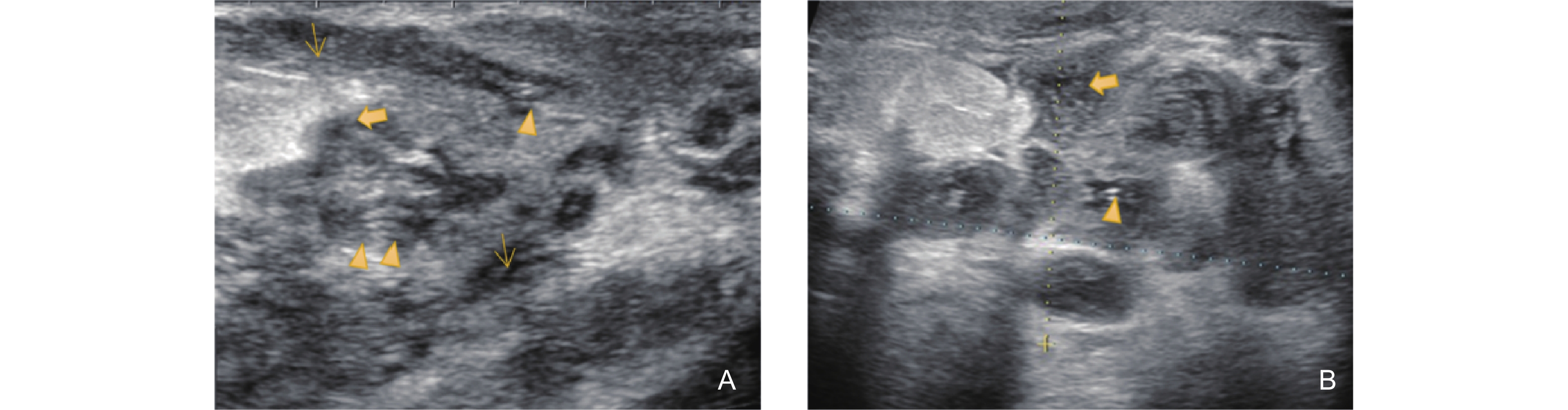
Figure 4. Ultrasonographic features of the Non-Mass Stage in GLM (type 4: Track Sign with Periductal Abscess Formation). (A) shows the ultrasonographic appearance of several ducts behind the areola with irregular dilation. The inner wall of the duct displays linear high echogenicity (triangle arrow), while the surrounding ductal wall is markedly thickened and presents as an extremely hypoechoic area (thin arrow). The stroma around the duct shows decreased echogenicity, slightly higher than that of the surrounding ductal wall (area marked with asterisks); (B) shows the same patient's ipsilateral breast, with a local quadrant demonstrating an irregular mixed echogenic area around the duct, which corresponds to fat necrosis and abscess formation (bold arrow).