From Non-Mass Stage to Complex Type: Ultrasonographic Assessment of Granulomatous Lobular Mastitis Across Six Clinical Stages
Yin Yuliana,1, Cheng Yifana,1, Zhou Liangmeib, Zhong Yuanyuana, Wang Binga, Wu Jingjinga, Ren Yajuanb, Gao Dongwenb, Chen Hongfenga, Ye Meinaa,*( ), Yin Haoqiangb,*()
), Yin Haoqiangb,*()
.
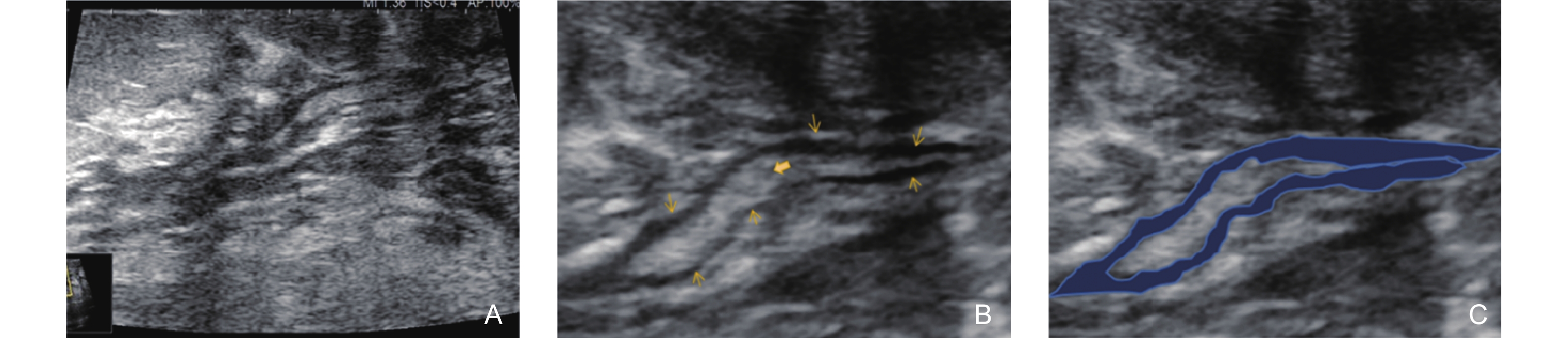
Figure 2. Ultrasonographic features of the Non-Mass Stage in GLM (type 2: Track Sign with Low Echogenicity on the Inner Wall). (A) shows the appearance of several ducts behind the areola with irregular dilation and thickening of the ductal walls; (B) a magnified image, reveals the thickened ductal walls with roughened inner walls. Dense lipid-containing secretions (bold arrow) are visible within the lumen, and a floating sign is observed upon probe compression. A thick, uneven hypoechoic area (thin arrow) surrounds the duct; (C) highlights the blue region, representing the hypoechoic track sign with uneven thickening around the duct.