), Yin Haoqiangb,*()
.
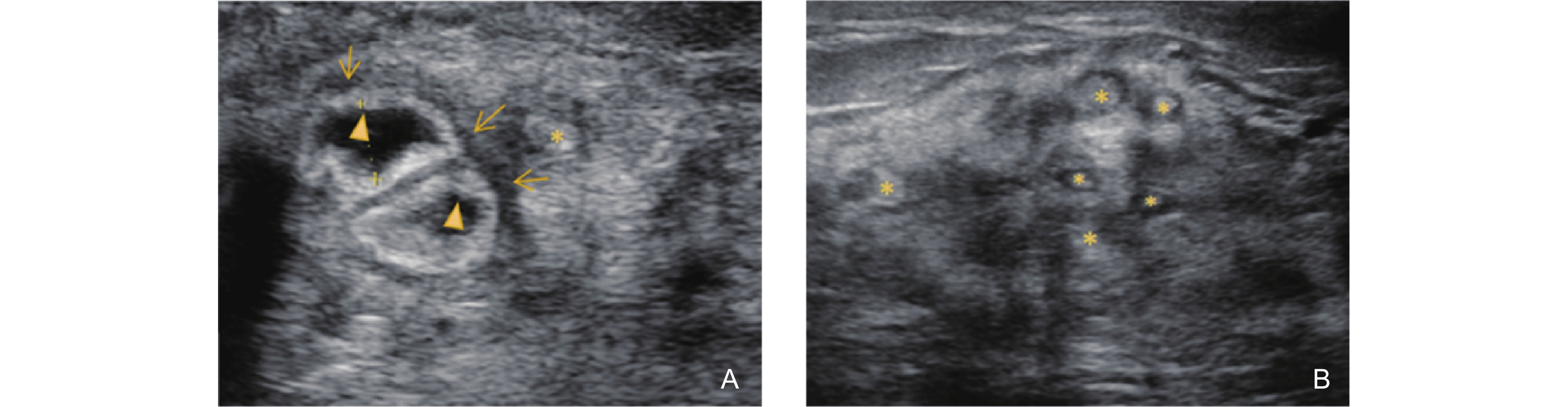
Figure 5. Ultrasonographic features of the Non-Mass Stage in GLM (type 5: coexistence of ductal inflammation, periductal Inflammation, and nodular Structures). (A) shows the sonographic appearance of several ducts behind the areola with irregular dilation. The ductal lumen is further expanded, with the inner walls thickened and rough, presenting as a ring-like high echogenicity pattern, consistent with the donut sign (triangle arrow). Two ducts merge with each other, and the surrounding ductal wall is markedly thickened, presenting as an extremely hypoechoic area (thin arrow). Another duct shows a relatively smaller diameter, with slight thickening of the ductal wall (area marked with an asterisk); (B) shows the same patient's ipsilateral breast, with a local quadrant revealing a nodular structure formed by the coexistence of dilated ducts and periductal fibrosis. Several ductal cross-sections with thickened walls are visible inside, showing high echogenicity, while the surrounding ductal area presents as hypoechoic (area marked with an asterisk).