From Non-Mass Stage to Complex Type: Ultrasonographic Assessment of Granulomatous Lobular Mastitis Across Six Clinical Stages
Yin Yuliana,1, Cheng Yifana,1, Zhou Liangmeib, Zhong Yuanyuana, Wang Binga, Wu Jingjinga, Ren Yajuanb, Gao Dongwenb, Chen Hongfenga, Ye Meinaa,*( ), Yin Haoqiangb,*()
), Yin Haoqiangb,*()
.
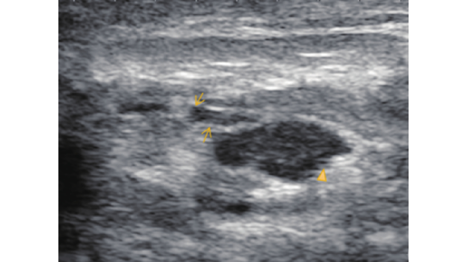
Figure 8. Ultrasonographic features of the Late Ulceration Stage in GLM.