
Advanced Ultrasound in Diagnosis and Therapy ›› 2026, Vol. 10 ›› Issue (3): 148-157.doi: 10.26599/AUDT.2026.260026
• REVIEW ARTICLE • Previous Articles Next Articles
He Xianga, Hu Ronga, Deng Xuejiab, Yang Fana,b,*( )
Received:2026-07-16
Revised:2026-09-24
Accepted:2026-09-30
Online:2026-09-30
Published:2026-10-12
Contact:
Yang Fan,
E-mail:jessica_cd@163.com
He Xiang, Hu Rong, Deng Xuejia, Yang Fan. Current Applications and Advances of Ultrasound in Predicting Adverse Pregnancy Outcomes in Pregnant Women with Pre-eclampsia. Advanced Ultrasound in Diagnosis and Therapy, 2026, 10(3): 148-157.
Figure 1
(A) A 31+4weeks pregnant patient with pre-eclampsia had an increased S/D ratio of the fetal umbilical artery; (B) A 25+2weeks pregnant patient with pre-eclampsia had a decreased PI of fetal middle cerebral artery. PSV, peak systolic velocity; EDV, end-diastolic velocity; HR, heart rate."
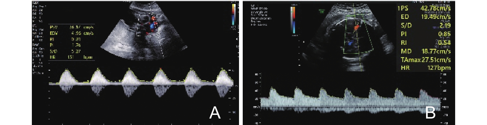
Figure 2
Three-dimensional power Doppler of placental blood flow in a 23+1weeks pregnant woman. VI, vascularization index; FI, flow index; VFI, vascularization-flow index."
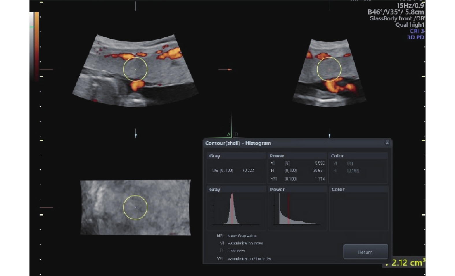
Figure 3
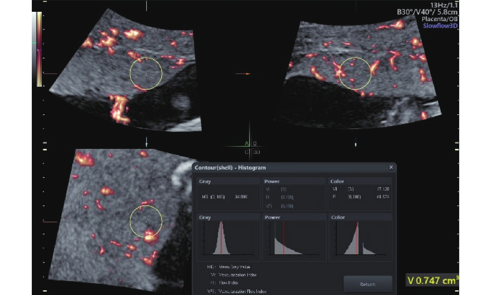
Placental microvascular blood flow in a 24+2weeks pregnant woman (VI: vascularization index, FI: flow index, VFI: vascularization-flow index)"
| [1] | Wu P, Green M, Myers JE. Hypertensive disorders of pregnancy. BMJ 2023; 381: e071653. |
| [2] |
Dimitriadis E, Rolnik DL, Zhou W, Estrada-Gutierrez G, Koga K, Francisco RPV, et al. Pre-eclampsia. Nat Rev Dis Primers 2023; 9: 8.
doi: 10.1038/s41572-023-00417-6 |
| [3] | Gestational hypertension and preeclampsia: ACOG Practice Bulletin, Number 222. Obstet Gynecol 2020;135:e237-e260. |
| [4] |
Heitkamp A, Meulenbroek A, van Roosmalen J, Gebhardt S, Vollmer L, de Vries JI, et al. Maternal mortality: near-miss events in middle-income countries, a systematic review. Bull World Health Organ 2021; 99: 693-707.
doi: 10.2471/BLT.21.285945 |
| [5] |
Chappell LC, Cluver CA, Kingdom J, Tong S. Pre-eclampsia. Lancet 2021; 398: 341-354.
doi: 10.1016/S0140-6736(20)32335-7 |
| [6] |
Magee LA, Brown MA, Hall DR, Gupte S, Hennessy A, Karumanchi SA, et al. The 2021 International Society for the Study of Hypertension in Pregnancy classification, diagnosis & management recommendations for international practice. Pregnancy Hypertens 2022; 27: 148-169.
doi: 10.1016/j.preghy.2021.09.008 |
| [7] |
Thangaratinam S, Gallos ID, Meah N, Usman S, Ismail KM, Khan KS. How accurate are maternal symptoms in predicting impending complications in women with preeclampsia? A systematic review and meta-analysis. Acta Obstet Gynecol Scand 2011; 90: 564-573.
doi: 10.1111/j.1600-0412.2011.01111.x |
| [8] |
Dröge LA, Perschel FH, Stütz N, Gafron A, Frank L, Busjahn A, et al. Prediction of preeclampsia-related adverse outcomes with the sFlt-1 (soluble fms-like tyrosine kinase 1)/PlGF (placental growth factor)-ratio in the clinical routine: a real-world study. Hypertension 2021; 77: 461-471.
doi: 10.1161/HYPERTENSIONAHA.120.15146 |
| [9] |
Vatish M, Powys VR, Cerdeira AS. Novel therapeutic and diagnostic approaches for preeclampsia. Curr Opin Nephrol Hypertens 2023; 32: 124-133.
doi: 10.1097/MNH.0000000000000870 |
| [10] |
Martini C, Saeed Z, Simeone P, Palma S, Ricci M, Arata A, et al. Preeclampsia: insights into pathophysiological mechanisms and preventive strategies. Am J Prev Cardiol 2025; 23: 101054.
doi: 10.1016/j.ajpc.2025.101054 |
| [11] | Gonser M, Vetter K. Diagnostic value and clinical value of Doppler ultrasound in obstetrics. Geburtshilfe Frauenheilkd 1995; 55: 605-615. |
| [12] | Bhide A, Acharya G, Bilardo CM, Brezinka C, Cafici D, Hernandez-Andrade E, et al. ISUOG practice guidelines: use of Doppler ultrasonography in obstetrics. Ultrasound Obstet Gynecol 2013; 41: 234-240. |
| [13] |
Mack LM, Mastrobattista JM, Gandhi R, Castro EC, Burgess APH, Lee W. Characterization of placental microvasculature using superb microvascular imaging. J Ultrasound Med 2019; 38: 2485-2491.
doi: 10.1002/jum.14919 |
| [14] | Kurjak A, Kupesic S, Banovic I, Hafner T, Kos M. The study of morphology and circulation of early embryo by three-dimensional ultrasound and power Doppler. J Perinat Med 1999; 27: 145-157. |
| [15] |
Hua Q, Yang FC, Zhou YD, Shi FL, You XY, Guo J, et al. Predictive models using machine learning to identify fetal growth restriction in patients with preeclampsia: development and evaluation study. J Med Internet Res 2025; 27: e70068.
doi: 10.2196/70068 |
| [16] | Yan MX, Li F, Jun S, Li LY, You WQ, Hu LP, et al. Predictive factors for fetal growth restriction in patients with preeclampsia: a clinical prediction study. Int J Gen Med 2025; 18: 2289-2301. |
| [17] | Caradeux J, Martinez-Portilla RJ, Peguero A, Sotiriadis A, Figueras F. Diagnostic performance of third-trimester ultrasound for the prediction of late-onset fetal growth restriction: a systematic review and meta-analysis. Am J Obstet Gynecol 2019; 220: 449.e1-e14. |
| [18] |
Miranda J, Rodriguez-Lopez M, Triunfo S, Sairanen M, Kouru H, Parra-Saavedra M, et al. Prediction of fetal growth restriction using estimated fetal weight vs a combined screening model in the third trimester. Ultrasound Obstet Gynecol 2017; 50: 603-611.
doi: 10.1002/uog.17393 |
| [19] |
Liu DJ, Gao Q, Wang YB, Xiong T. Placental dysfunction: the core mechanism for poor neurodevelopmental outcomes in the offspring of preeclampsia pregnancies. Placenta 2022; 126: 224-232.
doi: 10.1016/j.placenta.2022.07.014 |
| [20] |
Hall M, de Marvao A, Schweitzer R, Cromb D, Colford K, Jandu P, et al. Preeclampsia associated differences in the placenta, fetal brain, and maternal heart can be demonstrated antenatally: an observational cohort study using MRI. Hypertension 2024; 81: 836-847.
doi: 10.1161/HYPERTENSIONAHA.123.22442 |
| [21] |
Rogers CE, Smyser T, Smyser CD, Shimony J, Inder TE, Neil JJ. Regional white matter development in very preterm infants: perinatal predictors and early developmental outcomes. Pediatr Res 2016; 79: 87-95.
doi: 10.1038/pr.2015.172 |
| [22] | S C, F F, Z Cr, H Rz, Z Hj, D Xz. Clinical value of ultrasound in predicting adverse perinatal outcomes in fetuses with preeclampsia. J Clin Ultrasound Med 2025; 27: 384-389. |
| [23] | S C, L Hz. Application value of ultrasound in assessing fetal frontal lobe development in preeclampsia. Chin J Clin Med Imaging 2025; 36: 809-812. |
| [24] | Nielsen ST, Thomassen JQ, Kamstrup PR, Nordestgaard BG, Sillesen AS, Tybjaerg-Hansen A, et al. Impact of preeclampsia on cardiovascular risk factors in mothers and newborns. Circulation 2023; 147: a18-a18. |
| [25] |
Countouris ME, Bello NA. Advances in our understanding of cardiovascular diseases after preeclampsia. Circ Res 2025; 136: 583-593.
doi: 10.1161/CIRCRESAHA.124.325581 |
| [26] |
Roy R, Wang HN, Huang S, Minga I, Wang S, O'Hara M, et al. Association of preeclampsia with long-term coronary microvascular dysfunction utilizing cardiac stress magnetic resonance imaging. Circulation 2024; 150: a4143303-a4143303.
doi: 10.1161/circ.150.suppl_1.4143303 |
| [27] |
Orabona R, Vizzardi E, Sciatti E, Bonadei I, Valcamonico A, Metra M, et al. Insights into cardiac alterations after pre-eclampsia: an echocardiographic study. Ultrasound Obstet Gynecol 2017; 49: 124-133.
doi: 10.1002/uog.15983 |
| [28] |
Ajmi H, Abid D, Milouchi S, Louati D, Sghaier A, Choura D, et al. Interest of speckle tracking in the detection of cardiac involvement in pregnant women with hypertensive disorder. Pregnancy Hypertens 2018; 11: 136-141.
doi: 10.1016/j.preghy.2017.10.008 |
| [29] |
O'Driscoll JM, Giorgione V, Edwards JJ, Wiles JD, Sharma R, Thilaganathan B. Myocardial mechanics in hypertensive disorders of pregnancy: a systematic review and meta-analysis. Hypertension 2022; 79: 391-398.
doi: 10.1161/HYPERTENSIONAHA.121.18123 |
| [30] |
Packet B, Page AS, Bosteels J, Richter J. Peripartum fetal Doppler sonography and perinatal outcome: a systematic review and meta-analysis. BMC Pregnancy Childbirth 2025; 25: 545.
doi: 10.1186/s12884-025-07586-0 |
| [31] | Reis VFB, Fontes JHM, Medrado APB. Can middle cerebral artery Doppler predict adverse outcomes in growth-restricted fetuses? A systematic review and meta-analysis. Eur J Obstet Gynecol Reprod Biol 2026; 320: 11500. |
| [32] | Cao L, He BY, Zhou YQ, Chen TT, Gao YH, Yao BY. Utility of uterine artery Doppler ultrasound imaging in predicting preeclampsia during pregnancy: a meta-analysis. Med Ultrason 2024; 26: 197-204. |
| [33] | Kofinas AD, Penry M, Nelson LH, Meis PJ, Swain M. Uterine and umbilical artery flow velocity waveform analysis in pregnancies complicated by chronic hypertension or preeclampsia. South Med J 1990; 83: 150-155. |
| [34] |
De Paco C, Ventura W, Oliva R, Miguel M, Arteaga A, Nieto A, et al. Umbilical artery Doppler at 19 to 22 weeks of gestation in the prediction of adverse pregnancy outcomes. Prenat Diagn 2014; 34: 711-715.
doi: 10.1002/pd.4367 |
| [35] |
Lopez-Mendez MA, Martinez-Gaytan V, Cortes-Flores R, Ramos-Gonzalez RM, Ochoa-Torres MA, Garza-Veloz I, et al. Doppler ultrasound evaluation in preeclampsia. BMC Res Notes 2013; 6: 477.
doi: 10.1186/1756-0500-6-477 |
| [36] |
Spinillo A, Bergante C, Gardella B, Mainini R, Montanari L. Interaction between risk factors for fetal growth retardation associated with abnormal umbilical artery Doppler studies. Acta Obstet Gynecol Scand 2004; 83: 431-435.
doi: 10.1111/j.0001-6349.2004.00295.x |
| [37] |
Yoon BH, Lee CM, Kim SW. An abnormal umbilical artery waveform—a strong and independent predictor of adverse perinatal outcome in patients with preeclampsia. Am J Obstet Gynecol 1994; 171: 713-721.
doi: 10.1016/0002-9378(94)90087-6 |
| [38] |
Luo Y, Li Y, Zhang L. The combined use of ultrasound examination of hemodynamics in the umbilical artery and urine microalbumin levels can predict adverse pregnancy outcomes in patients with severe preeclampsia. J Obstet Gynaecol 2023; 43: 2208674.
doi: 10.1080/01443615.2023.2208674 |
| [39] |
El-Demiry NM, Maged AM, Gaafar HM, ElAnwary S, Shaltout A, Ibrahim S, et al. The value of fetal Doppler indices as predictors of perinatal outcome in women with preeclampsia with severe features. Hypertens Pregnancy 2020; 39: 95-102.
doi: 10.1080/10641955.2020.1732406 |
| [40] |
Spinillo A, Montanari L, Roccio M, Zanchi S, Tzialla C, Stronati M. Prognostic significance of the interaction between abnormal umbilical and middle cerebral artery Doppler velocimetry in pregnancies complicated by fetal growth restriction. Acta Obstet Gynecol Scand 2009; 88: 159-166.
doi: 10.1080/00016340802632358 |
| [41] | c WY, HNl, LWc, WHb. Predictive value of high-definition flow imaging in detecting systolic parameters at different locations of the M1 segment of the fetal middle cerebral artery for fetal distress in preeclampsia. J Nanjing Med Univ (Nat Sci Ed) 2024; 44: 1274-1282+1322. |
| [42] |
Vollgraff Heidweiller-Schreurs CA, De Boer MA, Heymans MW, Schoonmade LJ, Bossuyt PMM, Mol BWJ, et al. Prognostic accuracy of cerebroplacental ratio and middle cerebral artery Doppler for adverse perinatal outcome: systematic review and meta-analysis. Ultrasound Obstet Gynecol 2018; 51: 313-322.
doi: 10.1002/uog.18809 |
| [43] | Kumar A, Singh A, Kumari S, Saha SC, Singh T, Saini SS. Role of cerebroplacental ratio in predicting perinatal outcome. Cureus J Med Sci 2024; 16: 54861. |
| [44] |
Moawad EMI, Tammam ASF, Mosaad MM, El Sayed HM, Atef A. Evaluating the predictive value of fetal Doppler indices and neonatal outcome in late-onset preeclampsia with severe features: a cross-sectional study in a resource-limited setting. BMC Pregnancy Childbirth 2022; 22: 377.
doi: 10.1186/s12884-022-04704-0 |
| [45] |
Meler E, Figueras F, Mula R, Crispi F, Benassar M, Gómez O, et al. Prognostic role of uterine artery Doppler in patients with preeclampsia. Fetal Diagn Ther 2010; 27: 8-13.
doi: 10.1159/000258048 |
| [46] |
Agaoglu Z, Tanacan A, Ipek G, Peker A, Ozturk Agaoglu M, Ozkavak OO, et al. The role of the cerebro-placental-uterine ratio in predicting composite adverse perinatal outcomes in patients with pregnancy-induced hypertension. Pregnancy Hypertens 2024; 37: 101148.
doi: 10.1016/j.preghy.2024.101148 |
| [47] |
Oğuz Y, Ağaoğlu RT, Ulusoy CO, Öztürk Ö, Özgürlük İ, Soysal Ç, et al. A new Doppler index, cerebro-placental-uterine ratio, and fetal cardiac parameters in early onset preeclampsia. J Clin Ultrasound 2024; 52: 558-565.
doi: 10.1002/jcu.23659 |
| [48] | Pretorius DH, Nelson TR, Baergen RN, Pai E, Cantrell C. Imaging of placental vasculature using three-dimensional ultrasound and color power Doppler: a preliminary study. Ultrasound Obstet Gynecol 1998; 12: 45-49. |
| [49] |
Chen CY, Wang KG, Chen CP. Alteration of vascularization in preeclamptic placentas measured by three-dimensional power Doppler ultrasound. J Matern Fetal Neonatal Med 2013; 26: 1616-1622.
doi: 10.3109/14767058.2013.793661 |
| [50] |
Costa J, Rice H, Cardwell C, Hunter A, Ong S. An assessment of vascularity and flow intensity of the placenta in normal pregnancy and pre-eclampsia using three-dimensional ultrasound. J Matern Fetal Neonatal Med 2010; 23: 894-899.
doi: 10.3109/14767051003649862 |
| [51] |
Liu Y, Gao X, Shi D, Wang Y, Qi X, Wang N. Changes of placental three-dimensional power Doppler ultrasonography in third trimester among hypertensive disorders complicating pregnancy. Biomol Biomed 2023; 23: 894-901.
doi: 10.17305/bb.2023.9085 |
| [52] |
Surányi A, Altorjay A, Kaiser L, Nyári T, Németh G. Evaluation of placental vascularization by three-dimensional ultrasound examination in second and third trimester of pregnancies complicated by chronic hypertension, gestational hypertension or pre-eclampsia. Pregnancy Hypertens 2017; 8: 51-59.
doi: 10.1016/j.preghy.2017.03.004 |
| [53] |
Tingyu L, Meng Z, Huiqin Z, Yifei L, Yixi Z, Wen H, et al. Prognostic effect of left atrial function by three-dimensional speckle-tracking echocardiography on adverse pregnancy outcomes in patients with hypertensive disorders of pregnancy: a preliminary study. Echocardiography 2025; 42: e70287.
doi: 10.1111/echo.70287 |
| [54] |
Leung KY, Wan YL. Update on color flow imaging in obstetrics. Life (Basel) 2022; 12: 226.
doi: 10.3390/life12020226 |
| [55] |
Chen XL, Wei X, Zhao S, Huang H, Wang WY, Qiu JY, et al. Characterization of placental microvascular architecture by MV-Flow imaging in normal and fetal growth-restricted pregnancies. J Ultrasound Med 2021; 40: 1533-1542.
doi: 10.1002/jum.15531 |
| [56] |
Agaoglu Z, Bastemur AG, Bal AA, Kayaalp H, Serbetci H, Agaoglu MO, et al. Placental microvascular flow imaging as a predictor of disease severity and adverse perinatal outcomes in preeclampsia: an observational study. Int J Gynaecol Obstet 2026; 173: 1516-1527.
doi: 10.1002/ijgo.70771 |
| [57] | Song PF, Rubin JM, Lowerison MR. Super-resolution ultrasound microvascular imaging: is it ready for clinical use?. Z Med Phys 2023; 33: 309-323. |
| [58] |
Horgan R, Sinkovskaya E, Saade G, Kalafat E, Heeze E, Abuhamad A. Longitudinal assessment of intravillous arterioles in normal pregnancy using superb microvascular imaging. Ultrasound Obstet Gynecol 2025; 66: 331-336.
doi: 10.1002/uog.29308 |
| [59] | L C, Ng L, Sx W, Rh J, Tj Z, W L. Clinical significance of ultrasound microvascular imaging in detecting placental tissue microvessel density in patients with preeclampsia. Shandong Med J 2018; 58: 27-29. |
| [60] |
Bian XM, Biswas A, Huang XH, Lee KJ, Li TKT, Masuyama H, et al. Short-term prediction of adverse outcomes using the sFlt-1 (soluble fms-like tyrosine kinase 1)/PlGF (placental growth factor) ratio in Asian women with suspected preeclampsia. Hypertension 2019; 74: 164-172.
doi: 10.1161/HYPERTENSIONAHA.119.12760 |
| [61] |
Yamazaki T, Cerdeira AS, Teraoka Y, Oomori Y, Mukai Y, Sugimoto J, et al. Predictive accuracy of sFlt-1/PlGF ratio for preeclampsia and adverse outcomes: prospective, multicenter including primary, secondary, and tertiary care institutions, observational study in Japan. Hypertens Res 2025; 48: 2548-2557.
doi: 10.1038/s41440-025-02282-0 |
| [62] | Li XY, Ying JX, Xu BB, Cui XF, Tong QL. First-trimester β-hCG, PAPP-A, and NLR in relation to preeclampsia and perinatal outcomes: a case-control study. Sci Prog 2026; 109: 368504261430053. |
| [63] |
Conover CA, Oxvig C. The pregnancy-associated plasma protein-A (PAPP-A) story. Endocr Rev 2023; 44: 1012-1028.
doi: 10.1210/endrev/bnad017 |
| [64] |
Nolasco-Leaños AG, Ramírez-Valenzuela KL, Carrillo-Juárez RI, Molina-Pérez CJ, Isordia-Salas I, Leaños-Miranda A. Circulating angiogenic factors are associated with progression to preeclampsia and the occurrence of adverse outcomes in women with gestational hypertension. J Hypertens 2021; 39: 573-580.
doi: 10.1097/HJH.0000000000002669 |
| [65] |
Schmidt LJ, Rieger O, Neznansky M, Hackelöer M, Dröge LA, Henrich W, et al. A machine-learning-based algorithm improves prediction of preeclampsia-associated adverse outcomes. Am J Obstet Gynecol 2022; 227: 77.e1-77.e30.
doi: 10.1016/j.ajog.2022.01.026 |
| [66] |
Hoyler A, Rieger O, Hackelöer M, Neznansky M, Henrich W, Lorenz-Meyer L, et al. Validation of a machine-learning-based algorithm to predict preeclampsia-related adverse outcomes on a real-world dataset. Arch Gynecol Obstet 2026; 313: 81.
doi: 10.1007/s00404-025-08261-7 |
| [67] |
Montgomery-Csobán T, Kavanagh K, Murray P, Robertson C, Barry SJE, Ukah UV, et al. Machine learning-enabled maternal risk assessment for women with pre-eclampsia (the PIERS-ML model): a modelling study. Lancet Digit Health 2024; 6: e238-e250.
doi: 10.1016/S2589-7500(23)00267-4 |
| [1] | Oronsky Bryan, Li Dandan, Wang Morgan, Vo-Phamhi Jenny M., Caroen Scott, Abrouk Nacer, Reid Tony, Alizadeh Babak, Hristov Dimitre, Fisher George, Kaffas Ahmed El. 3D Dynamic Contrast-Enhanced Ultrasound for Personalized RRx-001 Dosing: A Preclinical and Clinical Pilot Study [J]. Advanced Ultrasound in Diagnosis and Therapy, 2026, 10(3): 169-177. |
| [2] | Huang Yunqian, Shi Junni, Chen Chuanjian, Li Jiawen, Yan Wei, Zheng Rui, Chen Man. Semiautomated Portable Hand-held 3D Ultrasound Imaging System for Carotid Atherosclerotic Plaque Scanning [J]. Advanced Ultrasound in Diagnosis and Therapy, 2026, 10(3): 193-202. |
| [3] | Ruby A.Usha, Chandran.J George Chellin. Efficient Fetal Ultrasound Image Segmentation with MFP-Unet: A Deep Learning Approach [J]. Advanced Ultrasound in Diagnosis and Therapy, 2026, 10(3): 216-222. |
| [4] | Cui Yuanjie, Guo Cuixia, Li Zhen, Zhang Juan, Wu Yuting, Wu Qingqing, Sun Lijuan. Use of Artificial Intelligence in Fetal Brain Ultrasound Examination: A review [J]. Advanced Ultrasound in Diagnosis and Therapy, 2026, 10(2): 90-97. |
| [5] | Li Wenqing, Jing Haoyu, Zhang Huiping, Bao Rui, Yang Zhen, Luo Yukun, Zhang Mingbo. RiskFactors for Bleeding in Patients Undergoing Ultrasound-guided Tube Thoracostomy: A Retrospective Study of 4073 Patients [J]. Advanced Ultrasound in Diagnosis and Therapy, 2026, 10(2): 98-105. |
| [6] | Ma Yutong, Chen Yingying, Jiancan Renqing, Ma Xiuzhu, Zhang Yun, Dang Xiaochu, Wang Xuejuan, Zhang Zhanlei, Lv Faqin. Quality Control Analysis of Normal Breast Imaging using a Remote Ultrasound Robot [J]. Advanced Ultrasound in Diagnosis and Therapy, 2026, 10(2): 106-111. |
| [7] | Mu Zhengqing, Feng Ru, Yang Muyang, Zhou Qin, Xu Changsong, Xie Rui. Water-Air Sequential Infusion Assisted Ultrasound-Guided Percutaneous Gastrostomy in a Patient with Complete Esophageal Obstruction [J]. Advanced Ultrasound in Diagnosis and Therapy, 2026, 10(2): 123-126. |
| [8] | Liu Taixia, Wang Hanxiang, Wang Dan, Zhang Yuke, Guo Yunyun, Liu Shuo, Zhang Minfeng, Nie Hongming, Shen Rui. The Role of Sonazoid-contrast-enhanced Ultrasound In Precision Diagnosis and Guidance for Radiofrequency Ablation Therapy of Hepatocellular Carcinoma: a Literature Review [J]. Advanced Ultrasound in Diagnosis and Therapy, 2026, 10(1): 20-28. |
| [9] | Fu Lijia, Li Na, Liao Ziling, Lin Yanping, Li Zhaojun, Li Fan. Advances in Breast Ultrasound Segmentation and Classification [J]. Advanced Ultrasound in Diagnosis and Therapy, 2026, 10(1): 29-41. |
| [10] | Li Na, Li Fan. Focused Ultrasound Combined with Microbubbles for Inducing Blood-Brain Barrier Opening: Cavitation Monitoring and Control [J]. Advanced Ultrasound in Diagnosis and Therapy, 2026, 10(1): 51-58. |
| [11] | Yin Yulian, Cheng Yifan, Zhou Liangmei, Zhong Yuanyuan, Wang Bing, Wu Jingjing, Ren Yajuan, Gao Dongwen, Chen Hongfeng, Ye Meina, Yin Haoqiang. From Non-Mass Stage to Complex Type: Ultrasonographic Assessment of Granulomatous Lobular Mastitis Across Six Clinical Stages [J]. Advanced Ultrasound in Diagnosis and Therapy, 2026, 10(1): 59-68. |
| [12] | Syahril Erlin, Abdullah Nusratuddin, Ilyas Muhammad, As’ad Suryani, Choridah Lina, Natzir Rosdiana, Kurniawan Liong Boy, Bahar Burhanuddin, Mappaware Nasrudin A., Mulyadi Farah Ekawati, Yacca Susdiaman Sudin. The Effects of Ajwa Dates Consumption (Phoenix dactylifera L), on Ovarian Follicular Features with Transvaginal Ultrasound in Perimenopausal Women [J]. Advanced Ultrasound in Diagnosis and Therapy, 2026, 10(1): 69-73. |
| [13] | Si Jiahao, Zhang Ye, Wang Shuaiyanga, Hao Liuwei, Duan Shaobo. Ultrasound-Guided Portal Vein Puncture for The Treatment of Hepatic Portal Venous Gas: A Case Report [J]. Advanced Ultrasound in Diagnosis and Therapy, 2026, 10(1): 74-78. |
| [14] | Guan Xin, Hu Xinyuan, Han Hong, Zhang Dezhi, Xu Huixiong. The Evolving Application of Ultrasound in the Precision Management of Small Hepatocellular Carcinoma [J]. Advanced Ultrasound in Diagnosis and Therapy, 2025, 9(4): 375-387. |
| [15] | Li Yanran, Cui Yuanjie, Wu Qingqing, Zhang Na. Current Applications of Artificial Intelligence in Obstetric Ultrasound [J]. Advanced Ultrasound in Diagnosis and Therapy, 2025, 9(4): 449-456. |
| Viewed | ||||||
|
Full text |
|
|||||
|
Abstract |
|
|||||
Share: WeChat
Copyright ©2018 Advanced Ultrasound in Diagnosis and Therapy
|
 Advanced Ultrasound in Diagnosis and Therapy (AUDT)
is licensed under a Creative Commons Attribution 4.0 International License.
