Advanced Ultrasound in Diagnosis and Therapy ›› 2026, Vol. 10 ›› Issue (1): 29-41.doi: 10.26599/AUDT.2026.250056
Previous Articles Next Articles
Fu Lijiaa, Li Nab, Liao Zilingc, Lin Yanpinga, Li Zhaojund, Li Fane,*( )
)
Received:2025-12-08
Revised:2026-01-07
Accepted:2026-01-18
Online:2026-03-31
Published:2026-03-30
Contact:
Department of Ultrasound, Shanghai Chest Hospital of Shanghai Jiao Tong University School of Medicine, 241 West Huaihai Road, Shanghai, 200030, China; Shanghai Jiao Tong University School of Medicine, 227 South Chongqing Road, Shanghai 200025, China, e-mail: medicineli@163.com(F L),
Fu Lijia, Li Na, Liao Ziling, Lin Yanping, Li Zhaojun, Li Fan. Advances in Breast Ultrasound Segmentation and Classification. Advanced Ultrasound in Diagnosis and Therapy, 2026, 10(1): 29-41.
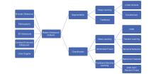
Figure 1
Schematic overview of the review structure. The framework connects the five primary ultrasound modalities (B-mode, Elastography, 3D Ultrasound, CEUS, and Color Doppler) to the downstream tasks of segmentation and classification, categorizing the key technological methodologies applied in recent CAD systems."

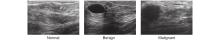
Figure 2
Three breast ultrasound images from the BUSI dataset [10]."
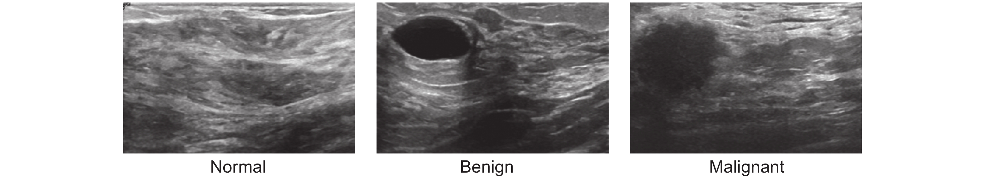
Figure 3
The upper image displays a SWE image indicating increased stiffness in the region surrounding the tumor, and the lower image is the corresponding B-mode ultrasound [11]."

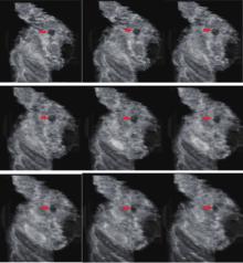
Figure 4
Somo-V generated coronal view of a body mass in 9 consecutive slices. Red arrows indicate the location of the mass [14]."
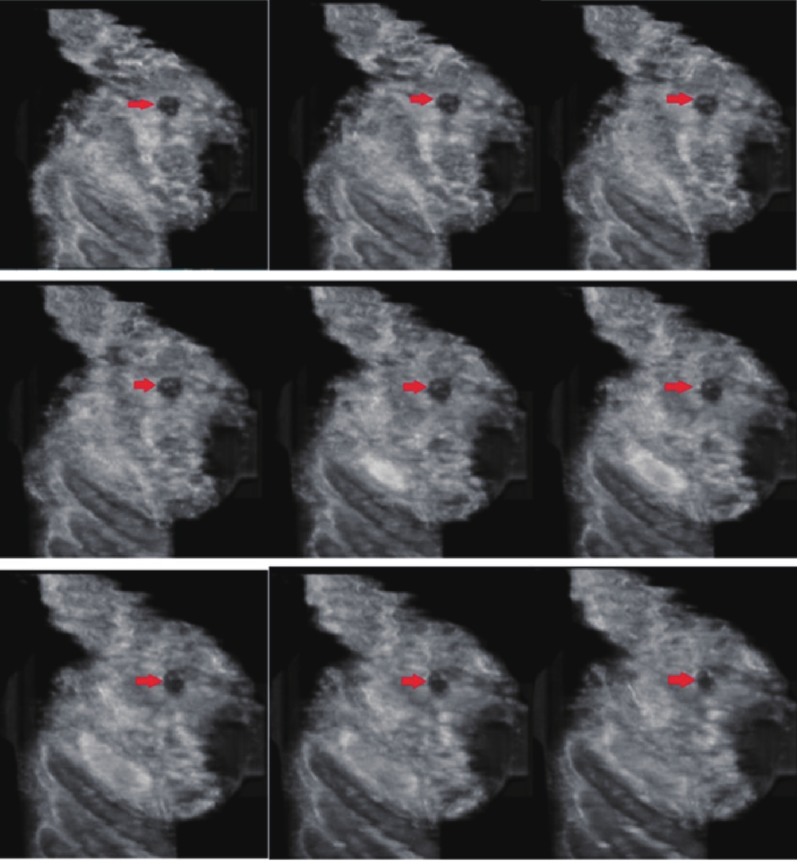
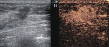
Figure 5
Images of breast tumors. The left image represents the US image and the right image shows the corresponding CEUS image [16]."

Figure 6
Schematic diagram of GG-Net [28]."
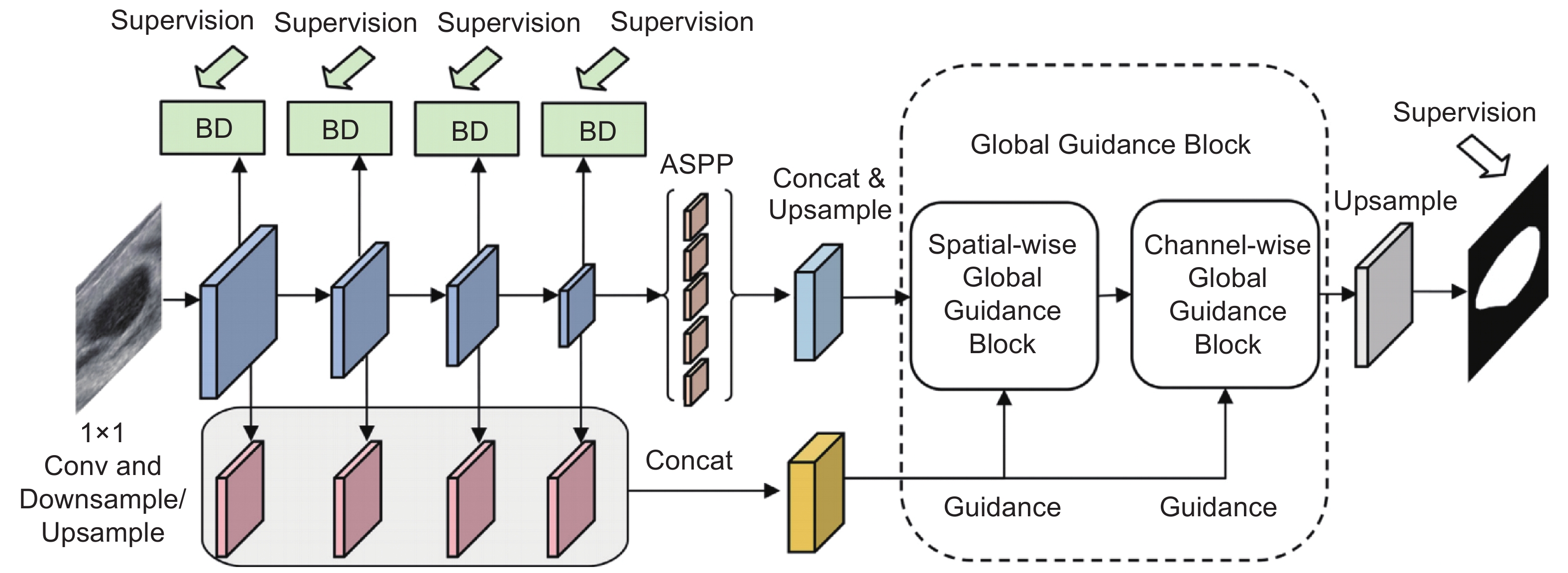
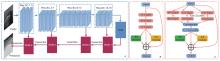
Figure 7
Schematic representation of the framework for the segmentation method utilizing a self-attention mechanism. (A) Network structure; (B) SC module integrated with ResNet; (C) SC module integrated with ResNeXt [36]."
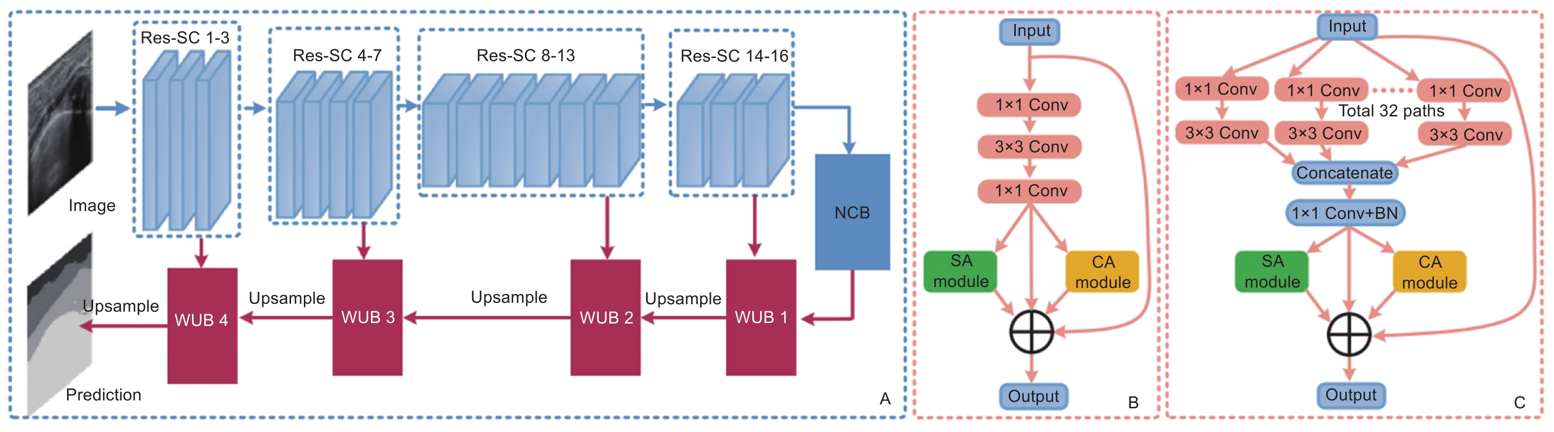
Table 1
Comparison of different breast ultrasound segmentation models."
| Author | Image type | Year | Method | Dataset | Performance (Dice) |
| Xue C et al. [ | B-mode | 2021 | GG-Net | BUSI/Private | 87.10%/82.10% |
| Ning Z et al. [ | B-mode | 2021 | SMU-Net | BUSI/UDIAT | 88.27%/87.03% |
| Chen G et al. [ | B-mode | 2022 | U-net + BAGNet + RFNet | BUSI | 79.35% |
| Zhai D et al. [ | B-mode | 2022 | ASSGAN | DBUI/SPDBUI/ADBUI | 86.90%/93.91%/76.44% |
| Chen G et al. [ | B-mode | 2022 | AAU-net | BUSI/UDIAT | 77.51%/78.14% |
| Huang R et al. [ | B-mode | 2022 | boundary-rendering | UDIAT | 89.40% |
| Li Y et al. [ | B-mode | 2022 | CAM-DLS | Private | 77.30% |
| Cho S W et al. [ | B-mode | 2022 | BTEC-Net + RFS-UNet | BUSI/UDIAT | 84.86%/85.37% |
| Lou M et al. [ | B-mode | 2022 | MCRNet | BUSI/UDIAT | 82.31%/90.05% |
| Iqbal A et al. [ | B-mode | 2022 | MDA-Net | UDIAT/BUSIS | 87.68%/91.85% |
| Wu H et al. [ | B-mode | 2023 | BUSSeg | BUSI/UDIAT | 85.77%/88.11% |
| Chen G et al. [ | B-mode | 2023 | NU-net | BUSI/UDIAT | 78.62%/80.80% |
| Lei B et al. [ | ABUS | 2020 | Self-attention + Res-SC + NCB | Private | 86.60% |
| Lei Y et al. [ | ABUS | 2021 | Mask scoring R-CNN | BUSI/UDIAT | 82.31%/90.05% |
| Meng Z et al. [ | US+CEUS | 2022 | CEUSegNet | Private | US: 91.05%, CEUS: 89.97% |
| Xie X et al. [ | US+CEUS | 2023 | IMAN | Private | US: 83.96%, CEUS: 81.16% |
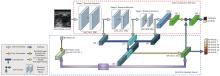
Figure 8
General schematic of the BVA Net. The BVA network consists of two parts, a main architecture (RA net) that applies spatial residual attention, and a BI-RADS attention branch that applies channel smart attention using BI-RADS hierarchy [44]."


Figure 9
Schematic diagram of the architecture of the CEUS video-based diagnostic model by Chen et al. [53]."
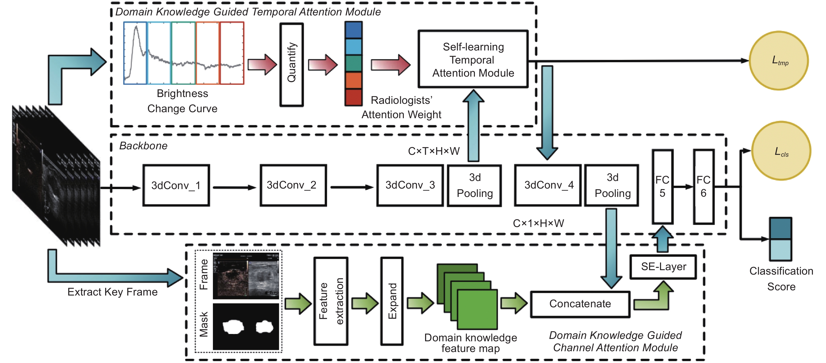
Table 2
Comparison of breast ultrasound classification models."
| Author | Image type | Year | Method | Dataset | Performance |
| Ciritsis A et al. [ | B-mode US | 2019 | dCNN | Private | AUC: 0.838 (Internal)/ 0.967 (External) |
| Al-Dhabyani W et al. [ | B-mode US | 2019 | NASNet + DAGAN | BUSI; UDIAT | ACC: 94% (BUSI); 99% (Combined) |
| Fujioka T et al. [ | SWE | 2020 | DenseNet169 | Private | AUC: 0.898; Sens: 85.7% |
| Li Y et al. [ | B-mode + SWE + CEUS | 2020 | RAB (Radiomics + Attribute Bagging) | Private | ACC: 84.1%; AUC: 0.919 |
| Moon W K et al. [ | B-mode US | 2020 | Ensemble CNN (VGG, ResNet, DenseNet) | Private; BUSI | ACC: 94.6%; (BUSI); AUC: 0.97 |
| Qian X et al. [ | B-mode + Color Doppler | 2020 | CNN | Private | AUC: 0.982; Spec: 88.7% |
| Wang J et al. [ | B-mode + Doppler + SWE + SE | 2020 | ResNet-18 | Private | ACC: 95.4%; Sens: 96.1% |
| Yang Z et al. [ | B-mode + CEUS | 2020 | TSDBN | Private | ACC: 90.2%; F1: 93.2% |
| Chen C et al. [ | CEUS | 2021 | DKG-C3D (3D-CNN + Attention) | Private | ACC: 86.3%; Sens: 97.2% |
| Xing J et al. [ | B-mode US | 2021 | BVA Net (ResNet-50 + Attention) | Private; UDIAT; BUSI | AUC: 0.91 (Private); AUC: 0.89 (BUSI) |
| Zhou Y et al. [ | 3D ABUS | 2021 | Faster R-CNN + Multi-view Analysis | Private | Sens: 95.1% |
| Bourouis S et al. [ | B-mode US | 2022 | GWO-WNN | Public | ACC: 98.0%; Sens: 98.8% |
| Gong X et al. [ | B-mode + CEUS | 2022 | BUS-Net (ResNet/R(2+1)D) | Private | ACC: 89.7%; AUC: 0.93 |
| Hejduk P et al. [ | 3D ABUS | 2022 | dCNN + Sliding Window | Private | ACC: 90.9% |
| Ragab M et al. [ | B-mode US | 2022 | Ensemble DL + CSO-MLP | BUSI | ACC: 97.1%; Spec: 97.7% |
| Shao Y et al. [ | S-WAVE (RF Data) | 2022 | SVM/Random Forest | Private | AUC: 95%; Sens: 95% |
| Xie L et al. [ | US + Peritumoral SWE | 2023 | EfficientNet-B0 | Private | AUC: 0.93; ACC: 91% |
| Yao Z et al. [ | Virtual EUS | 2023 | GAN (U-Net + Discriminator) | Private | V-EUS AUC: 0.75 |
| Atrey K et al. [ | US + Mammogram | 2024 | SVM (Cubic kernel) | Private | ACC: 98.8%; Spec: 99.3% |
| Guo D et al. [ | B-mode + CEUS | 2024 | KAMnet (Knowledge-Augmented) | Private | ACC: 88.2%; Sens: 90.9% |
| [1] |
Huang Q, Huang Y, Luo Y, Yuan F, Li X. Segmentation of breast ultrasound image with semantic classification of superpixels. Medical image analysis 2020; 61: 101657.
doi: 10.1016/j.media.2020.101657 |
| [2] | Sun Y S, Zhao Z, Yang Z N, Xu F, Lu H J, Zhu Z Y, et al. Risk factors and preventions of breast cancer. International journal of biological sciences 2017; 13: 1387. |
| [3] | Zheng R, Sun K, Zhang S, Zeng H, Zou X, Chen R, et al. Analysis of the prevalence of malignant tumors in China in 2015. Chin J Oncol 2019; 41: 19-28. |
| [4] |
Jesneck J L, Lo J Y, Baker J A. Breast mass lesions: computer-aided diagnosis models with mammographic and sonographic descriptors. Radiology 2007; 244: 390-398.
doi: 10.1148/radiol.2442060712 |
| [5] |
Lee J M, Ichikawa L E, Wernli K J, Bowles E, Specht J M, Kerlikowske K, et al. Digital mammography and breast tomosynthesis performance in women with a personal history of breast cancer, 2007–2016. Radiology 2021; 300: 290-300.
doi: 10.1148/radiol.2021204581 |
| [6] |
Houser M, Barreto D, Mehta A, Brem R F. Current and future directions of breast MRI. Journal of Clinical Medicine 2021; 10: 5668.
doi: 10.3390/jcm10235668 |
| [7] |
Comstock C E, Gatsonis C, Newstead G M, Snyder B S, Gareen I F, Bergin J T, et al. Comparison of abbreviated breast MRI vs digital breast tomosynthesis for breast cancer detection among women with dense breasts undergoing screening. Jama 2020; 323: 746-756.
doi: 10.1001/jama.2020.0572 |
| [8] |
Monticciolo D L, Newell M S, Moy L, Niell B, Monsees B, Sickles E A. Breast cancer screening in women at higher-than-average risk: recommendations from the ACR. Journal of the American College of Radiology 2018; 15: 408-414.
doi: 10.1016/j.jacr.2017.11.034 |
| [9] |
Xi X, Shi H, Han L, Wang T, Ding H Y, Zhang G, et al. Breast tumor segmentation with prior knowledge learning. Neurocomputing 2017; 237: 145-157.
doi: 10.1016/j.neucom.2016.09.067 |
| [10] |
Al Dhabyani W, Gomaa M, Khaled H, Fahmy A. Dataset of breast ultrasound images. Data in brief 2020; 28: 104863.
doi: 10.1016/j.dib.2019.104863 |
| [11] |
Zhou J, Zhan W, Chang C, Zhang X, Jia Y, Dong Y, et al. Breast lesions: evaluation with shear wave elastography, with special emphasis on the “stiff rim” sign. Radiology 2014; 272: 63-72.
doi: 10.1148/radiol.14130818 |
| [12] |
Bartolotta T V, Orlando A A M, Dimarco M, Zarcaro C, Ferraro F, Cirino A, et al. Diagnostic performance of 2D-shear wave elastography in the diagnosis of breast cancer: a clinical appraisal of cutoff values. La radiologia medica 2022; 127: 1209-1220.
doi: 10.1007/s11547-022-01546-w |
| [13] |
Rella R, Belli P, Giuliani M, Bufi E, Carlino G, Rinaldi P, et al. Automated breast ultrasonography (ABUS) in the screening and diagnostic setting: indications and practical use. Academic radiology 2018; 25: 1457-1470.
doi: 10.1016/j.acra.2018.02.014 |
| [14] |
Kozegar E, Soryani M, Behnam H, Salamati M, Tan T. Computer aided detection in automated 3-D breast ultrasound images: a survey. Artificial Intelligence Review 2020; 53: 1919-1941.
doi: 10.1007/s10462-019-09722-7 |
| [15] |
Wei X, Li Y, Zhang S, Ming G. Evaluation of thyroid cancer in Chinese females with breast cancer by vascular endothelial growth factor (VEGF), microvessel density, and contrast-enhanced ultrasound (CEUS). Tumor Biology 2014; 35: 6521-6529.
doi: 10.1007/s13277-014-1868-2 |
| [16] |
Li X, Li Y, Zhu Y, Fu L, Liu P. Association between enhancement patterns and parameters of contrast-enhanced ultrasound and microvessel distribution in breast cancer. Oncology letters 2018; 15: 5643-5649.
doi: 10.3892/ol.2018.8078 |
| [17] |
Lang I, Sklair Levy M, Spitzer H. Multi-scale texture-based level-set segmentation of breast B-mode images. Computers in biology and medicine 2016; 72: 30-42.
doi: 10.1016/j.compbiomed.2016.02.017 |
| [18] |
Shi P, Zhong J, Rampun A, Wang H. A hierarchical pipeline for breast boundary segmentation and calcification detection in mammograms. Computers in biology and medicine 2018; 96: 178-188.
doi: 10.1016/j.compbiomed.2018.03.011 |
| [19] |
Yang S C. A robust approach for subject segmentation of medical Images: Illustration with mammograms and breast magnetic resonance images. Computers & Electrical Engineering 2017; 62: 151-165.
doi: 10.1016/j.compeleceng.2016.12.022 |
| [20] |
Ning Z, Zhong S, Feng Q, Chen W, Zhang Y. SMU-Net: Saliency-guided morphology-aware U-Net for breast lesion segmentation in ultrasound image. IEEE transactions on medical imaging 2021; 41: 476-490.
doi: 10.1109/tmi.2021.3116087 |
| [21] |
Chen G, Dai Y, Zhang J. C-Net: Cascaded convolutional neural network with global guidance and refinement residuals for breast ultrasound images segmentation. Computer Methods and Programs in Biomedicine 2022; 225: 107086.
doi: 10.1016/j.cmpb.2022.107086 |
| [22] |
Huang K, Zhang Y, Cheng H D, Xing P, Zhang B. Semantic segmentation of breast ultrasound image with fuzzy deep learning network and breast anatomy constraints. Neurocomputing 2021; 450: 319-335.
doi: 10.1016/j.neucom.2021.04.012 |
| [23] |
Li Y, Liu Y, Huang L, Wang Z, Luo J. Deep weakly-supervised breast tumor segmentation in ultrasound images with explicit anatomical constraints. Medical image analysis 2022; 76: 102315.
doi: 10.1016/j.media.2021.102315 |
| [24] |
Zou H, Gong X, Luo J, Li T. A robust breast ultrasound segmentation method under noisy annotations. Computer Methods and Programs in Biomedicine 2021; 209: 106327.
doi: 10.1016/j.cmpb.2021.106327 |
| [25] |
Zhai D, Hu B, Gong X, Zou H, Luo J. ASS-GAN: Asymmetric semi-supervised GAN for breast ultrasound image segmentation. Neurocomputing 2022; 493: 204-216.
doi: 10.1016/j.neucom.2022.04.021 |
| [26] |
Huang R, Lin M, Dou H, Lin Z, Ying Q, Jia X, et al. Boundary-rendering network for breast lesion segmentation in ultrasound images. Medical image analysis 2022; 80: 102478.
doi: 10.1016/j.media.2022.102478 |
| [27] |
Wu H, Huang X, Guo X, Wen Z, Qin J. Cross-image dependency modeling for breast ultrasound segmentation. IEEE Transactions on Medical Imaging 2023; 42: 1619-1631.
doi: 10.1109/TMI.2022.3233648 |
| [28] |
Xue C, Zhu L, Fu H, Hu X, Li X, Zhang H, et al. Global guidance network for breast lesion segmentation in ultrasound images. Medical image analysis 2021; 70: 101989.
doi: 10.1016/j.media.2021.101989 |
| [29] |
Chen G, Li L, Dai Y, Zhang J, Yap M H. AAU-net: an adaptive attention U-net for breast lesions segmentation in ultrasound images. IEEE Transactions on Medical Imaging 2022; 42: 1289-1300.
doi: 10.1109/tmi.2022.3226268 |
| [30] |
Lou M, Meng J, Qi Y, Li X, Ma Y. MCRNet: Multi-level context refinement network for semantic segmentation in breast ultrasound imaging. Neurocomputing 2022; 470: 154-169.
doi: 10.1016/j.neucom.2021.10.102 |
| [31] |
Iqbal A, Sharif M. MDA-Net: Multiscale dual attention-based network for breast lesion segmentation using ultrasound images. Journal of King Saud University-Computer and Information Sciences 2022; 34: 7283-7299.
doi: 10.1016/j.jksuci.2021.10.002 |
| [32] |
Chen G, Li L, Zhang J, Dai Y. Rethinking the unpretentious U-net for medical ultrasound image segmentation. Pattern Recognition 2023; 142: 109728.
doi: 10.1016/j.patcog.2023.109728 |
| [33] |
Cho S W, Baek N R, Park K R. Deep Learning-based Multi-stage segmentation method using ultrasound images for breast cancer diagnosis. Journal of King Saud University-Computer and Information Sciences 2022; 34: 10273-10292.
doi: 10.1016/j.jksuci.2022.10.020 |
| [34] |
Gare G R, Li J, Joshi R, Magar R, Vaze M P, Yousefpour M, et al. W-Net: Dense and diagnostic semantic segmentation of subcutaneous and breast tissue in ultrasound images by incorporating ultrasound RF waveform data. Medical Image Analysis 2022; 76: 102326.
doi: 10.1016/j.media.2021.102326 |
| [35] |
Lee C Y, Chang T F, Chou Y H, Yang K C. Fully automated lesion segmentation and visualization in automated whole breast ultrasound (ABUS) images. Quantitative imaging in medicine and surgery 2020; 10: 568.
doi: 10.21037/qims.2020.01.12 |
| [36] |
Lei B, Huang S, Li H, Li R, Bian C, Chou Y H, et al. Self-co-attention neural network for anatomy segmentation in whole breast ultrasound. Medical image analysis 2020; 64: 101753.
doi: 10.1016/j.media.2020.101753 |
| [37] |
Zhou Y, Chen H, Li Y, Cao X, Wang S, Shen D. Cross-model attention-guided tumor segmentation for 3D automated breast ultrasound (ABUS) images. IEEE journal of biomedical and health informatics 2021; 26: 301-311.
doi: 10.1109/jbhi.2021.3081111 |
| [38] |
Lei Y, He X, Yao J, Wang T, Wang L, Li W, et al. Breast tumor segmentation in 3D automatic breast ultrasound using Mask scoring R‐CNN. Medical physics 2021; 48: 204-214.
doi: 10.1002/mp.14569 |
| [39] |
Ahmed S, Kamal U, Hasan M K. DSWE-Net: A deep learning approach for shear wave elastography and lesion segmentation using single push acoustic radiation force. Ultrasonics 2021; 110: 106283.
doi: 10.1016/j.ultras.2020.106283 |
| [40] | Meng Z, Zhu Y, Fan X, Tian J, Nie F, Wang K. CEUSegNet: A cross-modality lesion segmentation network for contrast-enhanced ultrasound. 2022 IEEE 19th International Symposium on Biomedical Imaging (ISBI) 2022; IEEE. p. 1-5. |
| [41] | Xie X, Chen C, Liu X, Wang Y, Wang R, Niu J. IMAN: An Iterative Mutual-Aid Network for Breast Lesion Segmentation on Multi-modal Ultrasound Images. 2023 IEEE International Conference on Bioinformatics and Biomedicine (BIBM) 2023; IEEE. p. 3954-3961. |
| [42] | Krithiga R, Geetha P. Breast cancer detection, segmentation and classification on histopathology images analysis: a systematic review. Archives of Computational Methods in Engineering 2021;28. |
| [43] |
Moon W K, Lee Y W, Ke H H, Lee S H, Huang C S, Chang R F. Computer‐aided diagnosis of breast ultrasound images using ensemble learning from convolutional neural networks. Computer methods and programs in biomedicine 2020; 190: 105361.
doi: 10.1016/j.cmpb.2020.105361 |
| [44] |
Xing J, Chen C, Lu Q, Cai X, Yu A, Xu Y, et al. Using BI-RADS stratifications as auxiliary information for breast masses classification in ultrasound images. IEEE Journal of Biomedical and Health Informatics 2020; 25: 2058-2070.
doi: 10.1109/jbhi.2020.3034804 |
| [45] |
Ciritsis A, Rossi C, Eberhard M, Marcon M, Becker A S, Boss A. Automatic classification of ultrasound breast lesions using a deep convolutional neural network mimicking human decision-making. European radiology 2019; 29: 5458-5468.
doi: 10.1007/s00330-019-06118-7 |
| [46] |
Al Dhabyani W, Gomaa M, Khaled H, Aly F. Deep learning approaches for data augmentation and classification of breast masses using ultrasound images. Int J Adv Comput Sci Appl 2019; 10: 1-11.
doi: 10.14569/ijacsa.2019.0100579 |
| [47] |
Bourouis S, Band S S, Mosavi A, Agrawal S, Hamdi M. Meta-heuristic algorithm-tuned neural network for breast cancer diagnosis using ultrasound images. Frontiers in Oncology 2022; 12: 834028.
doi: 10.3389/fonc.2022.834028 |
| [48] |
Ragab M, Albukhari A, Alyami J, Mansour R F. Ensemble deep-learning-enabled clinical decision support system for breast cancer diagnosis and classification on ultrasound images. Biology 2022; 11: 439.
doi: 10.3390/biology11030439 |
| [49] |
Yao Z, Luo T, Dong Y, Jia X, Deng Y, Wu G, et al. Virtual elastography ultrasound via generative adversarial network for breast cancer diagnosis. Nature communications 2023; 14: 788.
doi: 10.1038/s41467-023-36102-1 |
| [50] |
Fujioka T, Katsuta L, Kubota K, Mori M, Kikuchi Y, Kato A, et al. Classification of breast masses on ultrasound shear wave elastography using convolutional neural networks. Ultrasonic imaging 2020; 42: 213-220.
doi: 10.1177/0161734620932609 |
| [51] |
Shao Y, Hashemi H S, Gordon P, Warren L, Wang J, Rohling R, et al. Breast cancer detection using multimodal time series features from ultrasound shear wave absolute vibro-elastography. IEEE journal of biomedical and health informatics 2021; 26: 704-714.
doi: 10.1109/jbhi.2021.3103676 |
| [52] |
Xie L, Liu Z, Pei C, Liu X, Cui Y y, He N a, et al. Convolutional neural network based on automatic segmentation of peritumoral shear-wave elastography images for predicting breast cancer. Frontiers in Oncology 2023; 13: 1099650.
doi: 10.3389/fonc.2023.1099650 |
| [53] |
Chen C, Wang Y, Niu J, Liu X, Li Q, Gong X. Domain knowledge powered deep learning for breast cancer diagnosis based on contrast-enhanced ultrasound videos. IEEE Transactions on Medical Imaging 2021; 40: 2439-2451.
doi: 10.1109/TMI.2021.3078370 |
| [54] |
Yang Z, Gong X, Guo Y, Liu W. A temporal sequence dual-branch network for classifying hybrid ultrasound data of breast cancer. Ieee Access 2020; 8: 82688-82699.
doi: 10.1109/ACCESS.2020.2990683 |
| [55] |
Li Y, Liu Y, Zhang M, Zhang G, Wang Z, Luo J. Radiomics with attribute bagging for breast tumor classification using multimodal ultrasound images. Journal of Ultrasound in Medicine 2020; 39: 361-371.
doi: 10.1002/jum.15115 |
| [56] |
Gong X, Zhao X, Fan L, Li T, Guo Y, Luo J. BUS-net: a bimodal ultrasound network for breast cancer diagnosis. International Journal of Machine Learning and Cybernetics 2022; 13: 3311-3328.
doi: 10.1007/s13042-022-01596-6 |
| [57] |
Guo D, Lu C, Chen D, Yuan J, Duan Q, Xue Z, et al. A multimodal breast cancer diagnosis method based on knowledge-augmented deep learning. Biomedical Signal Processing and Control 2024; 90: 105843.
doi: 10.1016/j.bspc.2023.105843 |
| [58] |
Zhou Y, Chen H, Li Y, Wang S, Cheng L, Li J. 3D multi-view tumor detection in automated whole breast ultrasound using deep convolutional neural network. Expert Systems with Applications 2021; 168: 114410.
doi: 10.1016/j.eswa.2020.114410 |
| [59] |
Hejduk P, Marcon M, Unkelbach J, Ciritsis A, Rossi C, Borkowski K, et al. Fully automatic classification of automated breast ultrasound (ABUS) imaging according to BI-RADS using a deep convolutional neural network. European radiology 2022; 32: 4868-4878.
doi: 10.1007/s00330-022-08558-0 |
| [60] |
Qian X, Zhang B, Liu S, Wang Y, Chen X, Liu J, et al. A combined ultrasonic B-mode and color Doppler system for the classification of breast masses using neural network. European Radiology 2020; 30: 3023-3033.
doi: 10.1007/s00330-019-06610-0 |
| [61] | Wang J, Miao J, Yang X, Li R, Zhou G, Huang Y, et al. Auto-weighting for breast cancer classification in multimodal ultrasound. International Conference on Medical Image Computing and Computer-Assisted Intervention 2020;Springer. p. 190-199. |
| [62] |
Atrey K, Singh B K, Bodhey N K. Multimodal classification of breast cancer using feature level fusion of mammogram and ultrasound images in machine learning paradigm. Multimedia Tools and Applications 2024; 83: 21347-21368.
doi: 10.1007/s11042-023-16414-6 |
| [1] | Yin Yulian, Cheng Yifan, Zhou Liangmei, Zhong Yuanyuan, Wang Bing, Wu Jingjing, Ren Yajuan, Gao Dongwen, Chen Hongfeng, Ye Meina, Yin Haoqiang. From Non-Mass Stage to Complex Type: Ultrasonographic Assessment of Granulomatous Lobular Mastitis Across Six Clinical Stages [J]. Advanced Ultrasound in Diagnosis and Therapy, 2026, 10(1): 59-68. |
| [2] | Zhang Xiaoqian, Zhang Jingwen, Dong Yijie, Zhou Jianqiao. Research Progress and Clinical Translation of Photoacoustic–ultrasound Fusion Imaging in Breast Cancer Diagnosis and Therapy [J]. Advanced Ultrasound in Diagnosis and Therapy, 2025, 9(4): 467-482. |
| [3] | Shi Junni, Xu Jiatong, Chen Chuanjian, Xiang Guanghua, Zheng Wen, Chen Man. Ultrasound Viscoelasticity for Breast Tumor: High Diagnostic Performance at the Peritumoral Boundary [J]. Advanced Ultrasound in Diagnosis and Therapy, 2025, 9(3): 270-276. |
| [4] | An Zichen, Li Fan. Advancements in the Application of Convolutional Neural Networks in Ultrasound Imaging for Breast Cancer Diagnosis and Treatment [J]. Advanced Ultrasound in Diagnosis and Therapy, 2025, 9(1): 21-31. |
| [5] | Zhai Yue, Tan Dianhuan, Lin Xiaona, Lv Heng, Chen Yan, Li Yongbin, Luo Haiyu, Dan Qing, Zhao Chenyang, Xiang Hongjin, Zheng Tingting, Sun Desheng. Ultrasound Radiogenomics-based Prediction Models for Gene Mutation Status in Breast Cancer [J]. Advanced Ultrasound in Diagnosis and Therapy, 2025, 9(1): 10-20. |
| [6] | Raymond Sutjiadi, MS, Siti Sendari, PhD, Heru Wahyu Herwanto, PhD, Yosi Kristian, PhD. Deep Learning for Segmentation and Classification in Mammograms for Breast Cancer Detection: A Systematic Literature Review [J]. Advanced Ultrasound in Diagnosis and Therapy, 2024, 8(3): 94-105. |
| [7] | Leila Bayani, MD, Donya Goodarzi, BS, Reza Mardani, MD, Bita Eslami, PhD, Sadaf Alipour, MD. Localization of Nonpalpable Breast Lumps by Ultrasound Local Coordinates and Skin Inking: A Randomized Controlled Trial [J]. Advanced Ultrasound in Diagnosis and Therapy, 2023, 7(3): 267-271. |
| [8] | Changyan Wang, BS, Haobo Chen, MS, Jieyi Liu, BS, Changchun Li, BS, Weiwei Jiao, BS, Qihui Guo, BS, Qi Zhang, PhD. Deep Learning on Ultrasound Imaging for Breast Cancer Diagnosis and Treatment: Current Applications and Future Perspectives [J]. Advanced Ultrasound in Diagnosis and Therapy, 2023, 7(2): 91-113. |
| [9] | Won-Chul Bang, PhD, Vice President, Yeong Kyeong Seong, PhD, Jinyong Lee. The Impact of Deep Learning on Ultrasound in Diagnosis and Therapy: Enhancing Clinical Decision Support, Workflow Efficiency, Quantification, Image Registration, and Real-time Assistance [J]. Advanced Ultrasound in Diagnosis and Therapy, 2023, 7(2): 204-216. |
| [10] | Cancan Cui, MD, Zhaojun Li, PhD, Yanping Lin, PhD. Advances in Intelligent Segmentation and 3D/4D Reconstruction of Carotid Ultrasound Imaging [J]. Advanced Ultrasound in Diagnosis and Therapy, 2023, 7(2): 140-151. |
| [11] | Rui Chen, MM, Fangqi Guo, MM, Jia Guo, MD, Jiaqi Zhao, MD. Application and Prospect of AI and ABVS-based in Breast Ultrasound Diagnosis [J]. Advanced Ultrasound in Diagnosis and Therapy, 2023, 7(2): 130-135. |
| [12] | Priscilla Machado, MD, Ji-Bin Liu, MD, Flemming Forsberg, PhD. Sentinel Lymph Node Identification Using Contrast Lymphosonography: A Systematic Review [J]. Advanced Ultrasound in Diagnosis and Therapy, 2023, 7(1): 1-7. |
| [13] | Lanting Jia, MD, Jiaqi Zhao, MD, Qi Xu, PhD, Qian Pan, MD, Jianquan Zhang, MD. Quantitative Analysis of Textural Features Extracted from Sonograms of Biceps under Different Physiological States [J]. Advanced Ultrasound in Diagnosis and Therapy, 2020, 4(3): 183-188. |
| [14] | Ping Wang, MM, Junfan Li, Wensheng Yue, MM, Wenyan Li, MM, Yuqun Luo, MM. The Correlation between Traditional Ultrasound Features and the Expression of Estrogen Receptor, Progesterone Receptor, Human Epidermal Growth Factor Receptor-2, and Ki-67 in Breast Carcinoma [J]. Advanced Ultrasound in Diagnosis and Therapy, 2018, 2(3): 173-177. |
| [15] | Ping Wang, MM, Jian Liu, MD, Wensheng Yue, MM, Wenyan Li, MM, Yuqun Luo, MM, Jiao He, MM. The Value of Percutaneous Contrast-Enhanced Ultrasound in the Evaluation of Sentinel Lymph Node of Breast Cancer [J]. Advanced Ultrasound in Diagnosis and Therapy, 2018, 2(2): 113-118. |
| Viewed | ||||||
|
Full text |
|
|||||
|
Abstract |
|
|||||
Share: WeChat
Copyright ©2018 Advanced Ultrasound in Diagnosis and Therapy
|
 Advanced Ultrasound in Diagnosis and Therapy (AUDT)
is licensed under a Creative Commons Attribution 4.0 International License.
Advanced Ultrasound in Diagnosis and Therapy (AUDT)
is licensed under a Creative Commons Attribution 4.0 International License.