
Advanced Ultrasound in Diagnosis and Therapy ›› 2026, Vol. 10 ›› Issue (2): 90-97.doi: 10.26599/AUDT.2026.250061
Previous Articles Next Articles
Cui Yuanjiea, Guo Cuixiaa, Li Zhena, Zhang Juana, Wu Yutinga, Wu Qingqinga, Sun Lijuana,*( )
)
Received:2025-07-17
Revised:2025-10-12
Accepted:2025-12-08
Online:2026-06-30
Published:2026-07-03
Contact:
Department of Ultrasound,Beijing Obstetrics and Gynecology Hospital, Capital Medical University, No.251 Yaojiayuan Road, Chaoyang District, Beijing, China. (Lijuan Sun) e-mail: sunlijuan@ccmu.edu.cn (LJ S),
Cui Yuanjie, Guo Cuixia, Li Zhen, Zhang Juan, Wu Yuting, Wu Qingqing, Sun Lijuan. Use of Artificial Intelligence in Fetal Brain Ultrasound Examination: A review. Advanced Ultrasound in Diagnosis and Therapy, 2026, 10(2): 90-97.
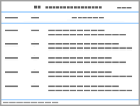
Table 1
Summary of artificial intelligence algorithms for fetal brain standard-plane detection"
| Paper | Model architecture | Plane | Dataset size | Performance metrics (For fetal brain) |
| FASP, fetal abdomen standard plane; FFSP, fetal facial standard plane; FFASP, fetal face axial standard plane; 4CH, four chamber view; FBSP, fetal brain standard plane; FFESP, fetal femur standard plane; CNN, convolutional neural network; 2D, two-dimensional ultrasound; ACC, accuracy; Rec, recall; Sen, sensitivity; Spe, specificity | ||||
| Zhang 2021 | Feature Extraction Network, Region Proposal Network and Class Prediction Network | FASP, FBSP, 4CH | 3280 2D | Prec = 0.95, Sen = 0.92, ACC = 0.95 |
| Qu 2020 | differential-CNN | FBSPs | 19142 2D | Prec = 0.93, Rec = 0.92, ACC = 0.93 |
| Cai 2020 | Temporal SonoEyeNet (TSEN) | FBSP, FASP, FFESP | 280 Videos | Vecsim = 0.98, Pre = 0.89, Rec = 0.85 |
| Kong 2018 | multi-scale dense networks (MSDNet) | FBSP, FASP, 4CH, FFSPs | 22715 2D | Pre = 1.00, Rec = 1.00 |
| Liang 2019 | SPRNet | 4CH, FASP, FBSP, FFASP, coronal FFSP | 22295 2D | ACC = 0.99, Sen = 0.99, Spe = 0.99 |
| Lin 2019 | MF R-CNN | FBSP | 1771 2D | ACC = 0.93, Prec = 0.89 Sen = 0.96 |
Table 2
Summary of artificial intelligence algorithms for biometric measurement of fetal brain"
| Paper | Model architecture | Biometry | Dataset size | Performance metrics |
| DSC, dice similarity coefficient; MSD, maximum symmetric contour distance; MAE, mean absolute error; ATC, average time consumed; RMSE, root-mean-squared error; SF, sylvian fissure; POF, parieto-occipital fissure; CLC, calcarine sulcus; HC, head circumference; BPD, biparietal diameter; AC, abdominal circumference; FL, femur length; TCD, transcerebellar diameter; CM, cisterna magna; Vp, posterior horn of the lateral ventricle; CNN, convolutional neural network; GA, gestation age; 2D, two-dimensional ultrasound; 3D, three-dimensional ultrasound. | ||||
| Foi 2014 | Difference of Gaussians revolved along Elliptical paths | BPD, HC, OFD | 90 2D | DSC = 0.98, MSD = 2.3 mm |
| Sinclair 2018 | Fully Convolutional Networks (FCN) | HC, BPD | HC18 | DSC = 0.98 |
| Zeng 2021 | DAG) V-Net | HC | HC18 | DSC = 0.98, MAE = 1.77 mm |
| Zhang 2022 | Regression CNN Model, CNN Segmentation Model | HC | HC18 | MAE = 1.08 mm (Segmentation) vs. 1.83 mm (Regression) |
| van den Heuvel 2018 | Random Forest Classifier, Hough transform, Dynamic Programming, Ellipse fit | HC | 1334 2D | DSC = 0.97, MAE = 2.8 mm |
| Płotka 2022 | U-Net | BPD, HC, AC, Fl | 750 videos | DSC = 0.96, MAE = 1.04, (HC), 0.27 (BPD) |
| Pluym 2021 | SonoCNS | BPD, HC, TCD, CM, Vp | 143 women | ICC of the automated approach compared to the manual measurements for BPD (0.81), HC (0.88), TCD (0.50), CM (0.23), Vp (0.26) |
| Chen 2020 | Mask R-CNN | lateral ventricles | 2900 2D | MAE = 1.8 mm, ATC = 0.13 s |
| Namburete 2015 | Regression forest | GA | 635 3D | RMSE = 6.10 days, r = 0.98 |
| Wyburd 2021 | CNN | SF, POF, CLC | 811 3D | MAE = 4.1 (SF), 5.1 (POF), 4.9 (CLC) |
Table 3
Summary of artificial intelligence algorithms for recognition of structures and 3D segmentation of fetal brain"
| Paper | Model architecture | Plane/Structure | Dataset size | Performance metrics |
| MSP, midsagittal plane; TT, transthalamic plane; TV, transventricular plane; TC, transcerebellar plane; TFc, transfrontal plane (coronal); TCaudc, transcaudate plane (coronal); TTc, transthalamic plane (coronal); TCc, transcerebellar plane (coronal); PSP, parasagittal plane; 3D, three-dimensional ultrasound; 2D, two-dimensional ultrasound; CSP, cavum septi pellucidi; MCA, middle cerebral artery; HC, head circumference | ||||
| Welp 2020 | 5DCNS+ | MSP, TT, TV, TC, TFc, TCaudc, TTc, TCc, PSP | 1019 3D | 999/1019 (98%) of the volumes, ≥ 8 planes were sufficiently visualized, 958/1019 (94%) all nine planes were sufficiently visualized |
| Wyburd 2020 | U-Net | cortical plate | 307 fetuses | DSC = 0.82 |
| Venturini 2019 | DAG V-Net | HC | 528 3D | DSC= 0.81 (thalamus), 0.82 (brainstem), 0.77 (cerebellum), 0.92 (white matter) |
| Wu 2020 | U-Net | CSP | 448 2D | DSC = 0.77, Pre = 0.79 |
| Wang 2018 | MCANet | MCA | 4005 2D | DSC = 0.77 |
Table 4
Summary of artificial intelligence algorithms for malformation diagnosis"
| Paper | Model architecture | Malformation | Dataset size | Performance metrics |
| IOU, intersection over union; 2D, two-dimensional; ACC, accuracy; Sen, sensitivity; Spe, specificity; DSC, dice similarity coefficient; AUPRC, area under precision-recall curve; AUROC, area under ROC curve | ||||
| Xie 2020 | CNN | Ventriculomegaly, hydrocephalus, Blake pouch cyst, Dandy–Walker malformations, cerebellar vermis hypoplasia | 54990 2D | DSC = 0.942 (craniocerebral region segmentation), average F1-score=0.96 on classification, average mean IOU of 0.497 on lesion localization |
| Xie 2020 | CNN | Binary classification as normal or abnormal | 29419 2D | ACC = 0.96, Sen = 0.97, Spe = 0.96, AUC = 0.99 |
| Lin 2022 | PAICS | Normal and 9 intracranial malformations | 43890 2D, 169 Video | Macro- and microaverage AUC = 0.93 and 0.98 (e internal validation image dataset), 0.90 and 0.90 (e external validation image dataset), 0.97 and 0.98 (real-time scan setting |
| Shirsat 2025 | CNN | Normal and 15 intracranial malformations | 1786 2D | ACC = 0.54 (Standard CNN), 0.63 (Separable CNN), 0.88 (Xception); Prec = 0.54 (Standard CNN), 0.56 (Separable CNN), 0.89 (Xception) |
| Olsen 2024 | DDPM-iNAAD | Intracranial malformations | 14760 2D | AUROC: 0.62 (Gaussian), 0.58 (Simplex), 0.57 (Pyramid) |
| Mykula 2024 | DDPM | Intracranial malformations | 252 2D | AUPRC = 0.79 (AnoDDPM with Simplex) and 0.80 (AutoDDPM with Gaussian) |
| [1] |
Koufos K, EI Haloui K, Dianati M, Higgins M, Elmirghani J, Imran MA, et al. Trends in intelligent communication systems: Review of standards, major research projects, and identification of research gaps. Journal of Sensor and Actuator Networks 2021; 104: 60
doi: 10.3390/jsan10040060 |
| [2] |
He F, Wang Y, Xiu Y, Zhang Y, Chen L. Artificial intelligence in prenatal ultrasound diagnosis. Frontiers in Medicine 2021; 8: 729978
doi: 10.3389/fmed.2021.729978 |
| [3] |
Suzuki K. Overview of deep learning in medical imaging. Radiological Physics and Technology 2017; 10: 257-273.
doi: 10.1007/s12194-017-0406-5 |
| [4] |
Litjens G, Kooi T, Bejnordi BE, Setio AAA, Ciompi F, Ghafoorian M, et al. A survey on deep learning in medical image analysis. Medical Image Analysis 2017; 42: 60-88.
doi: 10.1016/j.media.2017.07.005 |
| [5] | Liu S, Wang Y, Yang X, Lei B, Liu L, Li SX, et al. Deep learning in medical ultrasound analysis: a review. Engineering 2019; 52: 261-275. |
| [6] | Drukker L, Noble J, Papageorghiou A. Introduction to artificial intelligence in ultrasound imaging in obstetrics and gynecology. Ultrasound in Obstetrics & Gynecology 2020; 564: 498-505. |
| [7] | Shazly SA, Trabuco EC, Ngufor CG, Famuyide AO. Introduction to machine learning in obstetrics and gynecology. Obstetrics & Gynecology 2022; 1394: 669-679. |
| [8] | Ramirez Zegarra R, Ghi T. Use of artificial intelligence and deep learning in fetal ultrasound imaging. Ultrasound in Obstetrics & Gynecology 2023; 622: 185-194. |
| [9] |
Sreelakshmy R, Titus A, Sasirekha N, Logashanmugam E, Begam RB, Ramkumar G, et al. [Retracted] An automated deep learning model for the cerebellum segmentation from fetal brain images. BioMed Research International 2022; 20221: 8342767
doi: 10.1155/2023/9852080 |
| [10] |
Yousefpour Shahrivar R, Karami F, Karami E. Enhancing fetal anomaly detection in ultrasonography images: a review of machine learning-based approaches. Biomimetics 2023; 87: 519
doi: 10.3390/biomimetics8070519 |
| [11] | Salomon L, Alfirevic Z, Berghella V, Bilardo C, Chalouhi G, Costa FDS, et al. ISUOG Practice Guidelines updated): performance of the routine mid-trimester fetal ultrasound scan. Ultrasound in Obstetrics and Gynecology 2022; 596: 840-856. |
| [12] | Critchley M, Frederiks J, Vinken PJ, Bruyn G. Handbook of clinical neurology: North-Holland 1978. |
| [13] |
Cater SW, Boyd BK, Ghate SV. Abnormalities of the Fetal Central Nervous System: Prenatal US Diagnosis with Postnatal Correlation. Radiographics 2020; 405: 1458-1472.
doi: 10.1148/rg.2020200034 |
| [14] |
Griffiths PD, Bradburn M, Campbell MJ, Cooper CL, Graham R, Jarvis D, et al. Use of MRI in the diagnosis of fetal brain abnormalities in utero MERIDIAN): a multicentre, prospective cohort study. Lancet 2017; 38910068: 538-546.
doi: 10.1097/ogx.0000000000000454 |
| [15] | van Nisselrooij AEL, Teunissen AKK, Clur SA, Rozendaal L, Pajkrt E, Linskens IH, et al. Why are congenital heart defects being missed?. Ultrasound Obstet Gynecol 2020; 556: 747-757. |
| [16] |
Pinto NM, Keenan HT, Minich LL, Puchalski MD, Heywood M, Botto LD. Barriers to prenatal detection of congenital heart disease: a population-based study. Ultrasound Obstet Gynecol 2012; 404: 418-425.
doi: 10.1002/uog.10116 |
| [17] |
Fadda GM, Capobianco G, Balata A, Litta P, Ambrosini G, D'Antona D, et al. Routine second trimester ultrasound screening for prenatal detection of fetal malformations in Sassari University Hospital, Italy: 23 years of experience in 42,256 pregnancies. Eur J Obstet Gynecol Reprod Biol 2009; 1442: 110-114.
doi: 10.1016/j.ejogrb.2009.02.045 |
| [18] |
Rydberg C, Tunón K. Detection of fetal abnormalities by second‐trimester ultrasound screening in a non‐selected population. Acta obstetricia et gynecologica Scandinavica 2017; 962: 176-182.
doi: 10.1111/aogs.13037 |
| [19] | Zhang Q, Zhang S, Pan Y, Sun L, Li J, Qiao Y, et al. Deep learning to diagnose Hashimoto's thyroiditis from sonographic images. Nat Commun 2022; 131: 3759 |
| [20] | Gao Y, Zeng S, Xu X, Li H, Yao S, Song K, et al. Deep learning-enabled pelvic ultrasound images for accurate diagnosis of ovarian cancer in China: a retrospective, multicentre, diagnostic study. Lancet Digit Health 2022; 43: e179-e187. |
| [21] |
Fiorentino MC, Villani FP, Di Cosmo M, Frontoni E, Moccia S. A review on deep-learning algorithms for fetal ultrasound-image analysis. Med Image Anal 2023; 83: 102629
doi: 10.1016/j.media.2022.102629 |
| [22] |
Guo W, Li S, Yu X, Wen H, Yuan Y, Yang X. Artificial intelligence in prenatal ultrasound: clinical application and prospect. Advanced Ultrasound in Diagnosis & Therapy AUDT 2023; 72: 82-90.
doi: 10.37015/audt.2023.230024 |
| [23] | Malinger G, Paladini D, Haratz KK, Monteagudo A, Pilu GL, Timor-Tritsch IE. ISUOG Practice Guidelines updated: sonographic examination of the fetal central nervous system. Part 1: performance of screening examination and indications for targeted neurosonography. Ultrasound Obstet Gynecol 2020; 563: 476-484. |
| [24] | Paladini D, Malinger G, Birnbaum R, Monteagudo A, Pilu G, Salomon LJ, et al. ISUOG Practice Guidelines updated): sonographic examination of the fetal central nervous system. Part 2: performance of targeted neurosonography. Ultrasound Obstet Gynecol 2021; 574: 661-671. |
| [25] |
Zhang B, Liu H, Luo H, Li K. Automatic quality assessment for 2D fetal sonographic standard plane based on multitask learning. Medicine Baltimore 2021; 1004: e24427
doi: 10.1097/md.0000000000024427 |
| [26] |
Qu R, Xu G, Ding C, Jia W, Sun M. Standard plane identification in fetal brain ultrasound scans using a differential convolutional neural network. IEEE Access 2020; 8: 83821-83830.
doi: 10.1109/ACCESS.2020.2991845 |
| [27] |
Cai Y, Droste R, Sharma H, Chatelain P, Drukker L, Papageorghiou AT, et al. Spatio-temporal visual attention modelling of standard biometry plane-finding navigation. Med Image Anal 2020; 65: 101762
doi: 10.1016/j.media.2020.101762 |
| [28] | Xiao S, Zhang J, Zhu Y, Zhang Z, Cao H, Xie M, et al. Application and progress of artificial intelligence in fetal ultrasound. J Clin Med 2023; 129: 3298 |
| [29] | Liang J, Huang R, Kong P, Li S, Wang T, Lei B. SPRNet: Automatic fetal standard plane recognition network for ultrasound images. International Workshop on Preterm, Perinatal and Paediatric Image Analysis 2019; Proceedings: 38-46. |
| [30] | Kong P, Ni D, Chen S, Li S, Wang T, Lei B. Automatic and efficient standard plane recognition in fetal ultrasound images via multi-scale dense networks. International Workshop on Preterm, Perinatal and Paediatric Image Analysis; 2018; Proceedings: 160-168. |
| [31] |
Lin Z, Li S, Ni D, Liao Y, Wen H, Du J, et al. Multi-task learning for quality assessment of fetal head ultrasound images. Med Image Anal 2019; 58: 101548
doi: 10.1016/j.media.2019.101548 |
| [32] |
Salomon LJ, Alfirevic Z, Da Silva Costa F, Deter RL, Figueras F, Ghi T, et al. ISUOG Practice Guidelines: ultrasound assessment of fetal biometry and growth. Ultrasound Obstet Gynecol 2019; 536: 715-723.
doi: 10.1002/uog.20409 |
| [33] |
Foi A, Maggioni M, Pepe A, Rueda S, Noble JA, Papageorghiou AT, et al. Difference of Gaussians revolved along elliptical paths for ultrasound fetal head segmentation. Comput Med Imaging Graph 2014; 388: 774-784.
doi: 10.1016/j.compmedimag.2014.09.006 |
| [34] | Sinclair M, Baumgartner CF, Matthew J, Bai W, Martinez JC, Li Y, et al. Human-level performance on automatic head biometrics in fetal ultrasound using fully convolutional neural networks. 2018 40th Annual International Conference of the IEEE Engineering In Medicine And Biology Society EMBC 2018;714-717. |
| [35] |
Zeng Y, Tsui PH, Wu W, Zhou Z, Wu S. Fetal ultrasound image segmentation for automatic head circumference biometry using deeply supervised attention-gated v-net. J Digit Imaging 2021; 341: 134-148.
doi: 10.1007/s10278-020-00410-5 |
| [36] | Zhang J, Petitjean C, Ainouz S. Segmentation-based vs. regression-based biomarker estimation: a case study of fetus head circumference assessment from ultrasound images. J Imaging 2022; 8: 23 |
| [37] |
van den Heuvel TLA, de Bruijn D, de Korte CL, Ginneken BV. Automated measurement of fetal head circumference using 2D ultrasound images. PLoS One 2018; 138: e0200412
doi: 10.1371/journal.pone.0200412 |
| [38] | Płotka S, Klasa A, Lisowska A, Seliga-Siwecka J, Lipa M, Trzciński T, et al. Deep learning fetal ultrasound video model match human observers in biometric measurements. Phys Med Biol 2022;67. |
| [39] |
Pluym ID, Afshar Y, Holliman K, Kwan L, Bolagani A, Mok T, et al. Accuracy of automated three-dimensional ultrasound imaging technique for fetal head biometry. Ultrasound Obstet Gynecol 2021; 575: 798-803.
doi: 10.1002/uog.22171 |
| [40] |
Chen X, He M, Dan T, Wang N, Lin M, Zhang L, et al. Automatic measurements of fetal lateral ventricles in 2d ultrasound images using deep learning. Front Neurol 2020; 11: 526
doi: 10.3389/fneur.2020.00526 |
| [41] |
Namburete AI, Stebbing RV, Kemp B, Yaqub M, Papageorghiou AT, Alison Noble J. Learning-based prediction of gestational age from ultrasound images of the fetal brain. Med Image Anal 2015; 211: 72-86.
doi: 10.1016/j.media.2014.12.006 |
| [42] | Wyburd MK, Hesse LS, Aliasi M, Jenkinson M, Papageorghiou AT, Haak MC, et al. Assessment of regional cortical development through fissure based gestational age estimation in 3D fetal ultrasound. International Workshop on Uncertainty for Safe Utilization of Machine Learning in Medical Imaging 2021:12959;242-252. |
| [43] | Yang F, Leung KY, Lee YP, Chan HY, Tang MH. Fetal biometry by an inexperienced operator using two- and three-dimensional ultrasound. Ultrasound Obstet Gynecol 2010; 355: 566-571. |
| [44] |
Welp A, Gembicki M, Rody A, Weichert J. Validation of a semiautomated volumetric approach for fetal neurosonography using 5DCNS+ in clinical data from > 1100 consecutive pregnancies. Childs Nerv Syst 2020; 3612: 2989-2995.
doi: 10.1007/s00381-020-04607-5 |
| [45] |
Gembicki M, Welp A, Scharf JL, Dracopoulos C, Weichert J. A clinical approach to semiautomated three-dimensional fetal brain biometry-comparing the strengths and weaknesses of two diagnostic tools: 5DCNS+TM and SonoCNSTM. J Clin Med 2023; 12: 5334
doi: 10.3390/jcm12165334 |
| [46] |
Weichert J, Scharf JL. Advancements in artificial intelligence for fetal neurosonography: a comprehensive review. J Clin Med 2024; 13: 5626
doi: 10.3390/jcm13185626 |
| [47] |
Wyburd MK, Jenkinson M, Namburete AI. Cortical plate segmentation using CNNs in 3D fetal ultrasound. Annual Conference on Medical Image Understanding and Analysis 2020; 1248: 56-68.
doi: 10.1007/978-3-030-52791-4_5 |
| [48] |
Venturini L, Papageorghiou AT, Noble JA, Namburete AI. Multi-task CNN for structural semantic segmentation in 3D fetal brain ultrasound. Medical Image Understanding and Analysis 2020; 1065: 164-173.
doi: 10.1007/978-3-030-39343-4_14 |
| [49] | Wu Y, Shen K, Chen Z, Wu J, editors. Automatic measurement of fetal cavum septum pellucidum from ultrasound images using deep attention network. 2020 IEEE International Conference on image processing ICIP 2020:2511-2515. |
| [50] | Wang S, Hua Y, Cao Y, Song T, Xue Z, Gong X, et al., editors. Deep learning based fetal middle cerebral artery segmentation in large-scale ultrasound images. 2018 IEEE International Conference on Bioinformatics and Biomedicine BIBM 2018:532-539. |
| [51] | Xie B, Lei T, Wang N, Cai H, Xian J, He M, et al. Computer-aided diagnosis for fetal brain ultrasound images using deep convolutional neural networks. Int J Comput Assist Radiol Surg 2020; 158: 1303-1312. |
| [52] |
Xie HN, Wang N, He M, Zhang LH, Cai HM, Xian JB, et al. Using deep-learning algorithms to classify fetal brain ultrasound images as normal or abnormal. Ultrasound Obstet Gynecol 2020; 564: 579-587.
doi: 10.1002/uog.21967 |
| [53] | Lin M, He X, Guo H, He M, Zhang L, Xian J, et al. Use of real-time artificial intelligence in detection of abnormal image patterns in standard sonographic reference planes in screening for fetal intracranial malformations. Ultrasound Obstet Gynecol 2022; 593: 304-316. |
| [54] |
Lin M, Zhou Q, Lei T, Shang N, Zheng Q, He X, et al. Deep learning system improved detection efficacy of fetal intracranial malformations in a randomized controlled trial. NPJ Digit Med 2023; 61: 191
doi: 10.1038/s41746-023-00932-6 |
| [55] | Shirsat KP, Gawade S, Chopade S, Gujare RV, Bhandwalkar R. Fetal brain anomaly detection via ultrasound imaging using traditional and separable cnns with xception. Journal of Neonatal Surgery 2025; 1422S: 804-813. |
| [56] |
Olsen MDS, Ambsdorf J, Lin M, Taksøe-Vester C, Svendsen MBS, Christensen AN, et al. Unsupervised detection of fetal brain anomalies using denoising diffusion models. International Workshop on Advances in Simplifying Medical Ultrasound 2024; 15186: 209-219.
doi: 10.1007/978-3-031-73647-6_20 |
| [57] | Mykula H, Gasser L, Lobmaier S, Schnabel JA, Zimmer V, Bercea CI. Diffusion models for unsupervised anomaly detection in fetal brain ultrasound. International Workshop on Advances in Simplifying Medical Ultrasound 2024; 15186: 220-230. |
| [58] |
D'Alberti E, Patey O, Smith C, Šalović B, Hernandez-Cruz N, Noble JA, et al. Artificial intelligence-enabled prenatal ultrasound for the detection of fetal cardiac abnormalities: a systematic review and meta-analysis. EClinicalMedicine 2025; 84: 103250
doi: 10.1016/j.eclinm.2025.103250 |
| [59] | Day T, Matthew J, Budd S, Venturini L, Wright R, Farruggia A, et al. Interaction between clinicians and artificial intelligence to detect fetal atrioventricular septal defects on ultrasound: how can we optimize collaborative performance?. Ultrasound in Obstetrics & Gynecology 2024; 641: 28-35. |
| [60] |
Codaccioni C, Arthuis C, Deloison B, Bault JP, Henry C, Mahallati H, et al. Offline ultrasound–MRI fusion imaging for assessment of normal fetal brain development. Ultrasound in Obstetrics & Gynecology 2025; 664: 509-517.
doi: 10.1002/uog.29312 |
| [61] |
Salomon LJ, Bernard J-P, Millischer A-E, Sonigo P, Brunelle F, Boddaert N, et al. MRI and ultrasound fusion imaging for prenatal diagnosis. American Journal of Obstetrics and Gynecology 2013; 2092: 148
doi: 10.1097/01.ruq.0000450896.78339.b9 |
| [1] | Gao Yuanjing, Niu Zihan, Luo Yanwen, Zhou Mengyuan, Xiao Mengsu, Jiang Yuxin, Zhu Qingli. Research Progress on Ultrasound Radiomics in Preoperative Prediction of Axillary Lymph Node Metastasis in Breast Cancer [J]. Advanced Ultrasound in Diagnosis and Therapy, 2026, 10(2): 79-89. |
| [2] | Hu Xuelin, Zhu Ye, Zhang Zisang, Quan Yuanting, Chen Wenwen, Chen Leichong, Xu Guangyu, Qin Luning, Xie Mingxing, Zhang Li. Explainable Artificial Intelligence in Echocardiography [J]. Advanced Ultrasound in Diagnosis and Therapy, 2025, 9(4): 409-425. |
| [3] | Li Yanran, Cui Yuanjie, Wu Qingqing, Zhang Na. Current Applications of Artificial Intelligence in Obstetric Ultrasound [J]. Advanced Ultrasound in Diagnosis and Therapy, 2025, 9(4): 449-456. |
| [4] | Yu Xiao jie, Song Zheng lai, Chang Xue yong, Yu Jie, Liang Ping. Artificial Intelligence in Ultrasound Diagnosis of Liver Nodules: A Comprehensive Review of B-Mode and Contrast-enhanced Applications [J]. Advanced Ultrasound in Diagnosis and Therapy, 2025, 9(4): 326-346. |
| [5] | Zhong Xian, Xie Xiaoyan. Multimodal Ultrasound Radiomics in Liver Disease: Current Status and Future Directions [J]. Advanced Ultrasound in Diagnosis and Therapy, 2025, 9(4): 388-408. |
| [6] | Zhang Xiaoqian, Zhang Jingwen, Dong Yijie, Zhou Jianqiao. Research Progress and Clinical Translation of Photoacoustic–ultrasound Fusion Imaging in Breast Cancer Diagnosis and Therapy [J]. Advanced Ultrasound in Diagnosis and Therapy, 2025, 9(4): 467-482. |
| [7] | Jin Tong, Yu Xiaohu, Ai Zheng, Guo Hongcheng. Artificial Intelligence in Ultrasound Imaging: A Review of Progress from Machine Learning to Large Language Model [J]. Advanced Ultrasound in Diagnosis and Therapy, 2025, 9(4): 483-496. |
| [8] | Zheng Hairong, Meng Long, Li Fei, Niu Lili, Qiu Weibao, Ma Teng, Liu Chengbo, Zhu Xuefeng, Wan Liwen, Cai Feiyan. Advance in Ultrasound Super-resolution Imaging, Cell Manipulation and Inter-brain Communication [J]. Advanced Ultrasound in Diagnosis and Therapy, 2025, 9(4): 307-325. |
| [9] | Elkouahy Fatima Ezzahra, Bennis Ahmed, Merke Nicolas, Ouahid Hajar, Malali Hamid El, Taleb Lhoucine Ben, Mouhsen Azeddine. Advanced Diagnosis of Aortic Stenosis Disease Based on Ultrasound Images: A Novel Artificial Intelligence Approach [J]. Advanced Ultrasound in Diagnosis and Therapy, 2025, 9(3): 298-306. |
| [10] | Xu Jiale, Xia Shujun, Hua Qing, Mei Zihan, Hou Yiqing, Wei Minyan, Lai Limei, Yang Yixuan, Zhou Jianqiao. Performance of ChatGPT and Radiology Residents on Ultrasonography Board-Style Questions [J]. Advanced Ultrasound in Diagnosis and Therapy, 2024, 8(4): 250-254. |
| [11] | Yuhang Zheng, BS, Jianqiao Zhou, MD. Deep Learning in Ultrasound Localization Microscopy [J]. Advanced Ultrasound in Diagnosis and Therapy, 2024, 8(3): 86-92. |
| [12] | Raymond Sutjiadi, MS, Siti Sendari, PhD, Heru Wahyu Herwanto, PhD, Yosi Kristian, PhD. Deep Learning for Segmentation and Classification in Mammograms for Breast Cancer Detection: A Systematic Literature Review [J]. Advanced Ultrasound in Diagnosis and Therapy, 2024, 8(3): 94-105. |
| [13] | Yang Qi, MD, Dengsheng Sun, MD, Linyao Wang, MD, Jie Yu, MD, Ping Liang, MD. State-of-the-Art and Development Trend of Interventional Ultrasound in China [J]. Advanced Ultrasound in Diagnosis and Therapy, 2023, 7(4): 313-320. |
| [14] | Huihui Chai, MS, Xiaowan Bo, MD, Lehang Guo, MD, Chengzhong Peng, MD. Experience and Enlightenment of Handheld Ultrasound Applications in Multiple Scenarios Based on 5G Technology [J]. Advanced Ultrasound in Diagnosis and Therapy, 2023, 7(4): 356-365. |
| [15] | V. Merin Shobi, MCA, MPhil , ME, F. Ramesh Dhanaseelan, MSc, MTech , PhD. Review on Image Inpainting using Intelligence Mining Techniques [J]. Advanced Ultrasound in Diagnosis and Therapy, 2023, 7(4): 366-372. |
| Viewed | ||||||
|
Full text |
|
|||||
|
Abstract |
|
|||||
Share: WeChat
Copyright ©2018 Advanced Ultrasound in Diagnosis and Therapy
|
 Advanced Ultrasound in Diagnosis and Therapy (AUDT)
is licensed under a Creative Commons Attribution 4.0 International License.
Advanced Ultrasound in Diagnosis and Therapy (AUDT)
is licensed under a Creative Commons Attribution 4.0 International License.
