
Advanced Ultrasound in Diagnosis and Therapy ›› 2026, Vol. 10 ›› Issue (4): 277-297.doi: 10.26599/AUDT.2026.260041
• REVIEW ARTICLE • Previous Articles Next Articles
Li Moa,b,c, Jin Letiana,b,c, Xue Ged, Chen Jiyeb,c, Benariba Mohamed Aimenee, Wang Vicky Yangb,f,g, Teng Gaojunc,e,h, Xu Donga,b,c,e,f,g,*( )
Received:2026-09-18
Revised:2026-10-17
Accepted:2026-11-21
Online:2026-12-30
Published:2026-11-02
Contact:
Xu Dong,
E-mail:xudong@zjcc.org.cn
Li Mo, Jin Letian, Xue Ge, Chen Jiye, Benariba Mohamed Aimene, Wang Vicky Yang, Teng Gaojun, Xu Dong. Empowering Breast Cancer Precision Diagnosis with Advanced Ultrasound: From Radiomics to Radio-multiomics. Advanced Ultrasound in Diagnosis and Therapy, 2026, 10(4): 277-297.
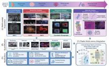
Figure 1
From multi-modal ultrasound acquisition to radio-multiomics in breast cancer. Four stages are linked across the organ (~cm), tissue (~mm), microvascular (~μm), and molecular (subcellular) scales. (A) Multi-modal acquisition covers structural imaging (B-mode, ABUS), blood flow imaging (CDFI, PDI, spectral Doppler, SMI, CEUS, SRUS), mechanical imaging (SE, SWE, viscoelastic elastography), and molecular biology (genome, transcriptome, digital pathology and IHC, spatial transcriptome, proteins); (B) Quantification yields morphological and radiomic features, hemodynamic and perfusion metrics, microvascular density, diameter and tortuosity, stiffness and viscoelasticity, and RF/QUS parameters; (C) AI extracts and fuses multi-modal, multi-scale image features that link imaging phenotypes to molecular subtypes and immune cell infiltration; (D) The model serves six clinical applications: screening, diagnosis, molecular and pathological typing, precision treatment planning, treatment response prediction, and personalized care. Abbreviations: ABUS, automated breast ultrasound; AI, artificial intelligence; B-mode, grayscale ultrasound; CDFI, color Doppler flow imaging; CEUS, contrast-enhanced ultrasound; ER, estrogen receptor; HER2, human epidermal growth factor receptor 2; IHC, immunohistochemistry; miRNA, microRNA; mRNA, messenger RNA; PDI, power Doppler imaging; PR, progesterone receptor; QUS, quantitative ultrasound; RF, radiofrequency; SE, strain elastography; SMI, superb microvascular imaging; SRUS, super-resolution ultrasound; SWE, shear wave elastography."

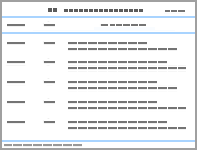
Table 1
Summary of representative AI/CAD models for breast ultrasound diagnosis"
| Author | Year | Country | Model and architecture | Study design | Centers | Input | No. of cases | Age (years) | Task | AUC | External‑ validation status |
| AI, artificial intelligence; AUC, area under the curve; BI-RADS, Breast Imaging Reporting and Data System; B-mode, grayscale ultrasound; BU-CAD, breast ultrasound computer-aided diagnosis; CAD, computer-aided diagnosis; CDFI, color Doppler flow imaging; DCNN, deep convolutional neural network; DL, deep learning; NR, not reported; SWE, shear wave elastography. ※Commercially available CAD system. | |||||||||||
| Wei et al. [ | 2022 | China | S-Detect (deep-learning CAD)※ | Prospective | 9 | B-mode | 901 | 45.2 ± 10.8 | Differentiation of benign and malignant breast masses | 0.906 | YES |
| Gu et al. [ | 2023 | China | DL-BI-RADS (RepVGG) | Prospective | 32 | B-mode | 5,012 | 44.1 ± 13.1 | Benign/malignant classification and 6-category BI-RADS | 0.980 | YES |
| Shen et al. [ | 2021 | United States | Breast ultrasound AI (attention mechanism) | Retrospective | 20 | B-mode + Color Doppler | 143,203 | 53.7 ± 13.7 | Breast cancer detection and triage of low-risk examinations | 0.976 | YES |
| Mango et al. [ | 2020 | United States | Koios DS※ | Retrospective | > 20 | B-mode | 900 | 53.6 | Lesion risk stratification and management recommendations | 0.88 | YES |
| Lai et al. [ | 2022 | Taiwan, China | BU-CAD (deep-learning CAD)※ | Retrospective | 1 | B-mode | 172 | 54.1 ± 12.0 | Lesion detection and diagnosis | 0.859 | NO |
| Chen et al. [ | 2025 | China | AINet | Retrospective | 7 | B-mode + SWE | 15,223 | N/A | Benign/malignant classification and BI-RADS grading | 0.942 | NO |
| Cai et al. [ | 2025 | International | AI-SWE (EfficientNetB1) | Retrospective | 12 | SWE | 1,294 | 46.5 ± 16.3 | Benign/malignant stratification of BI-RADS 3/4 lesions | 0.94/ 0.93 | YES |
| Qian et al. [ | 2024 | China | BMU-Net (ResNet-18 + Transformer) | Prospective | ≥ 3 | MMG + US (three-modal) + clinical | 5,025 | 48.8 | Coarse-/fine-grained breast cancer risk stratification | 0.948 | YES |
| Jiang et al. [ | 2021 | China | ResNet50-DCNN | Retrospective | 3 | B-mode | 2,120 | 49 | Breast cancer molecular subtype classification | 0.87/ 0.83 | YES |
| Zhou et al. [ | 2020 | China | Inception V3 | Retrospective | 2 | B-mode | 834 | 48 | Prediction of axillary lymph node metastasis | 0.89 | YES |
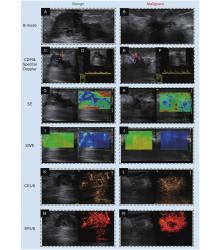
Figure 2
Comparison of multimodal ultrasound imaging features between benign and malignant breast lesions. (A, B) B-mode ultrasound; (C, E) color Doppler flow imaging; (D, F) spectral Doppler; (G, H) strain elastography; (I, J) shear wave elastography; (K, L) contrast-enhanced ultrasound; and (M, N) super-resolution ultrasound. The left column (A, C, D, G, I, K, M) shows the benign lesion, whereas the right column (B, E, F, H, J, L, N) shows the malignant lesion."

| [1] | Anderer S. Who releases global status report on cancer. Jama 2026; 336: 540 |
| [2] |
Kim J, Harper A, Mccormack V, Sung H, Houssami N, Morgan E, et al. Global patterns and trends in breast cancer incidence and mortality across 185 countries. Nat Med 2025; 31: 1154-1162.
doi: 10.1038/s41591-025-03502-3 |
| [3] |
Giuliano A E, Edge S B, Hortobagyi G N. Eighth edition of the ajcc cancer staging manual: breast cancer. Ann Surg Oncol 2018; 25: 1783-1785.
doi: 10.1245/s10434-018-6486-6 |
| [4] |
Guo L, Kong D, Liu J, Zhan L, Luo L, Zheng W, et al. Breast cancer heterogeneity and its implication in personalized precision therapy. Exp Hematol Oncol 2023; 12: 3.
doi: 10.1186/s40164-022-00363-1 |
| [5] |
Weinstein S P, Slanetz P J, Lewin A A, Battaglia T, Chagpar A B, Dayaratna S, et al. Acr appropriateness criteria® supplemental breast cancer screening based on breast density. J Am Coll Radiol 2021; 18: S456-s473.
doi: 10.1016/j.jacr.2021.09.002 |
| [6] |
Aristokli N, Polycarpou I, Themistocleous S C, Sophocleous D, Mamais I. Comparison of the diagnostic performance of magnetic resonance imaging (mri), ultrasound and mammography for detection of breast cancer based on tumor type, breast density and patient's history: a review. Radiography (Lond) 2022; 28: 848-856.
doi: 10.1016/j.radi.2022.01.006 |
| [7] |
Evans A, Trimboli R M, Athanasiou A, Balleyguier C, Baltzer P A, Bick U, et al. Breast ultrasound: recommendations for information to women and referring physicians by the european society of breast imaging. Insights Imaging 2018; 9: 449-461.
doi: 10.1007/s13244-018-0636-z |
| [8] |
Llinas-Bertran A, Butjosa-Espín M, Barberi V, Seoane J A. Multimodal data integration in early-stage breast cancer. Breast 2025; 80: 103892.
doi: 10.1016/j.breast.2025.103892 |
| [9] |
Kaplan S S. Clinical utility of bilateral whole-breast us in the evaluation of women with dense breast tissue. Radiology 2001; 221: 641-649.
doi: 10.1148/radiol.2213010364 |
| [10] |
Jacobson J A, Middleton W D, Allison S J, Dahiya N, Lee K S, Levine B D, et al. Ultrasonography of superficial soft-tissue masses: society of radiologists in ultrasound consensus conference statement. Radiology 2022; 304: 18-30.
doi: 10.1148/radiol.211101 |
| [11] |
Song H, Shu R, Uematsu T, Moon W K. Bi-rads v2025 for breast ultrasound: key updates and asian perspectives. Ultrasonography 2026; 45: 311-324.
doi: 10.14366/usg.26107 |
| [12] | Stanzani D, Chala L F, Barros N, Cerri G G, Chammas M C. Can doppler or contrast-enhanced ultrasound analysis add diagnostically important information about the nature of breast lesions?. Clinics (Sao Paulo) 2014; 69: 87-92. |
| [13] |
Watanabe T, Yamaguchi T, Okuno T, Konno S, Takaki R, Sato M, et al. Utility of b-mode, color doppler and elastography in the diagnosis of breast cancer: results of the cd-confirm multicenter study of 1351 breast solid masses. Ultrasound Med Biol 2021; 47: 3111-3121.
doi: 10.1016/j.ultrasmedbio.2021.07.009 |
| [14] |
Park A Y, Seo B K, Han M R. Breast ultrasound microvascular imaging and radiogenomics. Korean J Radiol 2021; 22: 677-687.
doi: 10.3348/kjr.2020.1166 |
| [15] |
Zhu J Y, He H L, Jiang X C, Bao H W, Chen F. Multimodal ultrasound features of breast cancers: correlation with molecular subtypes. BMC Med Imaging 2023; 23: 57.
doi: 10.1186/s12880-023-00999-3 |
| [16] |
Li M, Li Q, Yin Q, Wang Y, Shang J M, Wang L H. Evaluation of color doppler ultrasound combined with plasma mir-21 and mir-27a in the diagnosis of breast cancer. Clin Transl Oncol 2021; 23: 709-717.
doi: 10.1007/s12094-020-02501-9 |
| [17] |
Mehdikhani B, Benam M, Moradkhani A, Roostaee A, Bahman S S, Barmayoon P, et al. Evaluation of diagnostic value of doppler ultrasound in the diagnosis of malignant breast masses. Eur J Transl Myol 2024; 34: 12372.
doi: 10.4081/ejtm.2024.12372 |
| [18] |
Keshavarz E, Karimi M A, Valian N, Kalantari M, Valian K. Diagnostic value of spectral doppler ultrasound in detecting breast malignancies: an original article. Int J Cancer Manag 2018; 11: e8200.
doi: 10.5812/ijcm.8200 |
| [19] |
Zhang Q, Liang X, Zhang Y, Nie H, Chen Z. A review of contrast-enhanced ultrasound using sonovue® and sonazoid™ in non-hepatic organs. Eur J Radiol 2023; 167: 111060.
doi: 10.1016/j.ejrad.2023.111060 |
| [20] |
Janu E, Krikavova L, Little J, Dvorak K, Brancikova D, Jandakova E, et al. Prospective evaluation of contrast-enhanced ultrasound of breast bi-rads 3-5 lesions. BMC Med Imaging 2020; 20: 66.
doi: 10.1186/s12880-020-00467-2 |
| [21] |
Park A Y, Kwon M, Woo O H, Cho K R, Park E K, Cha S H, et al. A prospective study on the value of ultrasound microflow assessment to distinguish malignant from benign solid breast masses: association between ultrasound parameters and histologic microvessel densities. Korean J Radiol 2019; 20: 759-772.
doi: 10.3348/kjr.2018.0515 |
| [22] |
Chen C, Turco S, Kapetas P, Mann R, Wijkstra H, De Korte C, et al. Spatiotemporal analysis of contrast-enhanced ultrasound for differentiating between malignant and benign breast lesions. Eur Radiol 2024; 34: 4764-4773.
doi: 10.1007/s00330-023-10500-x |
| [23] |
Wang B, Yang D, Zhang X, Gong X, Xu T, Han J, et al. The diagnostic value of contrast-enhanced ultrasonography in breast ductal abnormalities. Cancer Imaging 2023; 23: 25.
doi: 10.1186/s40644-023-00539-w |
| [24] |
Li J K, Jin Z Y, Xu Y J, Fu N Q, Jiang Y, Li S Y, et al. Contrast-enhanced ultrasound to predict malignant upgrading of atypical ductal hyperplasia. Breast Cancer Res 2024; 26: 27.
doi: 10.1186/s13058-024-01772-2 |
| [25] |
Zhang X, Cheng F, Song X, Wang P, Tian S, Zhao X, et al. Superb microvascular imaging for evaluation of microvascularity in breast nodules compared with conventional doppler imaging. Quant Imaging Med Surg 2023; 13: 7029-7040.
doi: 10.21037/qims-23-136 |
| [26] |
Zhou F, Lin W, Yao J, Lu X, Yu L. Enhanced differentiation of breast lesions through integration of microvascular flow imaging and machine learning algorithms. Front Oncol 2026; 16: 1827050.
doi: 10.3389/fonc.2026.1827050 |
| [27] |
Lee E J, Chang Y W, Oh E, Hwang J, Kim H J, Hong S S. Reproducibility and diagnostic performance of the vascular index of superb microvascular imaging in real-time breast ultrasonography for evaluating breast masses. Ultrasonography 2021; 40: 398-406.
doi: 10.14366/usg.20153 |
| [28] |
Lee E J, Chang Y W. Prediction of complete response after neoadjuvant chemotherapy for invasive breast cancers: the utility of shear wave elastography and superb microvascular imaging in pretreatment breast ultrasound. Eur J Radiol 2024; 175: 111432.
doi: 10.1016/j.ejrad.2024.111432 |
| [29] |
Cai S, Wang H, Zhang X, Zhang L, Zhu Q, Sun Q, et al. Superb microvascular imaging technology can improve the diagnostic efficiency of the bi-rads system. Front Oncol 2021; 11: 634752.
doi: 10.3389/fonc.2021.634752 |
| [30] |
Zuo Y, Zhan Y, Zhou J, Xia H, Li T, Zhou F, et al. Prediction of pathological complete response to neoadjuvant chemotherapy for invasive breast cancers based on longitudinal ultrasound and superb microvascular imaging: a single-center retrospective study. PeerJ 2025; 13: e20171.
doi: 10.7717/peerj.20171 |
| [31] |
Zhang X Y, Cai S M, Zhang L, Zhu Q L, Sun Q, Jiang Y X, et al. Association between vascular index measured via superb microvascular imaging and molecular subtype of breast cancer. Front Oncol 2022; 12: 861151.
doi: 10.3389/fonc.2022.861151 |
| [32] |
Zhao G, Kong D, Xu X, Hu S, Li Z, Tian J. Deep learning-based classification of breast lesions using dynamic ultrasound video. Eur J Radiol 2023; 165: 110885.
doi: 10.1016/j.ejrad.2023.110885 |
| [33] |
Dencks S, Lowerison M, Hansen-Shearer J, Shin Y, Schmitz G, Song P, et al. Super-resolution ultrasound: from data acquisition and motion correction to localization, tracking, and evaluation. IEEE Trans Ultrason Ferroelectr Freq Control 2025; 72: 408-426.
doi: 10.1109/TUFFC.2025.3543322 |
| [34] |
Xia S, Zheng Y, Hua Q, Wen J, Luo X, Yan J, et al. Super-resolution ultrasound and microvasculomics: a consensus statement. Eur Radiol 2024; 34: 7503-7513.
doi: 10.1007/s00330-024-10796-3 |
| [35] |
Liu F, Xia S, Hua Q, Yuan C, Zheng Y, Tao R, et al. Evaluation of tibial nerve microcirculation in diabetes mellitus at superresolution us. Radiology 2026; 318: e250347.
doi: 10.1148/radiol.250347 |
| [36] |
Opacic T, Dencks S, Theek B, Piepenbrock M, Ackermann D, Rix A, et al. Motion model ultrasound localization microscopy for preclinical and clinical multiparametric tumor characterization. Nat Commun 2018; 9: 1527.
doi: 10.1038/s41467-018-03973-8 |
| [37] |
Hou X, Li Z, Liu Y, Gao J, Song T. Diagnostic value of super-resolution ultrasound imaging in differentiating benign and malignant bi-rads-4 breast lesions. Front Oncol 2025; 15: 1662492.
doi: 10.3389/fonc.2025.1662492 |
| [38] |
Hua Q, Xia S, Yuan C, Li Y, Zheng Y, Tao R, et al. The super resolution ultrasound in visualizing interstitial-lymphatic network in breast tumors: a clinical pilot application. Acad Radiol 2026; 33: 2899-2910.
doi: 10.1016/j.acra.2026.03.030 |
| [39] |
Li J, Wei C, Ying T, Liu Y, Wang R, Li M, et al. Differentiation of benign and malignant breast lesions by ultrasound localization microscopy. Insights Imaging 2025; 16: 128.
doi: 10.1186/s13244-025-02013-6 |
| [40] |
Xia S, Hua Q, Song Y, Yuan C, Zheng Y, Tao R, et al. Super-resolution ultrasound imaging of intranodal lymphatic sinuses for predicting sentinel lymph node metastasis in breast cancer: a preliminary study. Eur Radiol 2025; 35: 6079-6088.
doi: 10.1007/s00330-025-11520-5 |
| [41] |
Zhang M, Zhang B. Extracellular matrix stiffness: mechanisms in tumor progression and therapeutic potential in cancer. Exp Hematol Oncol 2025; 14: 54.
doi: 10.1186/s40164-025-00647-2 |
| [42] |
Kanagaraju V, Dhivya B, Devanand B, Maheswaran V. Utility of ultrasound strain elastography to differentiate benign from malignant lesions of the breast. J Med Ultrasound 2021; 29: 89-93.
doi: 10.4103/JMU.JMU_32_20 |
| [43] |
Barr R G, Nakashima K, Amy D, Cosgrove D, Farrokh A, Schafer F, et al. Wfumb guidelines and recommendations for clinical use of ultrasound elastography: part 2: breast. Ultrasound Med Biol 2015; 41: 1148-1160.
doi: 10.1016/j.ultrasmedbio.2015.03.008 |
| [44] |
Xu J, Zhang L, Wen W, He Y, Wei T, Zheng Y, et al. Evaluation of standard breast ultrasonography by adding two-dimensional and three-dimensional shear wave elastography: a prospective, multicenter trial. Eur Radiol 2024; 34: 945-956.
doi: 10.1007/s00330-023-10287-x |
| [45] |
Zhao Q Y, Zhao B, Wu G. Predictive value of shear wave elastography and molecular subtypes for postoperative efficacy in patients with early breast cancer. Ann Med 2025; 57: 2544879.
doi: 10.1080/07853890.2025.2544879 |
| [46] |
Huang J X, Lin S Y, Ou Y, Shi C G, Zhong Y, Wei M J, et al. Combining conventional ultrasound and sonoelastography to predict axillary status after neoadjuvant chemotherapy for breast cancer. Eur Radiol 2022; 32: 5986-5996.
doi: 10.1007/s00330-022-08751-1 |
| [47] | Tekcan Sanli D E, Yildirim D, Kandemirli S G, Sanli A N, Aribal E. Evaluation of multiparametric shear wave elastography indices in malignant and benign breast lesions. Acad Radiol 2022; 29 Suppl 1 S50-s61. |
| [48] |
Barr R G, Engel A, Kim S, Tran P, De Silvestri A. Improved breast 2d swe algorithm to eliminate false-negative cases. Invest Radiol 2023; 58: 703-709.
doi: 10.1097/RLI.0000000000000972 |
| [49] |
Osika M, Kijanka P. Ultrasound shear wave propagation modeling in general tissue-like viscoelastic materials. Ultrasound Med Biol 2024; 50: 627-638.
doi: 10.1016/j.ultrasmedbio.2024.01.008 |
| [50] |
Kumar V, Denis M, Gregory A, Bayat M, Mehrmohammadi M, Fazzio R, et al. Viscoelastic parameters as discriminators of breast masses: initial human study results. PLoS One 2018; 13: e0205717.
doi: 10.1371/journal.pone.0205717 |
| [51] |
Hossain M M, Gallippi C M. Viscoelastic response ultrasound derived relative elasticity and relative viscosity reflect true elasticity and viscosity: in silico and experimental demonstration. IEEE Trans Ultrason Ferroelectr Freq Control 2020; 67: 1102-1117.
doi: 10.1109/TUFFC.2019.2962789 |
| [52] | Woo E, Lee J Y, Seo B K, Bae M S, Park E K, Choi H, et al. Diagnostic performance of shear-wave dispersion imaging and shear-wave elastography for differentiating benign and malignant breast masses. Eur Radiol 2026; 36: 3378-3391. |
| [53] |
Jia W, Xia S, Jia X, Tang B, Cheng S, Nie M, et al. Ultrasound viscosity imaging in breast lesions: a multicenter prospective study. Acad Radiol 2024; 31: 3499-3510.
doi: 10.1016/j.acra.2024.03.017 |
| [54] |
Bae M S, Kim H Y, Oh H, Seo B K. Clinical applications of shear wave dispersion imaging for breast lesions: a pictorial essay. Ultrasonography 2023; 42: 589-599.
doi: 10.14366/usg.23079 |
| [55] |
Yang Z, Li X. A combined model of ultrasound viscoelasticity and inflammatory indices for differentiating benign and malignant breast lesions. BMC Med Imaging 2026; 26: 96.
doi: 10.1186/s12880-025-02117-x |
| [56] |
Tao J, Niu Z, Zhou M, Zhao J, Xiao M, Zhu Q. Viscoelastic mechanical properties assessed by ultrasound correlates with tumor proliferation in invasive ductal breast cancer: a preliminary study. Ultrasound Med Biol 2026; 52: 984-990.
doi: 10.1016/j.ultrasmedbio.2026.01.016 |
| [57] |
Xu J, Shi J, Huang Y, Chen C, Xiang G, Zheng W, et al. Diagnostic value of ultrasound peritumoral viscoelasticity parameters in breast cancer: enhancing bi-rads classification performance. BMC Med Imaging 2026; 26: 95.
doi: 10.1186/s12880-025-02144-8 |
| [58] |
Xiao X Y, Chen X, Guan X F, Wu H, Qin W, Luo B M. Superb microvascular imaging in diagnosis of breast lesions: a comparative study with contrast-enhanced ultrasonographic microvascular imaging. Br J Radiol 2016; 89: 20160546.
doi: 10.1259/bjr.20160546 |
| [59] |
Luo J, Li T, Liu Y. Diagnostic value of the vascular index measured by superb microvascular imaging for evaluating breast tumors: a meta-analysis. Med Ultrason 2023; 25: 415-422.
doi: 10.11152/mu-4124 |
| [60] |
Lee E J, Chang Y W. Combination of quantitative parameters of shear wave elastography and superb microvascular imaging to evaluate breast masses. Korean J Radiol 2020; 21: 1045-1054.
doi: 10.3348/kjr.2019.0765 |
| [61] |
Kurt S A, Kayadibi Y, Saracoglu M S, Ozturk T, Korkmazer B, Cerit M, et al. Prediction of molecular subtypes using superb microvascular imaging and shear wave elastography in invasive breast carcinomas. Acad Radiol 2023; 30: 14-21.
doi: 10.1016/j.acra.2022.04.017 |
| [62] |
Cui X W, Li K N, Yi A J, Wang B, Wei Q, Wu G G, et al. Ultrasound elastography. Endosc Ultrasound 2022; 11: 252-274.
doi: 10.4103/EUS-D-21-00151 |
| [63] | Golatta M, Pfob A, Büsch C, Bruckner T, Alwafai Z, Balleyguier C, et al. The potential of combined shear wave and strain elastography to reduce unnecessary biopsies in breast cancer diagnostics - an international, multicentre trial. Eur J Cancer 2022; 161: 1-9. |
| [64] |
Li J, Sun B, Li Y, Li S, Wang J, Zhu Y, et al. Correlation analysis between shear-wave elastography and pathological profiles in breast cancer. Breast Cancer Res Treat 2023; 197: 269-276.
doi: 10.1007/s10549-022-06804-z |
| [65] |
Bai L, You C, Zhou J, Xie L, Zhu X, Chang C, et al. Quantitative analysis of shear wave elastography and us-guided diffuse optical tomography for evaluating biological characteristics of breast cancer. Acad Radiol 2024; 31: 3489-3498.
doi: 10.1016/j.acra.2024.03.006 |
| [66] |
Liu H C, Lee H K, Urban M W, Zhou Q, Kijanka P. Acoustic radiation force-induced longitudinal shear wave for ultrasound-based viscoelastic evaluation. Ultrasonics 2024; 142: 107389.
doi: 10.1016/j.ultras.2024.107389 |
| [67] |
Liu Z, Liu W, Chen Q, Hu Y, Li Y, Zheng X, et al. Real-time nondestructive viscosity measurement of soft tissue based on viscoelastic response optical coherence elastography. Materials (Basel) 2023; 16: 6019.
doi: 10.3390/ma16176019 |
| [68] |
Zhang J, He Q, Xiao Y, Zheng H, Wang C, Luo J. Ultrasound image reconstruction from plane wave radio-frequency data by self-supervised deep neural network. Med Image Anal 2021; 70: 102018.
doi: 10.1016/j.media.2021.102018 |
| [69] |
Ouyang Y, Tsui P H, Wu S, Wu W, Zhou Z. Classification of benign and malignant breast tumors using h-scan ultrasound imaging. Diagnostics (Basel) 2019; 9: 182.
doi: 10.3390/diagnostics9040182 |
| [70] |
Wang Q, Jia X, Luo T, Yu J, Xia S. Deep learning algorithm using bispectrum analysis energy feature maps based on ultrasound radiofrequency signals to detect breast cancer. Front Oncol 2023; 13: 1272427.
doi: 10.3389/fonc.2023.1272427 |
| [71] |
Baek J, O'connell A M, Parker K J. Improving breast cancer diagnosis by incorporating raw ultrasound parameters into machine learning. Mach Learn Sci Technol 2022; 3: 045013.
doi: 10.1088/2632-2153/ac9bcc |
| [72] |
Xie Z, Sun Q, Han J, Sun P, Hu X, Ji N, et al. Spectral analysis enhanced net (sae-net) to classify breast lesions with bi-rads category 4 or higher. Ultrasonics 2024; 143: 107406.
doi: 10.1016/j.ultras.2024.107406 |
| [73] |
Sheibani-Asl N, Osapoetra L O, Czarnota G J, Sadeghi-Naini A. Ai-enabled rf data synthesis for breast ultrasound: efficacy in quantitative ultrasound tissue characterization. Sci Rep 2026; 16: 12544.
doi: 10.1038/s41598-026-43319-9 |
| [74] |
Wei Q, Yan Y J, Wu G G, Ye X R, Jiang F, Liu J, et al. The diagnostic performance of ultrasound computer-aided diagnosis system for distinguishing breast masses: a prospective multicenter study. Eur Radiol 2022; 32: 4046-4055.
doi: 10.1007/s00330-021-08452-1 |
| [75] | Gu Y, Xu W, Liu T, An X, Tian J, Ran H, et al. Ultrasound-based deep learning in the establishment of a breast lesion risk stratification system: a multicenter study. Eur Radiol 2023; 33: 2954-2964. |
| [76] |
Shen Y, Shamout F E, Oliver J R, Witowski J, Kannan K, Park J, et al. Artificial intelligence system reduces false-positive findings in the interpretation of breast ultrasound exams. Nat Commun 2021; 12: 5645.
doi: 10.1038/s41467-021-26023-2 |
| [77] |
Mango V L, Sun M, Wynn R T, Ha R. Should we ignore, follow, or biopsy? impact of artificial intelligence decision support on breast ultrasound lesion assessment. AJR Am J Roentgenol 2020; 214: 1445-1452.
doi: 10.2214/AJR.19.21872 |
| [78] |
Lai Y C, Chen H H, Hsu J F, Hong Y J, Chiu T T, Chiou H J. Evaluation of physician performance using a concurrent-read artificial intelligence system to support breast ultrasound interpretation. Breast 2022; 65: 124-135.
doi: 10.1016/j.breast.2022.07.009 |
| [79] |
Chen H, Li Y, Zhang J, Yang L, Sun Y, Chen Y, et al. An alignment and imputation network (ainet) for breast cancer diagnosis with multimodal multi-view ultrasound images. IEEE Trans Med Imaging 2026; 45: 1383-1394.
doi: 10.1109/TMI.2025.3625254 |
| [80] |
Cai L, Pfob A, Barr R G, Duda V, Alwafai Z, Balleyguier C, et al. Deep learning model for breast shear wave elastography to improve breast cancer diagnosis (inspired 006): An international, multicenter analysis. J Clin Oncol 2025; 43: 3482-3493.
doi: 10.1200/JCO-24-02681 |
| [81] | Qian X, Pei J, Han C, Liang Z, Zhang G, Chen N, et al. A multimodal machine learning model for the stratification of breast cancer risk. Nat Biomed Eng 2025; 9: 356-370. |
| [82] |
Jiang M, Zhang D, Tang S C, Luo X M, Chuan Z R, Lv W Z, et al. Deep learning with convolutional neural network in the assessment of breast cancer molecular subtypes based on us images: a multicenter retrospective study. Eur Radiol 2021; 31: 3673-3682.
doi: 10.1007/s00330-020-07544-8 |
| [83] |
Zhou L Q, Wu X L, Huang S Y, Wu G G, Ye H R, Wei Q, et al. Lymph node metastasis prediction from primary breast cancer us images using deep learning. Radiology 2020; 294: 19-28.
doi: 10.1148/radiol.2019190372 |
| [84] |
Nicholson W K, Silverstein M, Wong J B, Barry M J, Chelmow D, Coker T R, et al. Screening for breast cancer: us preventive services task force recommendation statement. Jama 2024; 331: 1918-1930.
doi: 10.1001/jama.2024.5534 |
| [85] |
Marcon M, Fuchsjäger M H, Clauser P, Mann R M. Esr essentials: Screening for breast cancer - general recommendations by eusobi. Eur Radiol 2024; 34: 6348-6357.
doi: 10.1007/s00330-024-10740-5 |
| [86] |
Ding R, Xiao Y, Mo M, Zheng Y, Jiang Y Z, Shao Z M. Breast cancer screening and early diagnosis in chinese women. Cancer Biol Med 2022; 19: 450-467.
doi: 10.20892/j.issn.2095-3941.2021.0676 |
| [87] |
Wang J, Zheng S, Ding L, Liang X, Wang Y, Greuter M J W, et al. Is ultrasound an accurate alternative for mammography in breast cancer screening in an asian population? a meta-analysis. Diagnostics (Basel) 2020; 10: 985.
doi: 10.3390/diagnostics10110985 |
| [88] |
Uematsu T, Izumori A, Moon W K. Overcoming the limitations of screening mammography in japan and korea: a paradigm shift to personalized breast cancer screening based on ultrasonography. Ultrasonography 2023; 42: 508-517.
doi: 10.14366/usg.23047 |
| [89] |
Payne N R, Hickman S E, Black R, Priest A N, Hudson S, Gilbert F J. Breast density effect on the sensitivity of digital screening mammography in a uk cohort. Eur Radiol 2025; 35: 177-187.
doi: 10.1007/s00330-024-10951-w |
| [90] |
Harada-Shoji N, Suzuki A, Ishida T, Zheng Y F, Narikawa-Shiono Y, Sato-Tadano A, et al. Evaluation of adjunctive ultrasonography for breast cancer detection among women aged 40-49 years with varying breast density undergoing screening mammography: a secondary analysis of a randomized clinical trial. JAMA Netw Open 2021; 4: e2121505.
doi: 10.1001/jamanetworkopen.2021.21505 |
| [91] |
Güldoğan N, Ulus S, Kovan Ö, Aksan A, Tokmakçıoğlu K, Camgöz Akdağ H, et al. Evaluating efficiency of time use and operational costs in a breast clinic workflow: a comparative analysis between automated breast ultrasound and handheld ultrasound. Eur J Breast Health 2023; 19: 311-317.
doi: 10.4274/ejbh.galenos.2023.2023-8-4 |
| [92] |
Klein Wolterink F, Ab Mumin N, Appelman L, Derks-Rekers M, Imhof-Tas M, Lardenoije S, et al. Diagnostic performance of 3d automated breast ultrasound (3d-abus) in a clinical screening setting-a retrospective study. Eur Radiol 2024; 34: 5451-5460.
doi: 10.1007/s00330-023-10568-5 |
| [93] |
Gatta G, Cappabianca S, La Forgia D, Massafra R, Fanizzi A, Cuccurullo V, et al. Second-generation 3d automated breast ultrasonography (prone abus) for dense breast cancer screening integrated to mammography: effectiveness, performance and detection rates. J Pers Med 2021; 11: 875.
doi: 10.3390/jpm11090875 |
| [94] |
Park C K S, Trumpour T, Aziz A, Bax J S, Tessier D, Gardi L, et al. Cost-effective, portable, patient-dedicated three-dimensional automated breast ultrasound for point-of-care breast cancer screening. Sci Rep 2023; 13: 14390.
doi: 10.1038/s41598-023-41424-7 |
| [95] |
Shen Y, He J, Liu M, Hu J, Wan Y, Zhang T, et al. Diagnostic value of contrast-enhanced ultrasound and shear-wave elastography for small breast nodules. PeerJ 2024; 12: e17677.
doi: 10.7717/peerj.17677 |
| [96] |
Li C, Wu M, Haiqing H. The application of multimodal ultrasound examination in the differential diagnosis of benign and malignant breast lesions of bi-rads category 4. Front Med (Lausanne) 2025; 12: 1596100.
doi: 10.3389/fmed.2025.1596100 |
| [97] |
Wang X, He Y, Wang L. Diagnostic value of shear wave elastography combined with super microvascular imaging for bi-rads 3-5 nodules. Front Oncol 2023; 13: 1192630.
doi: 10.3389/fonc.2023.1192630 |
| [98] |
Yang Z, Lyu Y, Yang Y, Xu C, Zhang K, Yang X, et al. Application of multimodal ultrasound combined with bi-rads in classification and diagnosis of non-mass breast lesions. J Clin Ultrasound 2026; 54: 609-617.
doi: 10.1002/jcu.70115 |
| [99] |
Niu Z, Gao Y, Xiao M, Mao F, Zhou Y, Zhu Q, et al. Contrast-enhanced lymphatic us can improve the preoperative diagnostic performance for sentinel lymph nodes in early breast cancer. Eur Radiol 2023; 33: 1593-1602.
doi: 10.1007/s00330-022-09139-x |
| [100] |
Zhuang L, Ming X, Liu J, Jia C, Jin Y, Wang J, et al. Comparison of lymphatic contrast-enhanced ultrasound and intravenous contrast-enhanced ultrasound in the preoperative diagnosis of axillary sentinel lymph node metastasis in patients with breast cancer. Br J Radiol 2022; 95: 20210897.
doi: 10.1259/bjr.20210897 |
| [101] |
Li S Y, Li Y M, Fang Y Q, Jin Z Y, Li J K, Zou X M, et al. Contrast-enhanced ultrasound radiomics model for predicting axillary lymph node metastasis and prognosis in breast cancer: a multicenter study. BMC Cancer 2025; 25: 1315.
doi: 10.1186/s12885-025-14632-9 |
| [102] |
Huang D, Shi Y, Cao W, Feng L, Luo Y, Zhao X, et al. Exempting axillary staging surgery in breast cancer using multimodal ultrasound imaging and radiomics of sentinel lymph nodes. EClinicalMedicine 2026; 92: 103782.
doi: 10.1016/j.eclinm.2026.103782 |
| [103] |
Xu S, Wang Q, Hong Z. The correlation between multi-mode ultrasonographic features of breast cancer and axillary lymph node metastasis. Front Oncol 2024; 14: 1433872.
doi: 10.3389/fonc.2024.1433872 |
| [104] |
Jiang B, Wu Y, Chen X, Jian C, Wang W. Artificial intelligence and multi-omics convergence in breast cancer: revolutionizing diagnosis, prognostication, and precision oncology. Crit Rev Oncol Hematol 2026; 220: 105160.
doi: 10.1016/j.critrevonc.2026.105160 |
| [105] |
Qi Y J, Su G H, You C, Zhang X, Xiao Y, Jiang Y Z, et al. Radiomics in breast cancer: current advances and future directions. Cell Rep Med 2024; 5: 101719.
doi: 10.1016/j.xcrm.2024.101719 |
| [106] |
Xi J, Sun D, Chang C, Zhou S, Huang Q. An omics-to-omics joint knowledge association subtensor model for radiogenomics cross-modal modules from genomics and ultrasonic images of breast cancers. Comput Biol Med 2023; 155: 106672.
doi: 10.1016/j.compbiomed.2023.106672 |
| [107] |
Peng X, Zhou X, Feng X, Fang N, Dong X, Hong W, et al. Artificial intelligence for triple-negative breast cancer from imaging to multi-omics. Front Oncol 2026; 16: 1834128.
doi: 10.3389/fonc.2026.1834128 |
| [108] |
Ran D, Li J, Zhao M, Du L, Zhang Y, Zhu J. Artificial intelligence integrates multi-omics data for precision stratification and drug resistance prediction in breast cancer. Front Oncol 2025; 15: 1612474.
doi: 10.3389/fonc.2025.1612474 |
| [109] |
Wu X, Dai W. Beyond imaging: integrating radiomics, genomics, and multi-omics for precision breast cancer management. Cancers (Basel) 2025; 17: 3408.
doi: 10.3390/cancers17213408 |
| [110] |
Yang Y, Long H, Feng Y, Tian S, Chen H, Zhou P. A multi-omics method for breast cancer diagnosis based on metabolites in exhaled breath, ultrasound imaging, and basic clinical information. Heliyon 2024; 10: e32115.
doi: 10.1016/j.heliyon.2024.e32115 |
| [111] |
Jiang L, You C, Xiao Y, Wang H, Su G H, Xia B Q, et al. Radiogenomic analysis reveals tumor heterogeneity of triple-negative breast cancer. Cell Rep Med 2022; 3: 100694.
doi: 10.1016/j.xcrm.2022.100694 |
| [112] |
Su G H, Xiao Y, You C, Zheng R C, Zhao S, Sun S Y, et al. Radiogenomic-based multiomic analysis reveals imaging intratumor heterogeneity phenotypes and therapeutic targets. Sci Adv 2023; 9: eadf0837.
doi: 10.1126/sciadv.adf0837 |
| [113] |
Guan J, Fan M, Li L. Mvnmf: Multiview nonnegative matrix factorization for radio-multigenomic analysis in breast cancer prognosis. Med Image Anal 2025; 103: 103566.
doi: 10.1016/j.media.2025.103566 |
| [114] |
Huang S C, Pareek A, Seyyedi S, Banerjee I, Lungren M P. Fusion of medical imaging and electronic health records using deep learning: a systematic review and implementation guidelines. NPJ Digit Med 2020; 3: 136.
doi: 10.1038/s41746-020-00341-z |
| [115] |
Picard M, Scott-Boyer M P, Bodein A, Périn O, Droit A. Integration strategies of multi-omics data for machine learning analysis. Comput Struct Biotechnol J 2021; 19: 3735-3746.
doi: 10.1016/j.csbj.2021.06.030 |
| [116] |
Chicco D, Cumbo F, Angione C. Ten quick tips for avoiding pitfalls in multi-omics data integration analyses. PLoS Comput Biol 2023; 19: e1011224.
doi: 10.1371/journal.pcbi.1011224 |
| [117] | Tao W, Jiang Q, Shan J, Yang Z, Zhang Y, Lu H, et al. A multimodal deep learning framework to predict breast cancer sentinel lymph node metastasis in ultrasound images with metabolomics interpretation. Current Genomics 2026. |
| [118] |
Liu J, Li Y, Tang W, Qian T, Dai L, Jia Z, et al. Cell-free DNA fragmentomics assay to discriminate the malignancy of breast nodules and evaluate treatment response. Genomics Proteomics Bioinformatics 2025; 23: qzaf028.
doi: 10.1093/gpbjnl/qzaf028 |
| [119] | Cui C, Yang H, Wang Y, Zhao S, Asad Z, Coburn L A, et al. Deep multimodal fusion of image and non-image data in disease diagnosis and prognosis: a review. Prog Biomed Eng (Bristol) 2023;5. |
| [120] |
Omran M M, Emam M, Gamaleldin M, Abushady A M, Elattar M A, El-Hadidi M. Comparative analysis of statistical and deep learning-based multi-omics integration for breast cancer subtype classification. J Transl Med 2025; 23: 709.
doi: 10.1186/s12967-025-06662-5 |
| [121] |
Tong L, Mitchel J, Chatlin K, Wang M D. Deep learning based feature-level integration of multi-omics data for breast cancer patients survival analysis. BMC Med Inform Decis Mak 2020; 20: 225.
doi: 10.1186/s12911-020-01225-8 |
| [1] | Cao Jiaying, Li Wenying, Qiu Yijie, Wei Li, Wang Ying, Huang Yunlin, Dong Yi, Dietrich Christoph F. Decoding Hepatocellular Carcinoma Heterogeneity with Multimodal Ultrasound and Acoustic Radiomics [J]. Advanced Ultrasound in Diagnosis and Therapy, 2026, 10(4): 334-345. |
| [2] | Cui Yuanjie, Guo Cuixia, Li Zhen, Zhang Juan, Wu Yuting, Wu Qingqing, Sun Lijuan. Use of Artificial Intelligence in Fetal Brain Ultrasound Examination: A review [J]. Advanced Ultrasound in Diagnosis and Therapy, 2026, 10(2): 90-97. |
| [3] | Fu Lijia, Li Na, Liao Ziling, Lin Yanping, Li Zhaojun, Li Fan. Advances in Breast Ultrasound Segmentation and Classification [J]. Advanced Ultrasound in Diagnosis and Therapy, 2026, 10(1): 29-41. |
| [4] | Hu Xuelin, Zhu Ye, Zhang Zisang, Quan Yuanting, Chen Wenwen, Chen Leichong, Xu Guangyu, Qin Luning, Xie Mingxing, Zhang Li. Explainable Artificial Intelligence in Echocardiography [J]. Advanced Ultrasound in Diagnosis and Therapy, 2025, 9(4): 409-425. |
| [5] | Li Yanran, Cui Yuanjie, Wu Qingqing, Zhang Na. Current Applications of Artificial Intelligence in Obstetric Ultrasound [J]. Advanced Ultrasound in Diagnosis and Therapy, 2025, 9(4): 449-456. |
| [6] | Yu Xiao jie, Song Zheng lai, Chang Xue yong, Yu Jie, Liang Ping. Artificial Intelligence in Ultrasound Diagnosis of Liver Nodules: A Comprehensive Review of B-Mode and Contrast-enhanced Applications [J]. Advanced Ultrasound in Diagnosis and Therapy, 2025, 9(4): 326-346. |
| [7] | Zhang Xiaoqian, Zhang Jingwen, Dong Yijie, Zhou Jianqiao. Research Progress and Clinical Translation of Photoacoustic–ultrasound Fusion Imaging in Breast Cancer Diagnosis and Therapy [J]. Advanced Ultrasound in Diagnosis and Therapy, 2025, 9(4): 467-482. |
| [8] | Jin Tong, Yu Xiaohu, Ai Zheng, Guo Hongcheng. Artificial Intelligence in Ultrasound Imaging: A Review of Progress from Machine Learning to Large Language Model [J]. Advanced Ultrasound in Diagnosis and Therapy, 2025, 9(4): 483-496. |
| [9] | Zheng Hairong, Meng Long, Li Fei, Niu Lili, Qiu Weibao, Ma Teng, Liu Chengbo, Zhu Xuefeng, Wan Liwen, Cai Feiyan. Advance in Ultrasound Super-resolution Imaging, Cell Manipulation and Inter-brain Communication [J]. Advanced Ultrasound in Diagnosis and Therapy, 2025, 9(4): 307-325. |
| [10] | Shi Junni, Xu Jiatong, Chen Chuanjian, Xiang Guanghua, Zheng Wen, Chen Man. Ultrasound Viscoelasticity for Breast Tumor: High Diagnostic Performance at the Peritumoral Boundary [J]. Advanced Ultrasound in Diagnosis and Therapy, 2025, 9(3): 270-276. |
| [11] | Elkouahy Fatima Ezzahra, Bennis Ahmed, Merke Nicolas, Ouahid Hajar, Malali Hamid El, Taleb Lhoucine Ben, Mouhsen Azeddine. Advanced Diagnosis of Aortic Stenosis Disease Based on Ultrasound Images: A Novel Artificial Intelligence Approach [J]. Advanced Ultrasound in Diagnosis and Therapy, 2025, 9(3): 298-306. |
| [12] | Zhai Yue, Tan Dianhuan, Lin Xiaona, Lv Heng, Chen Yan, Li Yongbin, Luo Haiyu, Dan Qing, Zhao Chenyang, Xiang Hongjin, Zheng Tingting, Sun Desheng. Ultrasound Radiogenomics-based Prediction Models for Gene Mutation Status in Breast Cancer [J]. Advanced Ultrasound in Diagnosis and Therapy, 2025, 9(1): 10-20. |
| [13] | An Zichen, Li Fan. Advancements in the Application of Convolutional Neural Networks in Ultrasound Imaging for Breast Cancer Diagnosis and Treatment [J]. Advanced Ultrasound in Diagnosis and Therapy, 2025, 9(1): 21-31. |
| [14] | Xu Jiale, Xia Shujun, Hua Qing, Mei Zihan, Hou Yiqing, Wei Minyan, Lai Limei, Yang Yixuan, Zhou Jianqiao. Performance of ChatGPT and Radiology Residents on Ultrasonography Board-Style Questions [J]. Advanced Ultrasound in Diagnosis and Therapy, 2024, 8(4): 250-254. |
| [15] | Raymond Sutjiadi, MS, Siti Sendari, PhD, Heru Wahyu Herwanto, PhD, Yosi Kristian, PhD. Deep Learning for Segmentation and Classification in Mammograms for Breast Cancer Detection: A Systematic Literature Review [J]. Advanced Ultrasound in Diagnosis and Therapy, 2024, 8(3): 94-105. |
| Viewed | ||||||
|
Full text |
|
|||||
|
Abstract |
|
|||||
Share: WeChat
Copyright ©2018 Advanced Ultrasound in Diagnosis and Therapy
|
 Advanced Ultrasound in Diagnosis and Therapy (AUDT)
is licensed under a Creative Commons Attribution 4.0 International License.
