Advanced Ultrasound in Diagnosis and Therapy ›› 2026, Vol. 10 ›› Issue (4): 334-345.doi: 10.26599/AUDT.2026.260028
• REVIEW ARTICLE • Previous Articles Next Articles
Cao Jiayinga, Li Wenyinga, Qiu Yijiea, Wei Lia, Wang Yinga, Huang Yunlina, Dong Yia,*(
), Dietrich Christoph Fa,b
Received:2026-08-10
Revised:2026-09-10
Accepted:2026-12-06
Online:2026-12-30
Published:2026-11-02
Contact:
Dong Yi,
E-mail:drdaisydong@hotmail.com
Cao Jiaying, Li Wenying, Qiu Yijie, Wei Li, Wang Ying, Huang Yunlin, Dong Yi, Dietrich Christoph F. Decoding Hepatocellular Carcinoma Heterogeneity with Multimodal Ultrasound and Acoustic Radiomics. Advanced Ultrasound in Diagnosis and Therapy, 2026, 10(4): 334-345.
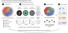
Figure 1
Conceptual framework for decoding intratumoral heterogeneity of hepatocellular carcinoma (HCC) using multimodal ultrasound. HCC is depicted as a spatially heterogeneous biological mosaic composed of distinct tumor habitats. Four complementary acoustic dimensions are interrogated by multimodal ultrasound: tumor perfusion using contrast-enhanced ultrasound (CEUS), biomechanical properties using shear-wave elastography (SWE), tissue microstructure using quantitative ultrasound and radiofrequency (QUS/RF) analysis, and vascular architecture using superb microvascular imaging (SMI) or ultrasound localization microscopy (ULM). Integration of these complementary imaging biomarkers through acoustic radiomics and artificial intelligence enables the generation of a multimodal acoustic heterogeneity signature, with potential applications in noninvasive prediction of microvascular invasion (MVI), histological grade, recurrence risk, treatment response, and longitudinal monitoring for precision management of HCC."
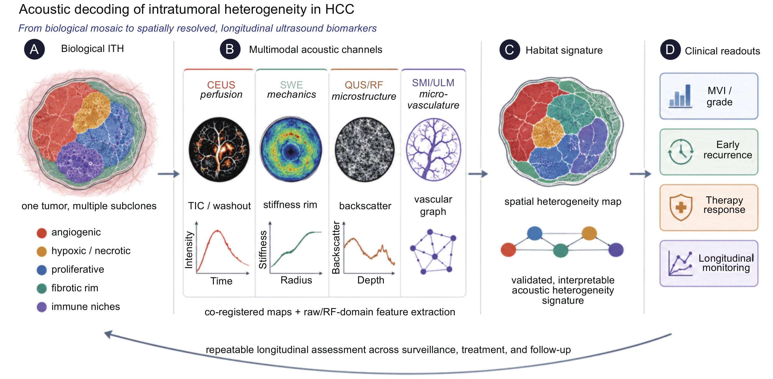
Figure 2
Whole-tumor versus habitat analysis for imaging intratumoral heterogeneity. (A) Conventional whole-tumor radiomics treats the lesion as a single region of interest, averaging quantitative imaging features across the entire tumor and thereby potentially obscuring spatial biological heterogeneity; (B) Habitat analysis preserves spatial information by clustering multiparametric imaging data into distinct imaging habitats that may correspond to biologically different tumor regions, including hyperperfused, transitional, hypoperfused, and necrotic compartments; (C) Conceptual extension to multimodal ultrasound, in which contrast-enhanced ultrasound (CEUS), shear-wave elastography (SWE), quantitative ultrasound/radiofrequency (QUS/RF) analysis, and superb microvascular imaging or ultrasound localization microscopy (SMI/ULM) are spatially integrated to generate region-specific acoustic habitats and a comprehensive multimodal acoustic heterogeneity signature for biological phenotyping of hepatocellular carcinoma. Schematic illustration."
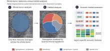
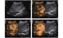
Figure 3
Representative contrast-enhanced ultrasound (CEUS) findings in hepatocellular carcinoma (HCC). A 72-year-old man with HCC confirmed by surgical histopathology (Edmondson–Steiner Grade II). (A) Grayscale ultrasound with color Doppler imaging demonstrates a heterogeneous hyperechoic liver lesion with peripheral vascular signals; (B, C) Following intravenous administration of 2.0 mL SonoVue®, the lesion exhibits heterogeneous arterial-phase hyperenhancement with a characteristic mosaic enhancement pattern; (D) Time-intensity curve analysis demonstrates peak enhancement at 23 s. Washout begins at approximately 45 s, followed by persistent hypoenhancement during the portal venous and late phases, consistent with the typical CEUS enhancement pattern of HCC. This case illustrates the marked intratumoral perfusion heterogeneity that can be visualized by CEUS and forms the basis for quantitative perfusion analysis and acoustic radiomics."
Table 1
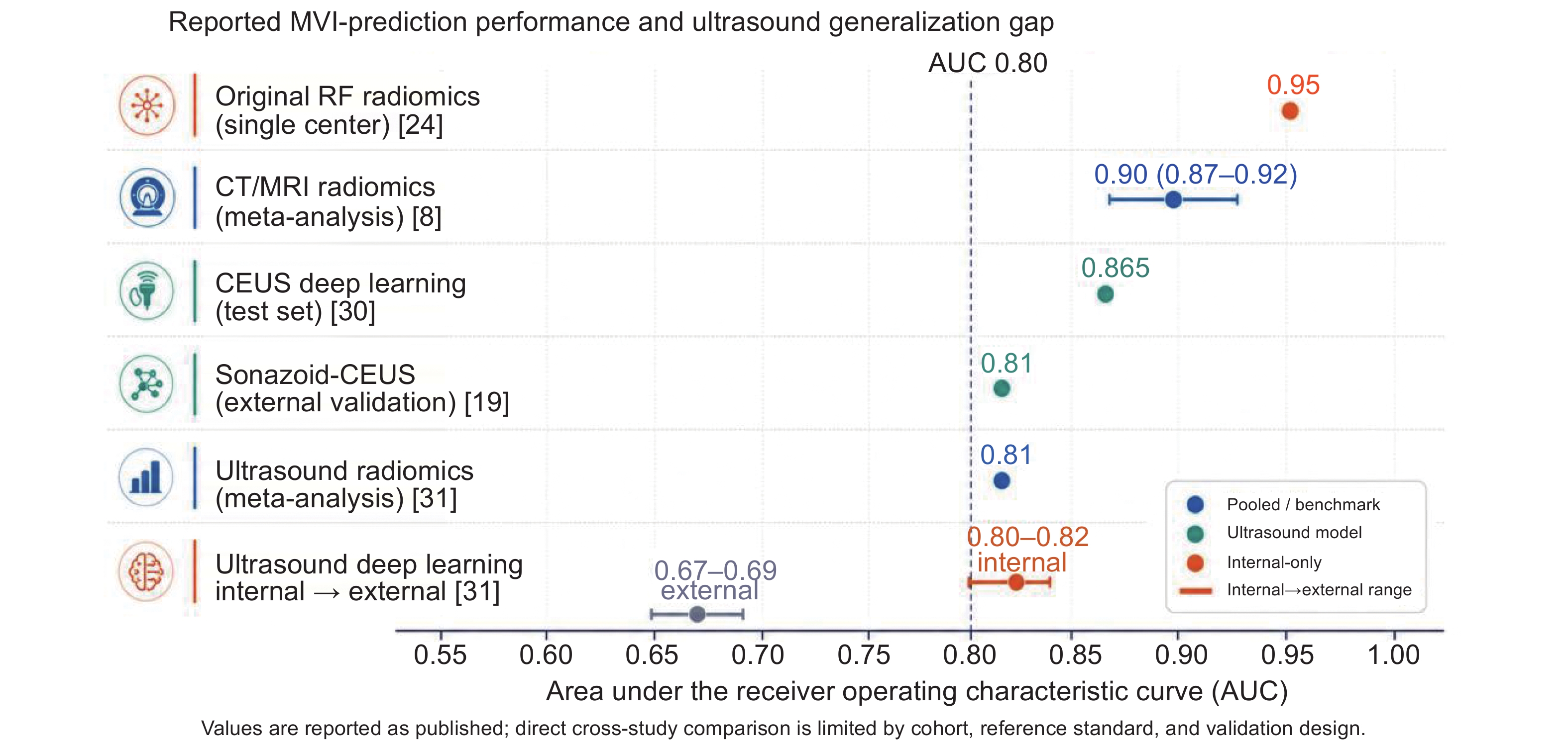
Representative reported performance of imaging biomarkers for preoperative prediction of microvascular invasion in hepatocellular carcinoma (non-head-to-head comparison)"
| Modality/model | Study design | AUROC (internal/external) | Principal limitation |
| AUROC, area under the receiver operating characteristic curve; CEUS, contrast-enhanced ultrasound; CT, computed tomography; MRI, magnetic resonance imaging; RF, radiofrequency. Bracketed numbers denote references. Values are reported as published; direct comparison across studies is limited by differences in cohort, reference standard, and validation design | |||
| CT/MRI radiomics | Meta-analysis | 0.90 (pooled) | I2 ≈ 94%; low Radiomics Quality Score [ |
| CEUS deep learning (spatiotemporal) | Single center | 0.865 (test) | Small independent test set [ |
| Sonazoid-CEUS | Prospective, multicenter | 0.81 (external) | Single nodule ≤ 5 cm [ |
| Ultrasound radiomics | Meta-analysis | 0.81 (pooled) | Small, partly overlapping samples [ |
| Ultrasound deep learning | Cross-institutional | 0.80-0.82/0.67-0.69 | Generalization gap on external data [ |
| Original-RF radiomics (current authors) | Single center | 0.95 (internal) | n = 42; no external validation [ |
Figure 4
Representative reported diagnostic performance of imaging biomarkers for the preoperative prediction of microvascular invasion (MVI) in hepatocellular carcinoma (HCC) (non-head-to-head comparison). Dots represent reported areas under the receiver operating characteristic curve (AUROCs) or pooled estimates from published studies, whereas horizontal bars indicate 95% confidence intervals or the range between internal and external validation when available. The dashed vertical line denotes an AUROC of 0.80, a commonly accepted threshold for good diagnostic discrimination. Although several imaging approaches-including CT/MRI radiomics, contrast-enhanced ultrasound (CEUS), ultrasound radiomics, deep learning, and radiofrequency (RF)-based analysis-have demonstrated promising diagnostic performance, the decline observed between internal and external validation of ultrasound deep-learning models highlights the persistent challenge of model generalizability. Due to differences in study design, patient populations, reference standards, and validation strategies, the parallel display in the figure is only used to illustrate the research background, and the ranking is based solely on reported values and does not represent the level of evidence."
| [1] | Sung H, Ferlay J, Siegel RL, Laversanne M, Soerjomataram I, Jemal A, et al. Global cancer statistics 2020: globocan estimates of incidence and mortality worldwide for 36 cancers in 185 countries. CA: a cancer journal for clinicians 2021; 71: 209-249. |
| [2] |
Finn RS, Qin S, Ikeda M, Galle PR, Ducreux M, Kim TY, et al. Atezolizumab plus bevacizumab in unresectable hepatocellular carcinoma. The New England journal of medicine 2020; 382: 1894-1905.
doi: 10.1056/NEJMoa1915745 |
| [3] |
Craig AJ, von Felden J, Garcia-Lezana T, Sarcognato S, Villanueva A. Tumour evolution in hepatocellular carcinoma. Nature reviews Gastroenterology & hepatology 2020; 17: 139-152.
doi: 10.1038/s41575-019-0229-4 |
| [4] |
Llovet JM, Kelley RK, Villanueva A, Singal AG, Pikarsky E, Roayaie S, et al. Hepatocellular carcinoma. Nature reviews Disease primers 2021; 7: 6.
doi: 10.1038/s41572-020-00240-3 |
| [5] |
Losic B, Craig AJ, Villacorta-Martin C, Martins-Filho SN, Akers N, Chen X, et al. Intratumoral heterogeneity and clonal evolution in liver cancer. Nature communications 2020; 11: 291.
doi: 10.1038/s41467-019-14050-z |
| [6] |
Rebouissou S, Nault JC. Advances in molecular classification and precision oncology in hepatocellular carcinoma. Journal of hepatology 2020; 72: 215-229.
doi: 10.1016/j.jhep.2019.08.017 |
| [7] |
Roayaie S, Blume IN, Thung SN, Guido M, Fiel MI, Hiotis S, et al. A system of classifying microvascular invasion to predict outcome after resection in patients with hepatocellular carcinoma. Gastroenterology 2009; 137: 850-855.
doi: 10.1053/j.gastro.2009.06.003 |
| [8] |
Li L, Wu C, Huang Y, Chen J, Ye D, Su Z. Radiomics for the preoperative evaluation of microvascular invasion in hepatocellular carcinoma: a meta-analysis. Frontiers in oncology 2022; 12: 831996.
doi: 10.3389/fonc.2022.831996 |
| [9] |
Banerjee S, Wang DS, Kim HJ, Sirlin CB, Chan MG, Korn RL, et al. A computed tomography radiogenomic biomarker predicts microvascular invasion and clinical outcomes in hepatocellular carcinoma. Hepatology (Baltimore, Md) 2015; 62: 792-800.
doi: 10.1002/hep.27877 |
| [10] |
Kitao A, Matsui O, Yoneda N, Kozaka K, Kobayashi S, Sanada J, et al. Hepatocellular carcinoma with β-catenin mutation: imaging and pathologic characteristics. Radiology 2015; 275: 708-717.
doi: 10.1148/radiol.14141315 |
| [11] |
Kang HJ, Kim H, Lee DH, Hur BY, Hwang YJ, Suh KS, et al. Gadoxetate-enhanced MRI features of proliferative hepatocellular carcinoma are prognostic after surgery. Radiology 2021; 300: 572-582.
doi: 10.1148/radiol.2021204352 |
| [12] |
Zhang X, Su GH, Chen Y, Gu YJ, You C. Decoding intratumoral heterogeneity: clinical potential of habitat imaging based on radiomics. Radiology 2023; 309: e232047.
doi: 10.1148/radiol.232047 |
| [13] |
Hoshida Y, Nijman SM, Kobayashi M, Chan JA, Brunet JP, Chiang DY, et al. Integrative transcriptome analysis reveals common molecular subclasses of human hepatocellular carcinoma. Cancer research 2009; 69: 7385-7392.
doi: 10.1158/0008-5472.CAN-09-1089 |
| [14] | Cancer Genome Atlas Research Network. Electronic address: wheeler@bcm.edu; Cancer Genome Atlas Research Network. Comprehensive and integrative genomic characterization of hepatocellular carcinoma. Cell 2017; 169: 1327-1341.e23. |
| [15] |
Montironi C, Castet F, Haber PK, Pinyol R, Torres-Martin M, Torrens L, et al. Inflamed and non-inflamed classes of HCC: a revised immunogenomic classification. Gut 2023; 72: 129-140.
doi: 10.1136/gutjnl-2021-325918 |
| [16] |
Fang JH, Xu L, Shang LR, Pan CZ, Ding J, Tang YQ, et al. Vessels that encapsulate tumor clusters (vetc) pattern is a predictor of sorafenib benefit in patients with hepatocellular carcinoma. Hepatology (Baltimore, Md) 2019; 70: 824-839.
doi: 10.1002/hep.30366 |
| [17] | Piscaglia F, Wilson SR, Lyshchik A, Cosgrove D, Dietrich CF, Jang HJ, et al. American college of radiology contrast enhanced ultrasound liver imaging reporting and data system (CEUS LI-RADS) for the diagnosis of hepatocellular carcinoma: a pictorial essay. Ultraschall in der Medizin (Stuttgart, Germany: 1980) 2017; 38: 320-324. |
| [18] |
Dietrich CF, Nolsøe CP, Barr RG, Berzigotti A, Burns PN, Cantisani V, et al. Guidelines and good clinical practice recommendations for contrast-enhanced ultrasound (CEUS) in the liver-update 2020 WFUMB in cooperation with EFSUMB, AFSUMB, AIUM, and FLAUS. Ultrasound in medicine & biology 2020; 46: 2579-2604.
doi: 10.1016/j.ultrasmedbio.2020.04.030 |
| [19] |
Yao J, Li K, Yang H, Lu S, Ding H, Luo Y, et al. Analysis of Sonazoid contrast-enhanced ultrasound for predicting the risk of microvascular invasion in hepatocellular carcinoma: a prospective multicenter study. European radiology 2023; 33: 7066-7076.
doi: 10.1007/s00330-023-09656-3 |
| [20] | Dong Y, Zuo D, Qiu YJ, Cao JY, Wang HZ, Wang WP. Prediction of histological grades and Ki-67 expression of hepatocellular carcinoma based on Sonazoid contrast enhanced ultrasound radiomics signatures. Diagnostics (Basel, Switzerland) 2022;12. |
| [21] |
Zhong X, Long H, Chen L, Xie Y, Shi Y, Peng J, et al. Stiffness on shear wave elastography as a potential microenvironment biomarker for predicting tumor recurrence in HBV-related hepatocellular carcinoma. Insights into imaging 2023; 14: 147.
doi: 10.1186/s13244-023-01505-7 |
| [22] |
Zhou Z, Gao A, Zhang Q, Wu W, Wu S, Tsui PH. Ultrasound backscatter envelope statistics parametric imaging for liver fibrosis characterization: a review. Ultrasonic imaging 2020; 42: 92-109.
doi: 10.1177/0161734620907886 |
| [23] |
Oelze ML, Mamou J. Review of quantitative ultrasound: envelope statistics and backscatter coefficient imaging and contributions to diagnostic ultrasound. IEEE transactions on ultrasonics, ferroelectrics, and frequency control 2016; 63: 336-351.
doi: 10.1109/TUFFC.2015.2513958 |
| [24] |
Dong Y, Wang QM, Li Q, Li LY, Zhang Q, Yao Z, et al. Preoperative prediction of microvascular invasion of hepatocellular carcinoma: radiomics algorithm based on ultrasound original radio frequency signals. Frontiers in oncology 2019; 9: 1203.
doi: 10.3389/fonc.2019.01203 |
| [25] | Yang F, Zhao J, Liu C, Mao Y, Mu J, Wei X, et al. Superb microvascular imaging technique in depicting vascularity in focal liver lesions: more hypervascular supply patterns were depicted in hepatocellular carcinoma. Cancer imaging: the official publication of the International Cancer Imaging Society 2019; 19: 92 |
| [26] |
Errico C, Pierre J, Pezet S, Desailly Y, Lenkei Z, Couture O, et al. Ultrafast ultrasound localization microscopy for deep super-resolution vascular imaging. Nature 2015; 527: 499-502.
doi: 10.1038/nature16066 |
| [27] | Huang C, Zhang W, Gong P, Lok UW, Tang S, Yin T, et al. Super-resolution ultrasound localization microscopy based on a high frame-rate clinical ultrasound scanner: an in-human feasibility study. Physics in medicine and biology 2021;66. |
| [28] |
Xia S, Zheng Y, Hua Q, Wen J, Luo X, Yan J, et al. Super-resolution ultrasound and microvasculomics: a consensus statement. European radiology 2024; 34: 7503-7513.
doi: 10.1007/s00330-024-10796-3 |
| [29] |
Zwanenburg A, Vallières M, Abdalah MA, Aerts H, Andrearczyk V, Apte A, et al. The image biomarker standardization initiative: standardized quantitative radiomics for high-throughput image-based phenotyping. Radiology 2020; 295: 328-338.
doi: 10.1148/radiol.2020191145 |
| [30] |
Zhang Y, Wei Q, Huang Y, Yao Z, Yan C, Zou X, et al. Deep learning of liver contrast-enhanced ultrasound to predict microvascular invasion and prognosis in hepatocellular carcinoma. Frontiers in oncology 2022; 12: 878061.
doi: 10.3389/fonc.2022.878061 |
| [31] | Zhang W, Guo Q, Zhu Y, Wang M, Zhang T, Cheng G, et al. Cross-institutional evaluation of deep learning and radiomics models in predicting microvascular invasion in hepatocellular carcinoma: validity, robustness, and ultrasound modality efficacy comparison. Cancer imaging: the official publication of the International Cancer Imaging Society 2024; 24: 142 |
| [32] | Selvaraju RR, Cogswell M, Das A, Vedantam R, Parikh D, Batra D, et al. Grad-CAM: visual explanations from deep networks via gradient-based localization. International Journal of Computer Vision 2019; 128: 336-359. |
| [33] |
Xiao Q, Zhu W, Tang H, Zhou L. Ultrasound radiomics in the prediction of microvascular invasion in hepatocellular carcinoma: A systematic review and meta-analysis. Heliyon 2023; 9: e16997.
doi: 10.1016/j.heliyon.2023.e16997 |

| [1] | Li Mo, Jin Letian, Xue Ge, Chen Jiye, Benariba Mohamed Aimene, Wang Vicky Yang, Teng Gaojun, Xu Dong. Empowering Breast Cancer Precision Diagnosis with Advanced Ultrasound: From Radiomics to Radio-multiomics [J]. Advanced Ultrasound in Diagnosis and Therapy, 2026, 10(4): 277-297. |
| [2] | Sui Yajuan, Meng Wenyi, Li Renjie, Li Lujing, Xu Zuofeng. Knowledge Mapping of Ultrasound-guided Thermal Ablation Combined with Immunotherapy for Hepatocellular Carcinoma: A Systematic Bibliometric Analysis [J]. Advanced Ultrasound in Diagnosis and Therapy, 2026, 10(4): 245-258. |
| [3] | Oronsky Bryan, Li Dandan, Wang Morgan, Vo-Phamhi Jenny M., Caroen Scott, Abrouk Nacer, Reid Tony, Alizadeh Babak, Hristov Dimitre, Fisher George, Kaffas Ahmed El. 3D Dynamic Contrast-Enhanced Ultrasound for Personalized RRx-001 Dosing: A Preclinical and Clinical Pilot Study [J]. Advanced Ultrasound in Diagnosis and Therapy, 2026, 10(3): 169-177. |
| [4] | Cui Yuanjie, Guo Cuixia, Li Zhen, Zhang Juan, Wu Yuting, Wu Qingqing, Sun Lijuan. Use of Artificial Intelligence in Fetal Brain Ultrasound Examination: A review [J]. Advanced Ultrasound in Diagnosis and Therapy, 2026, 10(2): 90-97. |
| [5] | Liu Taixia, Wang Hanxiang, Wang Dan, Zhang Yuke, Guo Yunyun, Liu Shuo, Zhang Minfeng, Nie Hongming, Shen Rui. The Role of Sonazoid-contrast-enhanced Ultrasound In Precision Diagnosis and Guidance for Radiofrequency Ablation Therapy of Hepatocellular Carcinoma: a Literature Review [J]. Advanced Ultrasound in Diagnosis and Therapy, 2026, 10(1): 20-28. |
| [6] | Hu Xuelin, Zhu Ye, Zhang Zisang, Quan Yuanting, Chen Wenwen, Chen Leichong, Xu Guangyu, Qin Luning, Xie Mingxing, Zhang Li. Explainable Artificial Intelligence in Echocardiography [J]. Advanced Ultrasound in Diagnosis and Therapy, 2025, 9(4): 409-425. |
| [7] | Guan Xin, Hu Xinyuan, Han Hong, Zhang Dezhi, Xu Huixiong. The Evolving Application of Ultrasound in the Precision Management of Small Hepatocellular Carcinoma [J]. Advanced Ultrasound in Diagnosis and Therapy, 2025, 9(4): 375-387. |
| [8] | Li Yanran, Cui Yuanjie, Wu Qingqing, Zhang Na. Current Applications of Artificial Intelligence in Obstetric Ultrasound [J]. Advanced Ultrasound in Diagnosis and Therapy, 2025, 9(4): 449-456. |
| [9] | Yu Xiao jie, Song Zheng lai, Chang Xue yong, Yu Jie, Liang Ping. Artificial Intelligence in Ultrasound Diagnosis of Liver Nodules: A Comprehensive Review of B-Mode and Contrast-enhanced Applications [J]. Advanced Ultrasound in Diagnosis and Therapy, 2025, 9(4): 326-346. |
| [10] | Zhong Xian, Xie Xiaoyan. Multimodal Ultrasound Radiomics in Liver Disease: Current Status and Future Directions [J]. Advanced Ultrasound in Diagnosis and Therapy, 2025, 9(4): 388-408. |
| [11] | Xu Junmei, Tahmasebi Aylin, Mohammed Amr, Pour Bahareh Kian, Liu Ji-Bin, Eisenbrey John R.. Contrast-enhanced Ultrasound LI-RADS for Nonradiation Treatment Response Assessment in Liver Tumor: A Pictorial Review Based on LR-TR v2024 [J]. Advanced Ultrasound in Diagnosis and Therapy, 2025, 9(4): 357-374. |
| [12] | Zhang Xiaoqian, Zhang Jingwen, Dong Yijie, Zhou Jianqiao. Research Progress and Clinical Translation of Photoacoustic–ultrasound Fusion Imaging in Breast Cancer Diagnosis and Therapy [J]. Advanced Ultrasound in Diagnosis and Therapy, 2025, 9(4): 467-482. |
| [13] | Jin Tong, Yu Xiaohu, Ai Zheng, Guo Hongcheng. Artificial Intelligence in Ultrasound Imaging: A Review of Progress from Machine Learning to Large Language Model [J]. Advanced Ultrasound in Diagnosis and Therapy, 2025, 9(4): 483-496. |
| [14] | Zheng Hairong, Meng Long, Li Fei, Niu Lili, Qiu Weibao, Ma Teng, Liu Chengbo, Zhu Xuefeng, Wan Liwen, Cai Feiyan. Advance in Ultrasound Super-resolution Imaging, Cell Manipulation and Inter-brain Communication [J]. Advanced Ultrasound in Diagnosis and Therapy, 2025, 9(4): 307-325. |
| [15] | Elkouahy Fatima Ezzahra, Bennis Ahmed, Merke Nicolas, Ouahid Hajar, Malali Hamid El, Taleb Lhoucine Ben, Mouhsen Azeddine. Advanced Diagnosis of Aortic Stenosis Disease Based on Ultrasound Images: A Novel Artificial Intelligence Approach [J]. Advanced Ultrasound in Diagnosis and Therapy, 2025, 9(3): 298-306. |
| Viewed | ||||||
|
Full text |
|
|||||
|
Abstract |
|
|||||
Share: WeChat
Copyright ©2018 Advanced Ultrasound in Diagnosis and Therapy
|
Advanced Ultrasound in Diagnosis and Therapy (AUDT) a>
is licensed under a Creative Commons Attribution 4.0 International License a>.