
Advanced Ultrasound in Diagnosis and Therapy ›› 2026, Vol. 10 ›› Issue (2): 79-89.doi: 10.26599/AUDT.2026.250066
Gao Yuanjinga, Niu Zihana, Luo Yanwenb, Zhou Mengyuana, Xiao Mengsua, Jiang Yuxina, Zhu Qinglia,*( )
)
Received:2025-07-21
Revised:2025-09-08
Accepted:2025-12-20
Online:2026-06-30
Published:2026-07-03
Contact:
Department of Ultrasound, Peking Union Medical College Hospital, Chinese Academy of Medical Sciences and Peking Union Medical College. (Qingli Zhu) e-mail: zqlpumch@126.com (QL Z)Gao Yuanjing, Niu Zihan, Luo Yanwen, Zhou Mengyuan, Xiao Mengsu, Jiang Yuxin, Zhu Qingli. Research Progress on Ultrasound Radiomics in Preoperative Prediction of Axillary Lymph Node Metastasis in Breast Cancer. Advanced Ultrasound in Diagnosis and Therapy, 2026, 10(2): 79-89.
Figure 1
Two technical pathways for predictive modeling. The upper panel depicts the radiomics approach, comprising: Imaging acquisition; Image preprocessing; ROI delineation; Construction of multidimensional parametric features based on pixel intensity distribution patterns and spatial topological relationships; Modeling and analysis; Prediction outcomes. The lower panel shows the deep learning approach, comprising: Imaging acquisition; Image preprocessing; End-to-end autonomous capture of abstract feature sets; Feature mapping; Prediction outcomes."
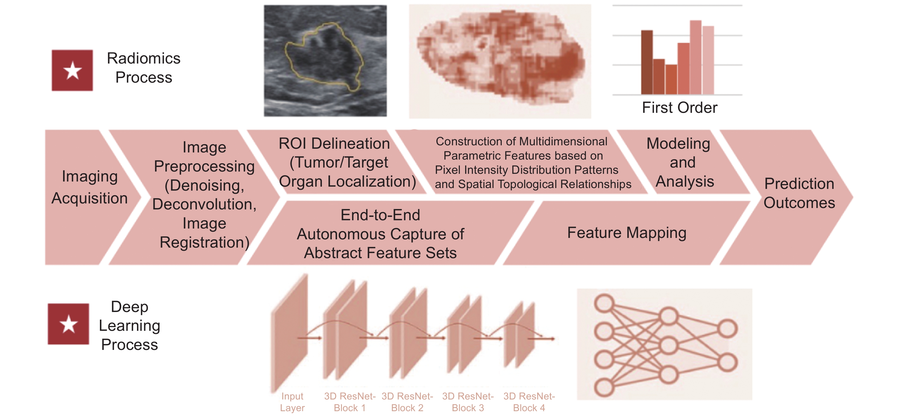
Table 1
Summary of ultrasound radiomics studies in predicting axillary lymph node(ALN) status"
| Author | Year | Image type | Number of participants | Number of Centers | AUC (dataset)←model name | ALN status | Parameters | Region of interest segmentation | Feature selection method | Model construction method | |
| >0 vs=0 | ≥3 vs. 1-2 | ||||||||||
| *means no seperated validation dataset. (final) means finally chosen method to construct the model. Training dataset: Used for model training; Validation dataset: A dataset randomly split from the same source as the training set, used for parameter tuning and model selection. In-test: internal dataset or independent dataset: Partitioned from the same-source data that was never involved in training. External dataset: dataset from other hospitals: Independently sourced data (e.g., collected from other hospitals/equipment). PL, Primary lesion; ALN, Axillary lymph node; val, Validation dataset; RAD, Radiomics; LR, Logistic regression; ICC, Intra-class correlation coefficient; LASSO, Least Absolute Shrinkage and Selection Operator; VIF, variance inflation factor; MRMR, Max-relevance and min-redundancy; ex-test, External dataset; LGOCV, leave group out cross-validation; PCA, principal component analysis; XGB/XGBoost, Extreme gradient boosting; SVM, support vector machine; DL, Deep learning; CNN, Convolutional Neural Network; RF, Random Forest; KNN, K-Nearest Neighbors; NB, naïve Bayesian; SWE, shear wave elastography; CEUS, Contrast enhanced ultrasound. | |||||||||||
| Only Grayscale US | |||||||||||
| Qiu [ | 2020 | Primary Lesion (PL) | 196 | 1 | 0.73 (validation dataset (val))← radiomics (radiomics(RAD)) 0.76 (val)←RAD + US-reported ALN | √ | RAD, US-reported ALN status | Manual | Elastic net logistic regression; univariate analyses | Logistic regression (LR) | |
| Yu [ | 2019 | PL | 426 | 1 | 0.71(val) ← RAD 0.81(val) ← combined model | √ | RAD + size + US-reported ALN status | Manual | Intra-class correlation coefficient (ICC); Least Absolute Shrinkage and Selection Operator (LASSO); variance inflation factor (VIF) | Multivariable LR | |
| Zhou [ | 2021 | PL | 192 | 2 | 0.65 (External dataset (ex-test)) | √ | RAD | Manual | Max-relevance and min-redundancy (MRMR); LASSO | LASSO; leave group out cross-validation (LGOCV) | |
| Gao [ | 2021 | PL | 343 | 1 | 0.73 (val) | √ | RAD,size, age | Manual | LASSO | LR | |
| Wu [ | 2024 | PL | 215 | 2 | 0.66 (ex-test) ← clinical model 0.72 (ex-test) ← RAD model | √ | RAD/clinicopathological features | Manual | ICC; Mann-Whitney U test; LASSO; principal component analysis (PCA) and Boruta (final) | Extreme gradient boosting (XGB/XGBoost)(final); LR; support vector machine (SVM) | |
| Zhang [ | 2023 | PL | 176 | 1 | 0.81(val)← Deep learning (DL)-RAD model 0.82 (val)←DL-nomogram | √ | RAD + ResNet-50; size, clinicopathological features | Manual | ICC, Independent sample T-test; random forest recursive elimination; LASSO | Univariate/multivariate LR analysis | |
| Lee [ | 2021 | PL + peritumoral area | 153 | 1 | 0.81 (val) | √ | DL features or handcrafted features (shape, textural) | Mask R- Convolutional Neural Network (CNN) | \; | DenseNet-121 (final); LR;SVM;XGBoost | |
| Wang [ | 2024 | PL+ peritumoral area | 321 | 1 | 0.78 (val)←clinical model 0.84 (val)←RAD model 0.91(val)←combined model | √ | RAD + conventional ultrasound & immunohistochemical indicators | Manual | Correlation analysis; Univariate analysis; MRMR; LASSO | LR; Decision Tree; SVM; XGB (final); Random Forest(RF); K-Nearest Neighbors (KNN) | |
| Zhang [ | 2024 | PL+ peritumoral area | 755 | 1 | Combined model: 0.87(val) ←presence/absence 0.91(val)←high/low tumor burden | √ | √ | Conventional ultrasound & clinical indicators | Manual | MRMR; LASSO; Pearson’s coefficient and VIF | Multivariate LR; |
| Coronado [ | 2019 | ALN | 105 | 2 | 0.94(train+val-k-fold*) | √ | Computed ROI features | Manual | A variation of Fisher Vector FV- CNN features Supervised machine learning | ||
| Sun [ | 2022 | ALN | 169 | 1 | 0.72 (test) | √ | CNN features | Manual | Custom CNN | ||
| Ozaki [ | 2022 | ALN | 300 | 1 | 0.97(val) | √ | DL features | Deep Analyzer | GoogleNet Xception; Supervised learning | ||
| Tang [ | 2023 | ALN | 147 | 2 | 0.92 (val) | √ | RAD | Manual | ICC; Spearman correlation analysis; LASSO | ||
| Chen [ | 2023 | ABUS | 310 | 1 | 0.79(val)← ABUS RAD 0.74(val)← ABUS features 0.85(val)← RAD+retraction phenomenon+ US reported ALN status | √ | RAD (ABUS) + ABUS imaging features +clinicopathologic features | Manual | Pearson correlation coefficient; Spearman’s rank correlation coefficient; LASSO | Univariate & Multivariate LR | |
| Li [ | 2023 | ABUS | 517 | 2 | 0.81(test) | √ | RAD (ABUS) + size+ US-reported LN status +retraction phenomenon | Manual | ICC; LASSO | Univariate & Multivariate LR | |
| Wang [ | 2023 | ABUS | 276 | 2 | 0.67(test)←RAD 0.83(test)←combined model | √ | RAD (ABUS) + US-reported LN status + convergence + erythrocyte distribution width | Manual | ICC;LASSO | univariate LR | |
| Li [ | 2023 | US video | 320 | 3 | 0.91(test)← video 0.86(test)← images | √ | CNN features | Normalize the data | Video: CNN with R2+1D, TIN (final), ResNet-3D Static images: ResNet50, InceptionV3, VGG19 | ||
| Zha [ | 2021 | PL | 452 | 1 | 0.77(val) ← RAD 0.83(val) ←combined model | √ | RAD+ MSKKCC nomogram | Manual | mRMR; LASSO Cox regression | LR | |
| Ouyang [ | 2024 | PL | 212 | 1 | 0.85 (val) | √ | RAD + size, tumor type, age, Ki67 | Manual | Pearson correlation Coefficients; LASSO | SVM, naïve Bayesian (NB), optimization method | |
| Yao [ | 2024 | PL | 278 | 1 | 0.92(val)← RAD 0.93(val)← combined model | √ | RAD + high tumor grade & positive LVI | Manual | The synthetic minority oversampling technique; PCA, recursive feature elimination | linear discriminant analysis; SVM, RF, decision tree; | |
| Zhao [ | 2023 | PL | 199 | 1 | 0.68 (val) ← RAD 0.69 (val) ← RAD+conventional US + peripheral blood T cell 0.79 (val) ← combined model | √ | RAD + peripheral blood T-cell subsets | Manual | LASSO | NB; LR (final), classification decision tree, SVM | |
| Qian [ | 2024 | PL | 364 | 1 | 0.89(val) ← RAD 0.74(val) ← clinical model 0.91(val) ← combined model | √ | DL (DenseNet-201) features + RAD + clinical features | Manual rectangle ROI | DL: Spearman correlation coefficient; RAD: LASSO; ICC; mRMR | Univariate analysis; Stepwise multivariable LR | |
| Zhou [ | 2019 | PL | 834 | 2 | 0.90(in-test) 0.89 (ex-test) | √ | DL features | / | Inception V3 (final); Inception-ResNet V2; ResNet-101 | ||
| Wang [ | 2024 | PL | 266 | 1 | 0.80 (in-test) | √ | DL features | DeepLabV3 + ResNet-101 | CNN | ||
| Liu [ | 2024 | PL | 883 | 4 | 0.91(ex-test1); 0.93 (ex-test2); 0.95 (ex-test3) | √ | DL features (ResNet50) + RAD+ clinical parameters | Manual | ResNet50; ICC; LASSO | Univariate & multivariate LR | |
| You [ | 2024 | PL | 792 | 3 | Combined model: 0.83 (ex-test1); 0.83(ex-test2) | √(received NAC) | DL features; Clinicopathologic data | You Only Look Once Version 5 | Progressive multi-granularity (PMG) classification network; Multivariable logistic regression; image super-resolution via iterative refinement (SR3) | ||
| Guo [ | 2020 | PL | 937 | 2 | 0.84(ex-test) ← SLN 0.81(ex-test) ←non-SLN | √ | Non-SLN metastasis | DL features + US-reported ALN status | Manual | DenseNet | |
| Multimodal US (Combined with Grayscale US) | |||||||||||
| Zheng [ | 2020 | PL (shear wave elastography (SWE) & grayscale) | 584 | 1 | Combined model: 0.902(in-test) ←presence/absence 0.91(in-test) ←high/low tumor burden | √ | √ | DL features of (grayscale & SWE) + clinicopathological parameters | Manual rectangle ROI | ResNet50 (final), ResNet101, Inception V3, and VGG19 | |
| Jiang [ | 2022 | PL (SWE) | 433 | 2 | C-index: 0.82(ex-test) ← presence/absence 0.81(ex-test) ← high/low tumor burden | √ | √ | RAD (grayscale/SWE) + molecular subtype + US-reported ALN status | Manual | Spearman’s correlation; LASSO; MRMR | Proportional odds ordinal LR |
| Wang [ | 2023 | PL (strain elastography) | 359 | 2 | 0.79(ex-test) ←PL (grayscale) 0.73 (ex-test) ←PL(color Doppler flow imaging (CDFI)); 0.88(ex-test) ←PL (elastography) | √ | DL (grayscale/CDFI/strain elastography) | Manual | ResNet-18 | ||
| Gong [ | 2025 | ALN (grayscale+ SWE) | 519 | 1 | 0.86 (in-test) ← ALN (grayscale) 0.83 (in-test) ←ALN (SWE); 0.93 (ex-test) ←ALN (SWE + grayscale) | √ | DAMF-former(grayscale/SWE) | Manual rectangle ROI | DAMF-former (core module: dual-modal adaptive fusion module) | ||
| Sun [ | 2024 | PL (grayscale, Contrast enhanced ultrasound (CEUS)) | 111 | 1 | 0.82 (val)← US-reported ALN 0.89 (val)← US-reported ALN+RAD (grayscale) 0.90 (val)← US-reported ALN+RAD (grayscale+CEUS) | √ | RAD (grayscale/CEUS), US-reported ALN status | Manual (grayscale); computer vision algorithm (CEUS) | VIF; MRMR; LASSO | LR | |
| Yan [ | 2025 | PL (grayscale, CEUS) | 282 | 2 | 0.81 (ex-test) | √ | RAD (grayscale),histologic type (only for nomogram) | Manual | RF, LASSO | Univariate & multivariate LR, NB, SVM; KNN; XGBoost (final); nomogram | |
| Other multimodal imaging (combined with grayscale US) | |||||||||||
| Tang [ | 2025 | PL (MRI + US) | 588 | 2 | 0.81(ex-test) ← DL (MRI + US + clinical parameters) | √ | DL features (MRI/US), age, MRI-derived tumor size, MRI-derived ALN status | Manual rectangle ROI | ConvNeXt (final), ResNet18, VGG16 → US 3D-ResNet (final), C3D, R (2+1)D → MRI; multivariable LR | ||
| Li [ | 2023 | (18F-FDG) PET/CT | 124 | 1 | 0.87(val)← T-stage+ US/PET reported ALN status+ RAD(PET) | √ | RAD (PET), T-stage, US/PET reported ALN status | Manual | LASSO | Univariate & multivariate LR | |
| [1] | Siegel RL, Miller KD, Fuchs HE, Jemal A. Cancer statistics, 2022. CA Cancer J Clin 2022; 72: 7-33. |
| [2] | Chen W, Zheng R, Baade PD, Zhang S, Zeng H, Bray F, et al. Cancer statistics in China, 2015. CA Cancer J Clin 2016; 66: 115-132. |
| [3] |
Sechopoulos I, Teuwen J, Mann R. Artificial intelligence for breast cancer detection in mammography and digital breast tomosynthesis: state of the art. Semin Cancer Biol 2021; 72: 214-225.
doi: 10.1016/j.semcancer.2020.06.002 |
| [4] |
Giuliano AE, Ballman KV, McCall L, Beitsch PD, Brennan MB, Kelemen PR, et al. Effect of axillary dissection vs no axillary dissection on 10-year overall survival among women with invasive breast cancer and sentinel node metastasis: the ACOSOG Z0011 (alliance) randomized clinical trial. JAMA 2017; 318: 918-926.
doi: 10.1001/jama.2017.11470 |
| [5] |
de Boniface J, Filtenborg Tvedskov T, Rydén L, Szulkin R, Reimer T, Kühn T, et al. Omitting axillary dissection in breast cancer with sentinel-node metastases. N Engl J Med 2024; 390: 1163-1175.
doi: 10.1056/NEJMoa2313487 |
| [6] | Lyman GH, Somerfield MR, Bosserman LD, Perkins CL, Weaver DL, Giuliano AE. Sentinel lymph node biopsy for patients with early-stage breast cancer: American society of clinical oncology clinical practice guideline update. J Clin Oncol 2017; 35: 561-564. |
| [7] |
Gentilini OD, Botteri E, Sangalli C, Galimberti V, Porpiglia M, Agresti R, et al. Sentinel lymph node biopsy vs no axillary surgery in patients with small breast cancer and negative results on ultrasonography of axillary lymph nodes: the sound randomized clinical trial. JAMA Oncol 2023; 9: 1557-1564.
doi: 10.1001/jamaoncol.2023.3759 |
| [8] |
Valente SA, Levine GM, Silverstein MJ, Rayhanabad JA, Weng-Grumley JG, Ji L, et al. Accuracy of predicting axillary lymph node positivity by physical examination, mammography, ultrasonography, and magnetic resonance imaging. Ann Surg Oncol 2012; 19: 1825-1830.
doi: 10.1245/s10434-011-2200-7 |
| [9] |
Alvarez S, Añorbe E, Alcorta P, López F, Alonso I, Cortés J. Role of sonography in the diagnosis of axillary lymph node metastases in breast cancer: a systematic review. AJR Am J Roentgenol 2006; 186: 1342-1348.
doi: 10.2214/AJR.05.0936 |
| [10] |
Cools-Lartigue J, Meterissian S. Accuracy of axillary ultrasound in the diagnosis of nodal metastasis in invasive breast cancer: a review. World J Surg 2012; 36: 46-54.
doi: 10.1007/s00268-011-1319-9 |
| [11] |
Chen X, He Y, Wang J, Huo L, Fan Z, Li J, et al. Feasibility of using negative ultrasonography results of axillary lymph nodes to predict sentinel lymph node metastasis in breast cancer patients. Cancer Med 2018; 7: 3066-3072.
doi: 10.1002/cam4.1606 |
| [12] | Zhu Y, Zhou W, Jia XH, Huang O, Zhan WW. Preoperative axillary ultrasound in the selection of patients with a heavy axillary tumor burden in early-stage breast cancer: what leads to false-positive results?. J Ultrasound Med 2018; 37: 1357-1365. |
| [13] |
Ahmed M, Jozsa F, Baker R, Rubio IT, Benson J, Douek M. Meta-analysis of tumour burden in pre-operative axillary ultrasound positive and negative breast cancer patients. Breast Cancer Res Treat 2017; 166: 329-336.
doi: 10.1007/s10549-017-4405-3 |
| [14] |
van Griethuysen JJM, Fedorov A, Parmar C, Hosny A, Aucoin N, Narayan V, et al. Computational radiomics system to decode the radiographic phenotype. Cancer Res 2017; 77: e104-e107.
doi: 10.1158/0008-5472.CAN-17-0339 |
| [15] |
Gillies RJ, Kinahan PE, Hricak H. Radiomics: Images are more than pictures, they are data. Radiology 2016; 278: 563-577.
doi: 10.1148/radiol.2015151169 |
| [16] |
Bevilacqua JL, Kattan MW, Fey JV, Cody HS, 3rd PI, Borgen Zee KJ. Doctor, what are my chances of having a positive sentinel node? A validated nomogram for risk estimation. J Clin Oncol 2007; 25: 3670-3679.
doi: 10.1200/JCO.2006.08.8013 |
| [17] |
Chue KM, Yong WS, Thike AA, Ahmed SS, Li HH, Wong CY, et al. Predicting the likelihood of additional lymph node metastasis in sentinel lymph node positive breast cancer: validation of the Memorial Sloan-Kettering Cancer Centre (MSKCC) nomogram. J Clin Pathol 2014; 67: 112-119.
doi: 10.1136/jclinpath-2013-201524 |
| [18] | Rouzier R, Uzan C, Rousseau A, Guillot E, Zilberman S, Meyer C, et al. Multicenter prospective evaluation of the reliability of the combined use of two models to predict non-sentinel lymph node status in breast cancer patients with metastatic sentinel lymph nodes: the MSKCC nomogram and the Tenon score. Results of the NOTEGS study. Br J Cancer 2017; 116: 1135-1140. |
| [19] |
Barranger E, Coutant C, Flahault A, Delpech Y, Darai E, Uzan S. An axilla scoring system to predict non-sentinel lymph node status in breast cancer patients with sentinel lymph node involvement. Breast Cancer Res Treat 2005; 91: 113-119.
doi: 10.1007/s10549-004-5781-z |
| [20] | Qiu X, Jiang Y, Zhao Q, Yan C, Huang M, Jiang T. Could ultrasound-based radiomics noninvasively predict axillary lymph node metastasis in breast cancer?. J Ultrasound Med 2020; 39: 1897-1905. |
| [21] |
Yu FH, Wang JX, Ye XH, Deng J, Hang J, Yang B. Ultrasound-based radiomics nomogram: A potential biomarker to predict axillary lymph node metastasis in early-stage invasive breast cancer. Eur J Radiol 2019; 119: 108658
doi: 10.1016/j.ejrad.2019.108658 |
| [22] |
Zhou WJ, Zhang YD, Kong WT, Zhang CX, Zhang B. Preoperative prediction of axillary lymph node metastasis in patients with breast cancer based on radiomics of gray-scale ultrasonography. Gland Surg 2021; 10: 1989-2001.
doi: 10.21037/gs-21-315 |
| [23] |
Gao Y, Luo Y, Zhao C, Xiao M, Ma L, Li W, et al. Nomogram based on radiomics analysis of primary breast cancer ultrasound images: prediction of axillary lymph node tumor burden in patients. Eur Radiol 2021; 31: 928-937.
doi: 10.1007/s00330-020-07181-1 |
| [24] | Wu J, Ge L, Guo Y, Xu D, Wang Z. Utilizing multiclassifier radiomics analysis of ultrasound to predict high axillary lymph node tumour burden in node-positive breast cancer patients: a multicentre study. Ann Med 2024; 56: 395061 |
| [25] | Zhang H, Zhao T, Zhang S, Sun J, Zhang F, Li X, et al. Prediction of axillary lymph node metastatic load of breast cancer based on ultrasound deep learning radiomics nomogram. Technol Cancer Res Treat 2023; 22: 15330338231166218 |
| [26] |
Lee YW, Huang CS, Shih CC, Chang RF. Axillary lymph node metastasis status prediction of early-stage breast cancer using convolutional neural networks. Comput Biol Med 2021; 130: 104206
doi: 10.1016/j.compbiomed.2020.104206 |
| [27] |
Wang SR, Cao CL, Du TT, Wang JL, Li J, Li WX, et al. Machine learning model for predicting axillary lymph node metastasis in clinically node positive breast cancer based on peritumoral ultrasound radiomics and shap feature analysis. J Ultrasound Med 2024; 43: 1611-1625.
doi: 10.1002/jum.16483 |
| [28] |
Zhang W, Wang S, Wang Y, Sun J, Wei H, Xue W, et al. Ultrasound-based radiomics nomogram for predicting axillary lymph node metastasis in early-stage breast cancer. Radiol Med 2024; 129: 211-221.
doi: 10.1007/s11547-024-01768-0 |
| [29] |
Coronado-Gutiérrez D, Santamaría G, Ganau S, Bargalló X, Orlando S, Oliva-Brañas ME, et al. Quantitative ultrasound image analysis of axillary lymph nodes to diagnose metastatic involvement in breast cancer. Ultrasound Med Biol 2019; 45: 2932-2941.
doi: 10.1016/j.ultrasmedbio.2019.07.413 |
| [30] |
Ozaki J, Fujioka T, Yamaga E, Hayashi A, Kujiraoka Y, Imokawa T, et al. Deep learning method with a convolutional neural network for image classification of normal and metastatic axillary lymph nodes on breast ultrasonography. Jpn J Radiol 2022; 40: 814-822.
doi: 10.1007/s11604-022-01261-6 |
| [31] |
Sun S, Mutasa S, Liu MZ, Nemer J, Sun M, Siddique M, et al. Deep learning prediction of axillary lymph node status using ultrasound images. Comput Biol Med 2022; 143: 105250
doi: 10.1016/j.compbiomed.2022.105250 |
| [32] |
Tang YL, Wang B, Ou-Yang T, Lv WZ, Tang SC, Wei A, et al. Ultrasound radiomics based on axillary lymph nodes images for predicting lymph node metastasis in breast cancer. Front Oncol 2023; 13: 1217309
doi: 10.3389/fonc.2023.1217309 |
| [33] |
Chen Y, Xie Y, Li B, Shao H, Na Z, Wang Q, et al. Automated breast ultrasound (ABUS)-based radiomics nomogram: an individualized tool for predicting axillary lymph node tumor burden in patients with early breast cancer. BMC Cancer 2023; 23: 340
doi: 10.1186/s12885-023-10743-3 |
| [34] | Li N, Song C, Huang X, Zhang H, Su J, Yang L, et al. Optimized radiomics nomogram based on automated breast ultrasound system: a potential tool for preoperative prediction of metastatic lymph node burden in breast cancer. Breast Cancer (Dove Med Press) 2023; 15: 121-132. |
| [35] |
Wang H, Yang XW, Chen F, Qin YY, Li XB, Ma SM, et al. Non-invasive assessment of axillary lymph node metastasis risk in early invasive breast cancer adopting automated breast volume scanning-based radiomics nomogram: a multicenter study. Ultrasound Med Biol 2023; 49: 1202-1211.
doi: 10.1016/j.ultrasmedbio.2023.01.006 |
| [36] |
Li WB, Du ZC, Liu YJ, Gao JX, Wang JG, Dai Q, et al. Prediction of axillary lymph node metastasis in early breast cancer patients with ultrasonic videos based deep learning. Front Oncol 2023; 13: 1219838
doi: 10.3389/fonc.2023.1219838 |
| [37] |
Zha HL, Zong M, Liu XP, Pan JZ, Wang H, Gong HY, et al. Preoperative ultrasound-based radiomics score can improve the accuracy of the Memorial Sloan Kettering Cancer Center nomogram for predicting sentinel lymph node metastasis in breast cancer. Eur J Radiol 2021; 135: 109512
doi: 10.1016/j.ejrad.2020.109512 |
| [38] |
Ouyang Q, Chen Q, Zhang L, Lin Q, Yan J, Sun H, et al. Construction of a risk prediction model for axillary lymph node metastasis in breast cancer based on gray-scale ultrasound and clinical pathological features. Front Oncol 2024; 14: 1415584
doi: 10.3389/fonc.2024.1415584 |
| [39] |
Yao J, Zhou W, Xu S, Jia X, Zhou J, Chen X, et al. Machine learning-based breast tumor ultrasound radiomics for pre-operative prediction of axillary sentinel lymph node metastasis burden in early-stage invasive breast cancer. Ultrasound Med Biol 2024; 50: 229-236.
doi: 10.1016/j.ultrasmedbio.2023.10.004 |
| [40] |
Zhao M, Zheng Y, Chu J, Liu Z, Dong F. Ultrasound-based radiomics combined with immune status to predict sentinel lymph node metastasis in primary breast cancer. Sci Rep 2023; 13: 16918
doi: 10.1038/s41598-023-44156-w |
| [41] |
Qian L, Liu X, Zhou S, Zhi W, Zhang K, Li H, et al. A cutting-edge deep learning-and-radiomics-based ultrasound nomogram for precise prediction of axillary lymph node metastasis in breast cancer patients ≥ 75 years. Front Endocrinol (Lausanne) 2024; 15: 1323452
doi: 10.3389/fendo.2024.1323452 |
| [42] |
Zhou LQ, Wu XL, Huang SY, Wu GG, Ye HR, Wei Q, et al. Lymph node metastasis prediction from primary breast cancer us images using deep learning. Radiology 2020; 294: 19-28.
doi: 10.1148/radiol.2019190372 |
| [43] |
Wang X, Nie L, Zhu Q, Zuo Z, Liu G, Sun Q, et al. Artificial intelligence assisted ultrasound for the non-invasive prediction of axillary lymph node metastasis in breast cancer. BMC Cancer 2024; 24: 910
doi: 10.1186/s12885-024-12619-6 |
| [44] |
Liu H, Zou L, Xu N, Shen H, Zhang Y, Wan P, et al. Deep learning radiomics based prediction of axillary lymph node metastasis in breast cancer. NPJ Breast Cancer 2024; 10: 22
doi: 10.1038/s41523-024-00628-4 |
| [45] |
You J, Huang Y, Ouyang L, Zhang X, Chen P, Wu X, et al. Automated and reusable deep learning (AutoRDL) framework for predicting response to neoadjuvant chemotherapy and axillary lymph node metastasis in breast cancer using ultrasound images: a retrospective, multicentre study. EClinicalMedicine 2024; 69: 102499
doi: 10.1016/j.eclinm.2024.102499 |
| [46] |
Guo X, Liu Z, Sun C, Zhang L, Wang Y, Li Z, et al. Deep learning radiomics of ultrasonography: Identifying the risk of axillary non-sentinel lymph node involvement in primary breast cancer. EBioMedicine 2020; 60: 103018
doi: 10.1016/j.ebiom.2020.103018 |
| [47] |
Zheng X, Yao Z, Huang Y, Yu Y, Wang Y, Liu Y, et al. Deep learning radiomics can predict axillary lymph node status in early-stage breast cancer. Nat Commun 2020; 11: 1236
doi: 10.1038/s41467-020-15027-z |
| [48] |
Jiang M, Li CL, Luo XM, Chuan ZR, Chen RX, Tang SC, et al. Radiomics model based on shear-wave elastography in the assessment of axillary lymph node status in early-stage breast cancer. Eur Radiol 2022; 32: 2313-2325.
doi: 10.1007/s00330-021-08330-w |
| [49] |
Wang C, Zhao Y, Wan M, Huang L, Liao L, Guo L, et al. Prediction of sentinel lymph node metastasis in breast cancer by using deep learning radiomics based on ultrasound images. Medicine (Baltimore) 2023; 102: e35868
doi: 10.1097/MD.0000000000035868 |
| [50] |
Gong C, Wu Y, Zhang G, Liu X, Zhu X, Cai N, et al. Computer-assisted diagnosis for axillary lymph node metastasis of early breast cancer based on transformer with dual-modal adaptive mid-term fusion using ultrasound elastography. Comput Med Imaging Graph 2025; 119: 102472
doi: 10.1016/j.compmedimag.2024.102472 |
| [51] |
Sun C, Gong X, Hou L, Yang D, Li Q, Li L, et al. A nomogram based on conventional and contrast-enhanced ultrasound radiomics for the noninvasively prediction of axillary lymph node metastasis in breast cancer patients. Front Oncol 2024; 14: 1400872
doi: 10.3389/fonc.2024.1400872 |
| [52] |
Yan M, He D, Sun Y, Huang L, Cai L, Wang C, et al. Comparative analysis of nomogram and machine learning models for predicting axillary lymph node metastasis in early-stage breast cancer: a study on clinically and ultrasound-negative axillary cases across two centers. Ultrasound Med Biol 2025; 51: 463-474.
doi: 10.1016/j.ultrasmedbio.2024.11.003 |
| [53] |
Tang X, Zhang H, Mao R, Zhang Y, Jiang X, Lin M, et al. Preoperative prediction of axillary lymph node metastasis in patients with breast cancer through multimodal deep learning based on ultrasound and magnetic resonance imaging images. Acad Radiol 2025; 32: 1-11.
doi: 10.1016/j.acra.2024.07.029 |
| [54] |
Li Y, Han D, Shen C, Duan X. Construction of a comprehensive predictive model for axillary lymph node metastasis in breast cancer: a retrospective study. BMC Cancer 2023; 23: 1028
doi: 10.1186/s12885-023-11498-7 |
| [55] |
Guo F, Sun S, Deng X, Wang Y, Yao W, Yue P, et al. Predicting axillary lymph node metastasis in breast cancer using a multimodal radiomics and deep learning model. Front Immunol 2024; 15: 1482020
doi: 10.3389/fimmu.2024.1482020 |
| [56] |
Wang Q, Lin Y, Ding C, Guan W, Zhang X, Jia J, et al. Multi-modality radiomics model predicts axillary lymph node metastasis of breast cancer using MRI and mammography. Eur Radiol 2024; 34: 6121-6131.
doi: 10.1007/s00330-024-10638-2 |
| [1] | Cui Yuanjie, Guo Cuixia, Li Zhen, Zhang Juan, Wu Yuting, Wu Qingqing, Sun Lijuan. Use of Artificial Intelligence in Fetal Brain Ultrasound Examination: A review [J]. Advanced Ultrasound in Diagnosis and Therapy, 2026, 10(2): 90-97. |
| [2] | Mo Sijie, Ding Zhimin, Huang Zhibin, Wang Mengyun, Xu Jinfeng, Zhang Hongan, Dong Fajin. Evaluating the Diagnostic Performance of Sound Touch Elastography, Visco-elastography, and Sound Attenuation in Chronic Liver Diseases Detection [J]. Advanced Ultrasound in Diagnosis and Therapy, 2026, 10(2): 112-122. |
| [3] | Shi Ming-Li, Liu Guo-Hui, Bao Fu-Xing. Advances in the Application of Ultrasound for Evaluating Placental Function in Fetal Growth Restriction (FGR) Pregnancy [J]. Advanced Ultrasound in Diagnosis and Therapy, 2026, 10(1): 42-50. |
| [4] | Hu Xuelin, Zhu Ye, Zhang Zisang, Quan Yuanting, Chen Wenwen, Chen Leichong, Xu Guangyu, Qin Luning, Xie Mingxing, Zhang Li. Explainable Artificial Intelligence in Echocardiography [J]. Advanced Ultrasound in Diagnosis and Therapy, 2025, 9(4): 409-425. |
| [5] | Li Yanran, Cui Yuanjie, Wu Qingqing, Zhang Na. Current Applications of Artificial Intelligence in Obstetric Ultrasound [J]. Advanced Ultrasound in Diagnosis and Therapy, 2025, 9(4): 449-456. |
| [6] | Zhong Xian, Xie Xiaoyan. Multimodal Ultrasound Radiomics in Liver Disease: Current Status and Future Directions [J]. Advanced Ultrasound in Diagnosis and Therapy, 2025, 9(4): 388-408. |
| [7] | Zhang Xiaoqian, Zhang Jingwen, Dong Yijie, Zhou Jianqiao. Research Progress and Clinical Translation of Photoacoustic–ultrasound Fusion Imaging in Breast Cancer Diagnosis and Therapy [J]. Advanced Ultrasound in Diagnosis and Therapy, 2025, 9(4): 467-482. |
| [8] | Jin Tong, Yu Xiaohu, Ai Zheng, Guo Hongcheng. Artificial Intelligence in Ultrasound Imaging: A Review of Progress from Machine Learning to Large Language Model [J]. Advanced Ultrasound in Diagnosis and Therapy, 2025, 9(4): 483-496. |
| [9] | Zhang Rong, Xie Liting, Jin Qijing, Zhang Chengyue, Guo Teng, Zhao Qiyu, Jiang Tian’an. Research Progress on the Application of Irreversible Electroporation Ablation in Cancers [J]. Advanced Ultrasound in Diagnosis and Therapy, 2025, 9(3): 229-244. |
| [10] | Zhang Minyu, Jia Baocheng, Huang Liuming. Imaging Diagnosis of Neonatal Umbilical Arteriovenous Malformation Complicated with Portosystemic Shunt [J]. Advanced Ultrasound in Diagnosis and Therapy, 2025, 9(2): 224-227. |
| [11] | An Zichen, Li Fan. Advancements in the Application of Convolutional Neural Networks in Ultrasound Imaging for Breast Cancer Diagnosis and Treatment [J]. Advanced Ultrasound in Diagnosis and Therapy, 2025, 9(1): 21-31. |
| [12] | Li Tiantian, Zhu Miao, Shen Dejuan, Qian Xiaoqin. The Diagnostic Pitfall of Lymphadenopathy: Ultrasonic Imaging Findings in a Case of Cat Scratch Lymphadenitis [J]. Advanced Ultrasound in Diagnosis and Therapy, 2025, 9(1): 92-95. |
| [13] | Xu Jiale, Xia Shujun, Hua Qing, Mei Zihan, Hou Yiqing, Wei Minyan, Lai Limei, Yang Yixuan, Zhou Jianqiao. Performance of ChatGPT and Radiology Residents on Ultrasonography Board-Style Questions [J]. Advanced Ultrasound in Diagnosis and Therapy, 2024, 8(4): 250-254. |
| [14] | Ge Xifeng, Liu Wenzheng, Chen Wen, Mei Fang, Cui Ligang. Gastroduodenal Intussusception Due to a Gastric Gastrointestinal Stromal Tumor in Adult from Sonographer's Perspective [J]. Advanced Ultrasound in Diagnosis and Therapy, 2024, 8(4): 255-258. |
| [15] | Shanqing Li, MM, Rong Hu, MM, Xijing Liu, MD, Fan Yang, MD. Update on the Genetics and Prenatal Ultrasound Features of Williams-Beuren Syndrome [J]. Advanced Ultrasound in Diagnosis and Therapy, 2024, 8(3): 79-85. |
| Viewed | ||||||
|
Full text |
|
|||||
|
Abstract |
|
|||||
Share: WeChat
Copyright ©2018 Advanced Ultrasound in Diagnosis and Therapy
|
 Advanced Ultrasound in Diagnosis and Therapy (AUDT)
is licensed under a Creative Commons Attribution 4.0 International License.
Advanced Ultrasound in Diagnosis and Therapy (AUDT)
is licensed under a Creative Commons Attribution 4.0 International License.
