Advanced Ultrasound in Diagnosis and Therapy ›› 2026, Vol. 10 ›› Issue (1): 42-50.doi: 10.26599/AUDT.2026.250064
Previous Articles Next Articles
Shi Ming-Lia, Liu Guo-Huib,*( ), Bao Fu-Xingb
Received:2025-07-19
Revised:2025-10-14
Accepted:2025-11-03
Online:2026-03-31
Published:2026-03-30
Contact:
Inner Mongolia Maternity and Child Health Care Hospital Department of Ultrasound, Hohhot, 010020. (Guo-Hui Liu), e-mail: Liuguohui0133@sina.com (GH L),
Shi Ming-Li, Liu Guo-Hui, Bao Fu-Xing. Advances in the Application of Ultrasound for Evaluating Placental Function in Fetal Growth Restriction (FGR) Pregnancy. Advanced Ultrasound in Diagnosis and Therapy, 2026, 10(1): 42-50.
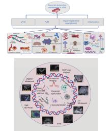
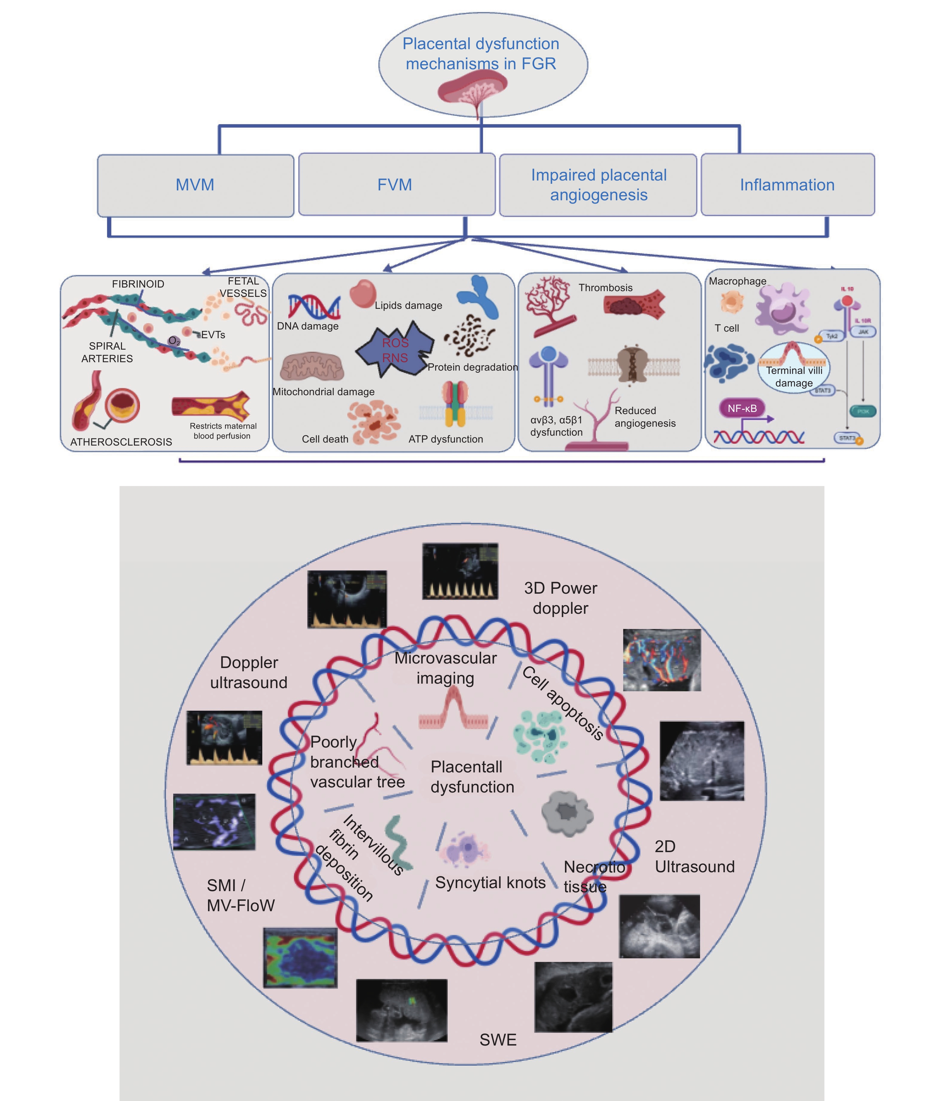
Figure 1
Correlation between ultrasound indicators and etiological mechanisms in placental dysfunction."
| [1] | Chu YY TH, Zhang JY, Xiong CQ, Huang HY, Liang RH, et al. The role of cerebral-placenta-uterine ratio in predicting late-onset fetal growth restriction. J Journal of Practical Obstetrics and Gynecology 2024; 40: 36-41. |
| [2] |
Odibo AO, Kayisli U, Lu Y, Kayisli O, Schatz F, Odibo L, et al. Longitudinal assessment of spiral artery and intravillous arteriole blood flow and adverse pregnancy outcome. J Ultrasound Obstet Gynecol 2022; 59: 350-357.
doi: 10.1002/uog.23760 |
| [3] |
Kooi EM W, Richter AE. Cerebral autoregulation in sick infants. J Clinics in Perinatology 2020; 47: 449-467.
doi: 10.1016/j.clp.2020.05.003 |
| [4] |
Umapathy A, Clark A, Sehgal A, Karanam V, Rajaraman G, Kalionis B, et al. Molecular regulators of defective placental and cardiovascular development in fetal growth restriction. J Clinical Science 2024; 138: 761-775.
doi: 10.1042/CS20220428 |
| [5] |
Choorakuttil RM, Satarkar SR, Sharma LK, Gupta A, Baghel A, Rajput E, et al. Diagnostic effectiveness of third-trimester fetal doppler studies in pregnancy to predict late-and-term stillbirth and neonatal mortality in the samrakshan program in india. Indian J Radiol Imaging 2023; 33: 28-35.
doi: 10.1055/s-0042-1759637 |
| [6] | Jiang H, Shi D, Cai L, Zhang Y, Sun Q, Jiang H, et al. Characteristics of maternal-placental vascular malperfusion and its correlation with neonatal adverse outcomes. J Journal of Reproductive Immunology 2025;168. |
| [7] |
Vedmedovska N, Rezeberga D, Teibe U, Melderis I, Donders GG. Placental pathology in fetal growth restriction. J Obstet Gynecol Reprod Biol 2011; 155: 36-40.
doi: 10.1016/j.ejogrb.2010.11.017 |
| [8] |
Zhang S, Regnault T, Barker P, Botting K, Mcmillen I, Mcmillan C, et al. Placental adaptations in growth restriction. J Nutrients 2015; 7: 360-389.
doi: 10.3390/nu7010360 |
| [9] |
Cindrova-davies T, Van patot M T, Gardner L, Jauniaux E, Burton G J, Charnock-jones D S. Energy status and HIF signalling in chorionic villi show no evidence of hypoxic stress during human early placental development. J MHR: Basic science of reproductive medicine 2015; 21: 296-308.
doi: 10.1093/molehr/gau105 |
| [10] |
Burton GJ, Jauniaux E, Charnock-jones DS. The influence of the intrauterine environment on human placental development. J The International Journal of Developmental Biology 2010; 54: 303-312.
doi: 10.1016/j.jri.2010.08.006 |
| [11] |
Burton GJ, Jauniaux E, Placenta DSC-JJ. Human early placental development: potential roles of the endometrial glands. J 2007; 28: S64-69.
doi: 10.1016/j.placenta.2007.01.007 |
| [12] |
Burton GJ, Watson AL, Hempstock J, Skepper JN, Jauniaux E. Uterine glands provide histiotrophic nutrition for the human fetus during the first trimester of pregnancy. J 2002; 87: 2954-2959.
doi: 10.1210/jc.87.6.2954 |
| [13] | Burton G J, Jauniaux E. What is the placenta? J American Journal of Obstetrics and Gynecology 2015;213: S6.e1-S6.e4. |
| [14] |
Jaiman S, Romero R, Bhatti G, Jung E, Gotsch F, Suksai M, et al. The role of the placenta in spontaneous preterm labor and delivery with intact membranes. J Journal of Perinatal Medicine 2022; 50: 553-566.
doi: 10.1515/jpm-2021-0681 |
| [15] |
Morley L C, Debant M, Walker J J, Beech D J, Simpson N A B. Placental blood flow sensing and regulation in fetal growth restriction. J Placenta 2021; 113: 23-28.
doi: 10.1016/j.placenta.2021.01.007 |
| [16] | Burton G J, Jauniaux E. Pathophysiology of placental-derived fetal growth restriction. J American Journal of Obstetrics and Gynecology 2018; 218: S745-S761. |
| [17] | Tairy D, Weiner E, Kovo M, Zamir A M, Gandelsman E, Levy M, et al. Fetal Growth Restriction in Hypertensive vs. Heavy Smoking Women—Placental Pathology, Ultrasound Findings, and Pregnancy Outcomes. J Reproductive Sciences 2020; 28: 819-827. |
| [18] |
Byrne J, Ranaei-zamani N, Hutchinson JC, Hillman S. A retrospective analysis of placental histopathological findings in gestational diabetes mellitus (GDM). J Placenta 2025; 162: 20-26.
doi: 10.1016/j.placenta.2025.02.008 |
| [19] |
Zur RL, Kingdom JC, Parks WT, Hobson SR. The placental basis of fetal growth restriction. J Obstetrics and Gynecology Clinics of North America 2020; 47: 81-98.
doi: 10.1016/j.ogc.2019.10.008 |
| [20] |
Yang H, Zhou M, Li H, Wei T, Tang C, Zhou Y, et al. effects of low-level lipid peroxidation on the permeability of nitroaromatic molecules across a membrane: a computational study. J ACS Omega 2020; 5: 4798-4806.
doi: 10.1021/acsomega.9b03462 |
| [21] |
Brooker I A, Fisher J J, SutherlanD J M, Pringle K G. Understanding the impact of placental oxidative and nitrative stress in pregnancies complicated by fetal growth restriction. J Placenta 2024; 158: 318-328.
doi: 10.1016/j.placenta.2024.11.005 |
| [22] |
Bruin C, Damhuis S, Gordijn S, Ganzevoort W. Evaluation and management of suspected fetal growth restriction. J Obstetrics and Gynecology Clinics of North America 2021; 48: 371-385.
doi: 10.1016/j.ogc.2021.02.007 |
| [23] | Guo Q Z Q. Relationship and management of hypercoagulation status and fetal growth restriction. J Journal of Practical Obstetrics and Gynecology 2022; 38: 247-250. |
| [24] | Zhou X, Wang W, Chen L, Yang Y, Wei X, Zhou J, et al. Investigation of potential protein biomarkers for the screening of placental-mediated fetal growth restriction disorders using targeted proteomics Olink technology J Frontiers in Immunology 2025;16 |
| [25] |
Qu L, Yin Y, Yin T, Zhang X, Zhou X, Sun L. NCOA2-induced secretion of leptin leads to fetal growth restriction via the NF-κB signaling pathway. J Annals of Translational Medicine 2023; 11: 166.
doi: 10.21037/atm-22-6444 |
| [26] | Rackaityte E, HALKIAS J. Mechanisms of fetal t cell tolerance and immune regulation. J Frontiers in Immunology 2020;11 |
| [27] | Joo E H, Kim Y R, Kim N, Jung J E, Han S H, Cho H Y. Effect of endogenic and exogenic oxidative stress triggers on adverse pregnancy outcomes: preeclampsia, fetal growth restriction, gestational diabetes mellitus and preterm birth. J International Journal of Molecular Sciences 2021;22 |
| [28] |
Feijo L S, Wolfsdorf K E, Canisso I F, Parry S, Felippe M J B. Application of blood parameters for the early diagnosis of natural ascending placentitis in pregnant mares. J Theriogenology 2024; 228: 37-53.
doi: 10.1016/j.theriogenology.2024.07.025 |
| [29] | Mekinian A, Kolanska K, Cheloufi M, Coulomb A, Cohen J, Abisror N, et al. Chronic villitis of unknown etiology (vue): obstetrical features, outcome and treatment. J Journal of Reproductive Immunology 2021;148 |
| [30] | Balcioglu H E, Van DE Water B, Danen E H J. Tumor-induced remote ECM network orientation steers angiogenesis. J Scientific Reports 2016;6 |
| [31] |
De palma M, Biziato D, Petrova T V. Microenvironmental regulation of tumour angiogenesis. J Nature Reviews Cancer 2017; 17: 457-474.
doi: 10.1038/nrc.2017.51 |
| [32] |
Ji S, Gumina D, Mcpeak K, Moldovan R, Post Miriam D, Su emily J. Human placental villous stromal extracellular matrix regulates fetoplacental angiogenesis in severe fetal growth restriction. J Clinical Science 2021; 135: 1127-1143.
doi: 10.1042/CS20201533 |
| [33] | Gumina DL, Ji S, Flockton A, Mcpeak K, Stich D, Moldovan R, et al. Dysregulation of integrin αvβ3 and α5β1 impedes migration of placental endothelial cells in fetal growth restriction. J Development 2022;149 |
| [34] |
Ji S, Xin H, Su emily J. Overexpression of the aryl hydrocarbon receptor nuclear translocator partially rescues fetoplacental angiogenesis in severe fetal growth restriction. J Clinical Science 2019; 133: 1353-1365.
doi: 10.1042/CS20190381 |
| [35] |
Hong J, Crawford K, Daly M, Clifton V, Da silva costa F, Perkins A V, et al. Utility of placental biomarkers and fetoplacental dopplers in predicting likely placental pathology in early and late fetal growth restriction – A prospective study. J Placenta 2024; 156: 20-29.
doi: 10.1016/j.placenta.2024.08.016 |
| [36] | Sun C, Groom KM, Oyston C, Chamley LW, Clark AR, James JL. The placenta in fetal growth restriction: What is going wrong? J Placenta 2020;96:10-18. |
| [37] |
Cindrova-davies T, Sferruzzi-perri AN. Human placental development and function. J Seminars in Cell & Developmental Biology 2022; 131: 66-77.
doi: 10.1016/j.semcdb.2022.03.039 |
| [38] | Rizzo G, Obstetrics D AJM. Diagnosis and management of late fetal growth restriction: an update. J 2021; 73: 389-391. |
| [39] |
Nemec S F, Schwarz‐nemec U, Prayer D, Weber M, Bettelheim D, Kasprian G. Femur development in fetal growth restriction as observed on prenatal magnetic resonance imaging. J Ultrasound in Obstetrics & Gynecology 2023; 61: 601-609.
doi: 10.1002/uog.26133 |
| [40] | Sadiku E, Sun L, Macgowan C K, Seed M, Morrison J L. Advanced magnetic resonance imaging in human placenta: insights into fetal growth restriction and congenital heart disease. J Frontiers in Cardiovascular Medicine 2024;11 |
| [41] | Yehuda B, Rabinowich A, Zilberman A, Wexler Y, Haratz K K, Miller E, et al. Reduced gyrification in fetal growth restriction with prenatal magnetic resonance images. J Cerebral Cortex 2024;34 |
| [42] |
Hong J, Crawford K, Cavanagh E, Clifton V, Da silva costa F, Perkins A V, et al. Placental biomarker and fetoplacental Doppler abnormalities are strongly associated with placental pathology in pregnancies with small‐for‐gestational‐age fetus: prospective study. J Ultrasound in Obstetrics & Gynecology 2025; 65: 749-760.
doi: 10.1002/uog.29237 |
| [43] |
Macdonald TM, Hui L, Robinson AJ, Dane KM, Middleton AL, Tong S, et al. Cerebral–placental–uterine ratio as novel predictor of late fetal growth restriction: prospective cohort study. J Ultrasound in Obstetrics & Gynecology 2019; 54: 367-375.
doi: 10.1002/uog.20150 |
| [44] | Júnior EA, Zamarian AC, Caetano AC, Peixoto AB, Obstetrics LM. Physiopathology of late-onset fetal growth restriction. J 2021; 73: 392-408. |
| [45] |
Graupner O, Kuschel B, Axt-fliedner R, Enzensberger C. New Markers for Placental Dysfunction at Term – Potential for More. J Geburtshilfe und Frauenheilkunde 2022; 82: 719-726.
doi: 10.1055/a-1761-1337 |
| [46] | Yan L. Application of umbilical artery blood flow monitoring in obstetrics. J Chinese Journal of Practical Gynecology and Obstetrics 2022; 38: 392-395. |
| [47] |
Meler E, Martínez J, Boada D, Mazarico E, Figueras F. Doppler studies of placental function. J Placenta 2021; 108: 91-96.
doi: 10.1016/j.placenta.2021.03.014 |
| [48] | Liu H, Zhang L, Luo X, Li J, Huang S, Qi H. Prediction of late-onset fetal growth restriction by umbilical artery velocities at 37 weeks of gestation: a cross-sectional study J BMJ Open 2022;12 |
| [49] |
Shmueli A, Mor L, Blickstein O, Sela R, Weiner E, Gonen N, et al. Placental pathology in pregnancies with late fetal growth restriction and abnormal cerebroplacental ratio. J Placenta 2023; 138: 83-87.
doi: 10.1016/j.placenta.2023.05.010 |
| [50] |
Lei B, Yao Y, Chen S, Li S, Li W, Ni D, et al. Discriminative Learning for Automatic Staging of Placental Maturity via Multi-layer Fisher Vector. J 2015; 5: 12818.
doi: 10.1038/srep12818 |
| [51] |
Alanjari AWE, Keating S, Ryan G, Kingdom J. Prenatal diagnosis, clinical outcomes, and associated pathology in pregnancies complicated by massive subchorionic thrombohematoma (Breus’ mole). J Prenat Diagn 2013; 33: 973-978.
doi: 10.1002/pd.4176 |
| [52] | Boulis TS RB, Williamson AK. Massive subchorionic placental cyst and poor fetal growth: a case report. J Reprod Med 2015; 60: 458-460. |
| [53] |
Srinivasan V, Melbourne A, Oyston C, James JL, Clark AR. Multiscale and multimodal imaging of utero-placental anatomy and function in pregnancy. J Placenta 2021; 112: 111-122.
doi: 10.1016/j.placenta.2021.07.290 |
| [54] | Cao W. Evaluation of placental function in fetal growth restriction and hypertensive disorder complicating pregnancy by three-dimensional power Doppler ultrasound J 2020: 000035 |
| [55] | And Alternative Medicine EC. Retracted: The application value of three‐dimensional power doppler ultrasound in fetal growth restriction. J Evidence-Based Complementary and Alternative Medicine 2023;2023 |
| [56] |
Menon A, Meena J, Manchanda S, Sinhal S, Shivhare S, Kumar S. Role of Placental Vascularization Indices and Shear Wave Elastography in Fetal Growth Restriction. J J Obstet Gynaecol India 2023; 73: 75-82.
doi: 10.1007/s13224-023-01826-z |
| [57] |
Shiina T, Nightingale K R, Palmeri M L, Hall T J, Bamber J C, Barr R G, et al. WFUMB guidelines and recommendations for clinical use of ultrasound elastography: part 1: basic principles and terminology. J Ultrasound in Medicine & Biology 2015; 41: 1126-1147.
doi: 10.1016/j.ultrasmedbio.2015.03.009 |
| [58] |
Anuk AT TA, Erol SA, et al. Value of shear-wave elastography and cerebral–placental–uterine ratio in women diagnosed with preeclampsia and fetal growth restriction in prediction of adverse perinatal outcomes. J The Journal of Maternal-Fetal & Neonatal Medicine 2022; 35: 10001-10009.
doi: 10.1080/14767058.2022.2081804 |
| [59] |
Kılıç F, Kayadibi Y, Yüksel M A, Adaletli İ, Ustabaşıoğlu F E, Öncül M, et al. Shear wave elastography of placenta: in vivo quantitation of placental elasticity in preeclampsia. J 2015; 21: 202-207.
doi: 10.5152/dir.2014.14338 |
| [60] |
Chen Q, Zhu Y M, Zhang Y K, Gui L P, Fan H R, Zhang Y, et al. The role of shear wave elastography in the assessment of placentas in healthy pregnancy women during third-trimester: A preliminary study. J J Gynecol Obstet Hum Reprod 2022; 51: 102292.
doi: 10.1016/j.jogoh.2021.102292 |
| [61] | Y. Y L H H B L J D L. Clinical value of multi-parameter combined ultrasound in placental function evaluation in patients with gestational diabetes mellitus during second trimester. J Chinese Journal of Ultrasound in Medicine 2023; 39: 56-59. |
| [62] |
Mcaleavey S A, Parker K J, Ormachea J, Wood R W, Stodgell C J, Katzman P J, et al. Shear wave elastography in the living, perfused, post-delivery placenta. J Ultrasound in Medicine & Biology 2016; 42: 1282-1288.
doi: 10.1016/j.ultrasmedbio.2016.01.009 |
| [63] |
Quibel T, Deloison B, Chammings F, Chalouhi G E, SIAUVE N, Alison M, et al. Placental elastography in a murine intrauterine growth restriction model. J Prenat Diagn 2015; 35: 1106-1111.
doi: 10.1002/pd.4654 |
| [64] | Sun L, Li N, Jia L, Zhang C, Wang S, Jiao R, et al. Comparison of superb microvascular imaging and conventional doppler imaging techniques for evaluating placental microcirculation: a prospective study J Medical Science Monitor 2020;26 |
| [65] |
Mack LM, Mastrobattista JM, Gandhi R, Castro EC, Burgess APH, Lee W. Characterization of placental microvasculature using superb microvascular imaging. J Journal of Ultrasound in Medicine 2019; 38: 2485-2491.
doi: 10.1002/jum.14919 |
| [66] |
Furuya N, Hasegawa J, Homma C, Kawahara T, Iwahata Y, Iwahata H, et al. Novel ultrasound assessment of placental pathological function using superb microvascular imaging. J The Journal of Maternal-Fetal & Neonatal Medicine 2020; 35: 3036-3039.
doi: 10.1080/14767058.2020.1795120 |
| [67] |
Hasegawa J S N. SMI for imaging of placental infarction. J Placenta 2016; 47: 96-98.
doi: 10.1016/j.placenta.2016.08.092 |
| [68] |
Garcia-jimenez R, Borrero gonzález C, García-mejido JA, Fernández-palacín A, Robles A, Sosa F, et al. Assessment of late on-set fetal growth restriction using SMI (superb microvascular imaging) doppler. J Quantitative Imaging in Medicine and Surgery 2023; 13: 4305-4312.
doi: 10.21037/qims-22-807 |
| [69] |
Chen X, Wei X, Zhao S, Huang H, Wang W, Qiu J, et al. Characterization of placental microvascular architecture by mv‐flow imaging in normal and fetal growth–restricted pregnancies. J Journal of Ultrasound in Medicine 2020; 40: 1533-1542.
doi: 10.1002/jum.15531 |
| [70] |
Furuya N, Hasegawa J, Doi M, Koike J, Suzuki N. Accuracy of prenatal ultrasound in evaluating placental pathology using superb microvascular imaging: a prospective observation study. J Ultrasound Med Biol 2022; 48: 27-34.
doi: 10.1016/j.ultrasmedbio.2021.09.002 |
| [1] | Zhang Rong, Xie Liting, Jin Qijing, Zhang Chengyue, Guo Teng, Zhao Qiyu, Jiang Tian’an. Research Progress on the Application of Irreversible Electroporation Ablation in Cancers [J]. Advanced Ultrasound in Diagnosis and Therapy, 2025, 9(3): 229-244. |
| [2] | Zhang Minyu, Jia Baocheng, Huang Liuming. Imaging Diagnosis of Neonatal Umbilical Arteriovenous Malformation Complicated with Portosystemic Shunt [J]. Advanced Ultrasound in Diagnosis and Therapy, 2025, 9(2): 224-227. |
| [3] | An Zichen, Li Fan. Advancements in the Application of Convolutional Neural Networks in Ultrasound Imaging for Breast Cancer Diagnosis and Treatment [J]. Advanced Ultrasound in Diagnosis and Therapy, 2025, 9(1): 21-31. |
| [4] | Li Tiantian, Zhu Miao, Shen Dejuan, Qian Xiaoqin. The Diagnostic Pitfall of Lymphadenopathy: Ultrasonic Imaging Findings in a Case of Cat Scratch Lymphadenitis [J]. Advanced Ultrasound in Diagnosis and Therapy, 2025, 9(1): 92-95. |
| [5] | Xu Jiale, Xia Shujun, Hua Qing, Mei Zihan, Hou Yiqing, Wei Minyan, Lai Limei, Yang Yixuan, Zhou Jianqiao. Performance of ChatGPT and Radiology Residents on Ultrasonography Board-Style Questions [J]. Advanced Ultrasound in Diagnosis and Therapy, 2024, 8(4): 250-254. |
| [6] | Ge Xifeng, Liu Wenzheng, Chen Wen, Mei Fang, Cui Ligang. Gastroduodenal Intussusception Due to a Gastric Gastrointestinal Stromal Tumor in Adult from Sonographer's Perspective [J]. Advanced Ultrasound in Diagnosis and Therapy, 2024, 8(4): 255-258. |
| [7] | Shanqing Li, MM, Rong Hu, MM, Xijing Liu, MD, Fan Yang, MD. Update on the Genetics and Prenatal Ultrasound Features of Williams-Beuren Syndrome [J]. Advanced Ultrasound in Diagnosis and Therapy, 2024, 8(3): 79-85. |
| [8] | Guiwu Chen, MS, Zhizhong He, BS, Wenqin Liu, MS, Junjun Chen, BS, Xiaomin Liao, MS, Yuhuan Xie, BS. Multimodal Sonographic Findings of Embryonal Carcinoma in the Testis: A Case Report and Literature Review [J]. Advanced Ultrasound in Diagnosis and Therapy, 2024, 8(2): 74-77. |
| [9] | Siqi Zheng, MM, Min Bai, MM. Application Progress of Ultrasound Elastography in the Evaluation of Diabetic Peripheral Neuropathy [J]. Advanced Ultrasound in Diagnosis and Therapy, 2024, 8(1): 1-6. |
| [10] | Guoying Zhang, MD, Ying Tang, BS, Mingyang Wang, MD, Weina Kong, MD. Diagnostic Utility of Superb Microvascular Imaging of ultrasound Examinations to Evaluate Hepatic Ischemia-reperfusion Injury [J]. Advanced Ultrasound in Diagnosis and Therapy, 2024, 8(1): 15-21. |
| [11] | Hui Li, MD, Nan Zheng, MD, Penglin Zou, MD, Chao Jia, MD, Long Liu, MD, Gang Li, MD, Ziqi Wang, MD, Rong Wu, MD, Lianfang Du, MD, Qiusheng Shi, MD. The Role of Ultrasonography in the Diagnosis of Systemic Sarcoidosis: a Case Report and Literature Review [J]. Advanced Ultrasound in Diagnosis and Therapy, 2024, 8(1): 32-38. |
| [12] | Sebastián Eustaquio Martín Pérez, MSc, Raúl Hernández García, PT, Alberto Brito Lorenzo, PT, Carlos Daniel Sabater Cruz, PT, Mario Herrera Pérez, PhD, Fidel Rodríguez Hernández, PhD, Kristin Briem, PhD, Isidro Miguel Martín Pérez, MD. Ultrasonographic Identification of Muscle Atrophy in Hamstring Muscles after Anterior Cruciate Ligament Repair among Soccer Players: A Case-control Study [J]. Advanced Ultrasound in Diagnosis and Therapy, 2023, 7(4): 381-389. |
| [13] | Lujing Li, MD, Zuofeng Xu, MD. The Value of VTTQ Combined with B-mode US for Distinguishing Benign from Malignant Breast Masses by Comparing with SE: A Clinical Research [J]. Advanced Ultrasound in Diagnosis and Therapy, 2023, 7(4): 394-400. |
| [14] | Rodanthi Sfakiotaki, MS, Sergia Liasi, BM, Eleni Papaiakovou, BM, Irene Vraka, PhD, Marina Vakaki, PhD, Chrysoula Koumanidou, PhD. Juvenile Granulosa Cell Tumor of the Testis: A Preoperative Approach of the Diagnosis with Ultrasound [J]. Advanced Ultrasound in Diagnosis and Therapy, 2023, 7(4): 409-411. |
| [15] | Yiming Li, BM, Jing Xiao, MD, Fang Xie, MD, Yu Lin, BM, Mingbo Zhang, MD, Yukun Luo, MD. The Value of CEUS in the Diagnosis and Treatment of Thyroid Primary Squamous Cell Carcinoma: A Case Report [J]. Advanced Ultrasound in Diagnosis and Therapy, 2023, 7(4): 412-415. |
| Viewed | ||||||
|
Full text |
|
|||||
|
Abstract |
|
|||||
Share: WeChat
Copyright ©2018 Advanced Ultrasound in Diagnosis and Therapy
|
 Advanced Ultrasound in Diagnosis and Therapy (AUDT)
is licensed under a Creative Commons Attribution 4.0 International License.
Advanced Ultrasound in Diagnosis and Therapy (AUDT)
is licensed under a Creative Commons Attribution 4.0 International License.