Advanced Ultrasound in Diagnosis and Therapy ›› 2023, Vol. 7 ›› Issue (2): 61-72.doi: 10.37015/AUDT.2023.230020
• Review Articles • Previous Articles Next Articles
Siyi Xun, MAa, Wei Ke, PhDa, Mingfu Jiang, MAa, Huachao Chen, BAa, Haoming Chen, BAa, Chantong Lam, PhDa, Ligang Cui, MDb,*( ), Tao Tan, PhDa,*()
Received:2023-03-29
Revised:2023-04-07
Accepted:2023-04-22
Online:2023-06-30
Published:2023-04-27
Contact:
Ligang Cui, MD, Tao Tan, PhD,
E-mail:ligangcui@pku.edu.cn;taotan@mpu.edu.mo
Siyi Xun, MA, Wei Ke, PhD, Mingfu Jiang, MA, Huachao Chen, BA, Haoming Chen, BA, Chantong Lam, PhD, Ligang Cui, MD, Tao Tan, PhD. Current Status, Prospect and Bottleneck of Ultrasound AI Development: A Systemic Review. Advanced Ultrasound in Diagnosis and Therapy, 2023, 7(2): 61-72.
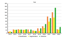
Figure 1
The results of AI-based ultrasound image processing research in classification, segmentation and detection in the past ten years."
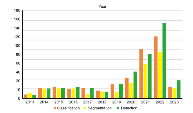
Figure 2
Keyword cluster analysis results of papers on the application of AI in ultrasound in the last decade. Different colors represent different classes, and the number label represents the size of the class. The smaller the number, the larger the corresponding class, and the higher the frequency of the keyword in papers we reviewed. In the figure, the three largest categories are: “#0 breast cancer”, “#1 computer-aided diagnosis system”, “#2 using carotid ultrasound”."
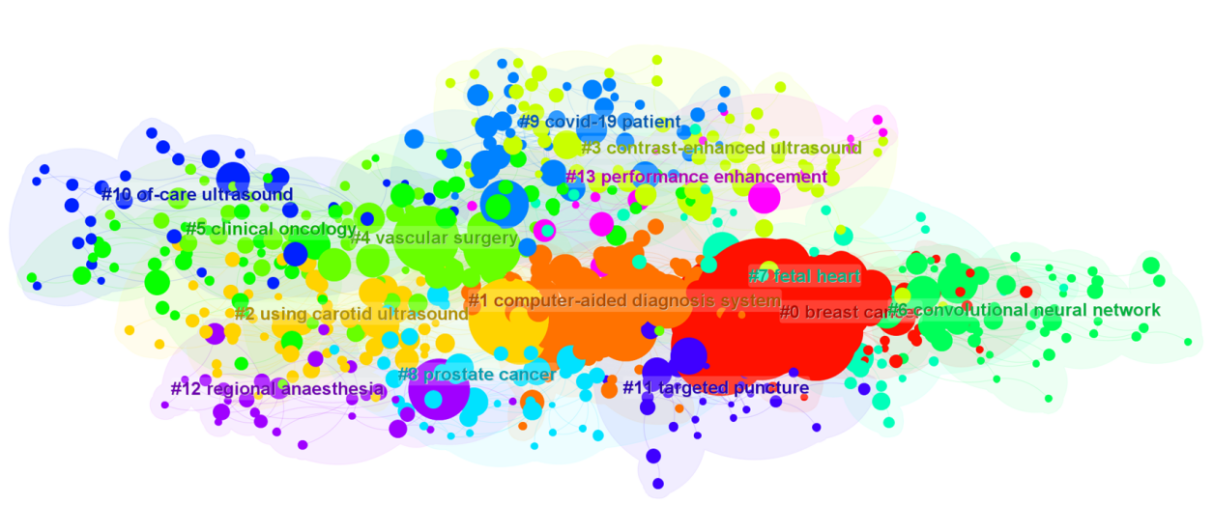
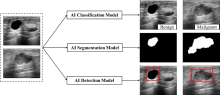
Figure 3
Application flow of AI model in ultrasound image classification, segmentation and detection. Take breast ultrasound images as an example, the image data is from Al-Dhabyani et al. [6]."
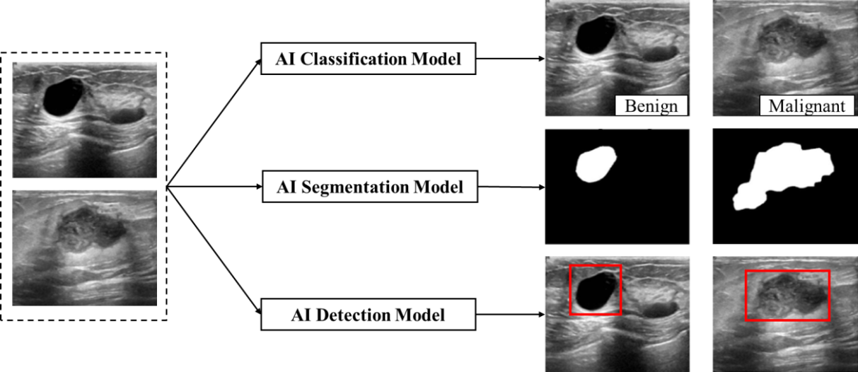
Table 1
Summary the areas of interest, methods, data sets and performance of major researches on AI in the field of medical ultrasound classification in recent years"
| Paper | Region | Method | Dataset | Performance |
|---|---|---|---|---|
| Cao et al. [ | Breast lesions | DenseNet | Private dataset Benign = 579 Malignant = 464 | APR = 0.9689 ARR = 0.6723 F1 = 0.7938 |
| Al-Dhabyani et al. [ | Breast lesions | CNN Transfer learning | BUSI dataset Total = 780 Normal = 133 Benign = 437 Malignant = 210 | Acc = 0.94 |
| Han et al. [ | Breast lesions | DDSTN | Private dataset Total = 106 Benign = 54 Malignant = 51 | Acc = 0.8679 ± 0.0154 Sen = 0.8645 ± 0.0144 Spe = 0.8731 ± 0.0437 |
| Zhou et al. [ | Breast lesions | Multi-task learning | Private dataset Total = 170 | Acc = 0.741 Rec = 0.798 Pre = 0.826 FPR = 0.392 F1 = 0.811 |
| Badawy et al. [ | Breast lesions | FCM U-Net | Private dataset Total = 1200 | Acc = 0.9544 F1 = 0.6807 |
| Bourouis et al. [ | Breast lesions | GWO-WNN | Private dataset Total = 346 Benign = 97 Malignant = 249 | Acc = 0.98 Sen = 0.988 Spe = 0.959 |
| Jabeen et al. [ | Breast lesions | CNN DarkNet53 | BUSI dataset Total = 780 Normal = 133 Benign = 437 Malignant = 210 | Acc = 0.991 |
| Ragab et al. [ | Breast lesions | VGG-16 VGG-19 SqueezeNet | BUSI dataset Total = 780 Normal = 133 Benign = 437 Malignant = 210 | Acc = 0.9709 |
| Gheflati et al. [ | Breast lesions | ViT | BUSI dataset Total = 780 Normal = 133 Benign = 437 Malignant = 210 | Acc = 0.867 AUC = 0.95 |
| Ayana et al. [ | Breast lesions | MSTL | Mendeley dataset Total = 250 Benign = 100 Malignant = 150 | Acc = 0.999 Sen = 1 Spe = 0.98 AUC = 0.999 F1 = 0.989 |
| Liu et al. [ | Thyroid nodules | Multi-branch classification network | Private dataset1 Benign = 2551 Malignant = 5139 Private dataset2 Benign = 128 Malignant = 322 | Acc = 0.971 Sen = 0.982 Spe = 0.951 |
| Kuo et al. [ | Kidney | ResNet | Private dataset Total = 4505 | Acc = 0.856 |
| Roy et al. [ | Lung | CNN | ICLUS-DB video Total = 277 | F1 = 0.61 ± 0.12 Pre = 0.70 ± 0.19 Rec = 0.60 ± 0.07 |
| Xie et al. [ | Brain | CNN | Private dataset Standard = 15372 Abnormal = 14047 | Acc = 0.963 Sen = 0.969 Spe = 0.959 |
| Sanagala et al. [ | Carotid | DCNN Transfer learning | Private dataset Total = Unknown | AUC = 0.8333, 0.9566 |
Table 2
Summary the region of interest, method, dataset, and performance of the major researches on AI in medical ultrasound segmentation in recent years"
| Paper | Region | Method | Dataset | Performance |
|---|---|---|---|---|
| Chen et al. [ | Anatomical structures | Iterative multi-domain regularized deep learning | Private dataset Train = Unknown Test = Unknown | DSC = 0.927 |
| Cui et al. [ | Ultrasound | SegNet | Private dataset Train = Unknown Test = Unknown | Unknown |
| Dangoury et al. [ | Ultrasound | V-net | Private dataset Train = 5635 Test = 5508 | DSC = 0.8501 Sen = 0.8556 Spe = 0.9987 Acc = 0.9992 |
| Yap et al. [ | Breast | CNN | Inbreast Total = 410 | AUC = 0.94 |
| Sharifzadeh et al. [ | Breast | Shift-Invariant Segmentation | US breast images dataset Train = 100 Validation = 30 Test = 33 | DSC = 0.94 JI = 0.91 |
| Gare et al. [ | Subcutaneous, Breast | W-Net | Private dataset Train = 450 Test = 50 | DSC = 0.883 |
| Yin et al. [ | Kidney | Pixelwise Classification Networks | Private dataset Total = 918 | DSC = 0.959 |
| Torres et al. [ | Kidney | BEAS framework | Private dataset Total = 45 | DSC = 0.93 |
| Chen et al. [ | Kidney | SDFNet | Private dataset Train = 450 Test = 50 | DSC = 0.941 |
| Valente et al. [ | Kidney | Deep learning method | Private dataset Training = 2166 Validation = 193 Testing = 358 | DSC = 0.94 |
| Leclerc et al. [ | heart | Deep learning | CAMUS Total = 2000 | DSC = 0.92 |
| Pu et al. [ | Fetal heart | MobileUNet-FPN | Private dataset Train = 575 Validation = 102 Test = 207 | DSC = 0.935 |
| Ma et al. [ | Thyroid | CNN | Private dataset Total = 352 | DSC = 0.901 |
| Li et al. [ | Ovary Follicle | Cr-UNet | Private dataset Train = 2509 Test = 695 | DSC = 0.9601 |
| Qiu et al. [ | Mouse Embryo | Deep Learning | Private dataset Train = Unknown Test = Unknown | ACC = 0.98 |
Table 3
Summary the region of interest, method, dataset, and performance of the major researches on AI in medical ultrasound detection in recent years"
| Paper | Region | Method | Dataset | Performance |
|---|---|---|---|---|
| Kim et al. [ | Breast cancer | Weakly-supervised deep learning method | Private dataset Train = 1000 Test = 400 | AUC = 0.86-0.96 |
| Shen et al. [ | Breast cancer | Deep learning model | NYU Breast Ultrasound Dataset Train = 3265744 Test = 1632872 Validation = 544291 | AUC = 0.976 Sen = 0.918 PPV = 0.38 |
| Niu et al. [ | Breast lesions | Grey level gradient cooccurrence Matrix analysis | Private dataset Total = 206 | BI-RADS 4A: Acc > 0.9 |
| Zhang et al. [ | Breast lesions | Lightweight neural network | Private dataset Train = 5030 Test = Unknown Validation = 1830 | Sen = 0.8925 Spe = 0.9633 Average Pre = 0.85 |
| Qian et al. [ | Breast cancer | Deep-learning system | Private dataset Train = 10815 Test = 912 Validation = Unknown | Bimodal US images: AUC= 0.880 MultimodalUS images: AUC = 0.920 |
| Baloescu et al. [ | Lung lesions | Deep learning automated algorithm | Private dataset Train = 1847 Test = 100 Validation = 468 | Sen = 0.93 Spe = 0. 96 |
| Diaz-Escobar et al. [ | Lung lesions, COVID-19 | Deep learning architectures | POCUS dataset Train = 2661 Test = Unknown Validation = 665 | Average Acc = 0.891 Balanced Acc = 0.893 COVID-19 detection AUC = 0.971 |
| Fang et al. [ | Lung lesions | CNN architectures based on transfer learning | Private dataset Train = 916 Test = Unknown Validation = Unknown | Clinical diagnosis, CXR, Chest CT: Kappa values = 0.943, 0.837, 0.835 |
| Kulhare et al. [ | Lung lesions | Single Shot CNN Model | Private dataset Train = 18713 Test = 444 Validation = Unknown | Acc = 0.89 |
| Abdel-Basset et al. [ | Lung lesions, COVID-19 | CNN | POCUS dataset Total = 3234 | Acc = 0. 934 F1 = 0.931 AUC = 0.97 |
| Choi et al. [ | Thyroid nodule | CAD system using AI | Private dataset Train = Unknown Test = Unknown Validation = Unknown | Sen = 0.907 |
| Wei et al. [ | Thyroid nodule | CNN | Private dataset Train = 5000 Test = 2214 | Acc = 0.92 |
| Wang et al. [ | Thyroid nodule | YOLOv2 neural network | Private dataset Train = 5007 Test = Unknown Validation = 351 | ROC = 0.902 Sen = 0.905 PPV = 0.9522 NPV = 0.8099 Acc = 0.9031 Spe = 0.8991 |
| Jassal et al. [ | Thyroid nodule | AI model | Private dataset Train = 857 Test = 198 Validation = Unknown | Acc = 0.89 Sen = 0.89 Spe = 0.83 F1 = 0.94 AUC = 0.86 |
| Deng et al. [ | Thyroid nodule | ResNet50 Random forest | Private dataset Train = 366 Test = 122 Validation = 122 | Sen = 0.8587 Spe = 0.9718 Acc = 0.9377 AUC = 0.982 |
| [1] |
Reddy UM, Filly RA, Copel JA. Prenatal imaging: ultrasonography and magnetic resonance imaging. Obstet Gynecol 2008; 112:145-157.
doi: 10.1097/01.AOG.0000318871.95090.d9 pmid: 18591320 |
| [2] |
Anas EMA, Seitel A, Rasoulian A, John PS, Pichora D, Darras K, et al. Bone enhancement in ultrasound using local spectrum variations for guiding percutaneous scaphoid fracture fixation procedures. Int J CARS 2015; 10:959-969.
doi: 10.1007/s11548-015-1181-6 |
| [3] |
Noble JA, Boukerroui D. Ultrasound image segmentation: a survey. IEEE Trans Med Imaging 2006; 25:987-1010.
doi: 10.1109/TMI.2006.877092 |
| [4] |
Pereira F, Bueno A, Rodriguez A, Perrin D, Marx G, Cardinale M, et al. Automated detection of coarctation of aorta in neonates from two dimensional echocardiograms. J Med Imaging 2017; 4:014502.
doi: 10.1117/1.JMI.4.1.014502 |
| [5] |
Chen C. CiteSpace II: Detecting and visualizing emerging trends and transient patterns in scientific literature. Journal of the American Society for Information Science and Technology 2006; 57:359-377.
doi: 10.1002/(ISSN)1532-2890 |
| [6] |
Al-Dhabyani W, Gomaa M, Khaled H, Fahmy A. Dataset of breast ultrasound images. Data Brief 2020; 28:104863.
doi: 10.1016/j.dib.2019.104863 |
| [7] |
Cao Z, Duan L, Yang G, Yue T, Chen Q. An experimental study on breast lesion detection and classification from ultrasound images using deep learning architectures. BMC medical imaging 2019; 19:1-9.
doi: 10.1186/s12880-018-0301-5 |
| [8] |
Al-Dhabyani W, Gomaa M, Khaled H, Fahmy A. Deep learning approaches for data augmentation and classification of breast masses using ultrasound images. Int. J. Adv. Comput. Sci. Appl 2019; 10:1-11.
doi: 10.3390/app10010001 |
| [9] | Han X, Wang J, Zhou W, Chang C, Ying S, Shi J. Deep doubly supervised transfer network for diagnosis of breast cancer with imbalanced ultrasound imaging modalities//Medical Image Computing and Computer Assisted Intervention-MICCAI 2020: 23rd International Conference, Lima, Peru, October 4-8, 2020, Proceedings, Part VI 23. Springer International Publishing; 2020:141-149. |
| [10] |
Zhou Y, Chen H, Li Y, Liu Q, Xu X, Wang S, et al. Multi-task learning for segmentation and classification of tumors in 3D automated breast ultrasound images. Medical Image Analysis 2021; 70:101918.
doi: 10.1016/j.media.2020.101918 |
| [11] | Badawy SM, Mohamed AENA, Hefnawy AA, Zidan HE, GadAllah MT, El-Banby GM. Automatic semantic segmentation of breast tumors in ultrasound images based on combining fuzzy logic and deep learning—a feasibility study. PLoS One 2021; 16:e0251899. |
| [12] |
Bourouis S, Band SS, Mosavi A, Agrawal S, Ahila A, Poongodi M, et al. Meta-heuristic algorithm-tuned neural network for breast cancer diagnosis using ultrasound images. Frontiers in Oncology 2022; 12:834028.
doi: 10.3389/fonc.2022.834028 |
| [13] |
Jabeen K, Khan MA, Alhaisoni M, Tariq U, Zhang YD, Hamza A, et al. Breast cancer classification from ultrasound images using probability-based optimal deep learning feature fusion. Sensors 2022; 22:807.
doi: 10.3390/s22030807 |
| [14] |
Ragab M, Albukhari A, Alyami J, Mansour RF. Ensemble deep-learning-enabled clinical decision support system for breast cancer diagnosis and classification on ultrasound images. Biology 2022; 11:439.
doi: 10.3390/biology11030439 |
| [15] | Gheflati B, Rivaz H. Vision transformers for classification of breast ultrasound images. Annu Int Conf IEEE Eng Med Biol Soc 2022:480-483. |
| [16] |
Ayana G, Park J, Jeong JW, Choe S. A novel multistage transfer learning for ultrasound breast cancer image classification. Diagnostics 2022; 12:135.
doi: 10.3390/diagnostics12010135 |
| [17] |
Liu T, Guo Q, Lian C, Ren X, Liang S, Yu J, et al. Automated detection and classification of thyroid nodules in ultrasound images using clinical-knowledge-guided convolutional neural networks. Medical image analysis 2019; 58:101555.
doi: 10.1016/j.media.2019.101555 |
| [18] |
Kuo CC, Chang CM, Liu KT, Lin WK, Chiang HY, Chung CW, et al. Automation of the kidney function prediction and classification through ultrasound-based kidney imaging using deep learning. NPJ digital medicine 2019; 2:29.
doi: 10.1038/s41746-019-0104-2 |
| [19] |
Roy S, Menapace W, Oei S, Luijten B, Fini E, Saltori. Deep learning for classification and localization of COVID-19 markers in point-of-care lung ultrasound. IEEE Trans Med Imaging 2020; 39:2676-2687.
doi: 10.1109/TMI.42 |
| [20] |
Xie HN, Wang N, He M, Zhang LH, Cai HM, Xian B J, et al. Using deep-learning algorithms to classify fetal brain ultrasound images as normal or abnormal. Ultrasound Obstet Gynecol 2020; 56:579-587.
doi: 10.1002/uog.21967 pmid: 31909548 |
| [21] | Skandha SS, Gupta SK, Saba L, Koppula VK, Johri AM, Khanna NN, et al. 3-D optimized classification and characterization artificial intelligence paradigm for cardiovascular/stroke risk stratification using carotid ultrasound-based delineated plaque: AtheromaticTM 2.0. Comput Biol Med 2020;125103958. |
| [22] | Chen H, Zheng Y, Park JH, Heng PA, Zhou SK, et al. Iterative multi-domain regularized deep learning for anatomical structure detection and segmentation from ultrasound images. IEEE Transactions on Medical Imaging 2019; 38:2514-2524. |
| [23] |
Cui W, Meng D, Lu K, Wang L, Zhou X, Li Y, et al. Automatic segmentation of ultrasound images using SegNet and local Nakagami distribution fitting model. Biomedical Signal Processing and Control 2023; 81:104431.
doi: 10.1016/j.bspc.2022.104431 |
| [24] | Dangoury S, Sadik M, Alali A, Fail A, Khoury R, Nassar J. V-net performances for 2D ultrasound image segmentation. IEEE 18th International Colloquium on Signal Processing & Applications (CSPA) 2022:96-100. |
| [25] | Yap MH, Pons G, Martí J, Ganau S, Sentís M, Zwiggelaar R, et al. Automated breast ultrasound lesions detection using convolutional neural networks. IIEEE J Biomed Health Inform 2018; 22:1218-1226. |
| [26] | Sharifzadeh M, Benali H, Rivaz H. Shift-invariant segmentation in breast ultrasound images. IEEE International Ultrasonics Symposium (IUS) 2021:1-4. |
| [27] |
Gare GR, Li J, Joshi R, Patel S, Kumar V, Shah P, et al. W-Net: Dense and diagnostic semantic segmentation of subcutaneous and breast tissue in ultrasound images by incorporating ultrasound RF waveform data. Medical Image Analysis 2022; 76:102326.
doi: 10.1016/j.media.2021.102326 |
| [28] |
Yin S, Peng Q, Li H, Gao X, Liu L, Feng S, et al. Automatic kidney segmentation in ultrasound images using subsequent boundary distance regression and pixelwise classification networks. Medical Image Analysis 2020; 60:101602.
doi: 10.1016/j.media.2019.101602 |
| [29] | Torres HR, Queirós S, Morais P, Fonseca JC, Vilaça JL, Oliveira C, et al. Kidney segmentation in 3-D ultrasound images using a fast phase-based approach. IEEE Trans Ultrason Ferroelectr Freq Control 2020;68:1521-1531. |
| [30] |
Chen G, Dai Y, Li R, Wang J, Wu X, Zhang L, et al. SDFNet: Automatic segmentation of kidney ultrasound images using multi-scale low-level structural feature. Expert Systems with Applications 2021; 185:115619.
doi: 10.1016/j.eswa.2021.115619 |
| [31] |
Valente S, Ferreira HA, Silva M, Oliveira R, Santos J, Gomes P, et al. A deep learning method for kidney segmentation in 2D ultrasound images. Annu Int Conf IEEE Eng Med Biol Soc 2022: 2022:3911-3914.
doi: 10.1109/EMBC48229.2022.9871748 pmid: 36086291 |
| [32] | Leclerc S, Smistad E, Pedrosa J, Østvik A, Cervenansky F, Espinosa F, et al. Deep learning for segmentation using an open large-scale dataset in 2D echocardiography. IEEE Trans Med Imaging 2019;38:2198-2210. |
| [33] |
Pu B, Wang Q, Zhang Y, Zhao H, Liu Y, Chen X, et al. MobileUNet-FPN: A semantic segmentation model for fetal ultrasound four-chamber segmentation in edge computing environments. IEEE J Biomed Health Inform 2022; 26:5540-5550.
doi: 10.1109/JBHI.2022.3182722 |
| [34] |
Ma J, Wu F, Jiang T, Zhao Q, Kong D. Ultrasound image-based thyroid nodule automatic segmentation using convolutional neural networks. Int J Comput Assist Radiol Surg 2018; 13:1685-1697.
doi: 10.1007/s11548-018-1815-6 |
| [35] |
Li H, Fang J, Liu S, Liang X, Yang X, Mai Z, et al. Cr-UNet: a composite network for ovary and follicle segmentation in ultrasound images. IEEE J Biomed Health Inform 2019; 24:974-983.
doi: 10.1109/JBHI.6221020 |
| [36] | Qiu Z, Qin Y, Duan X, Shi Y, Yang X, Qin J, et al. Automatic mouse embryo brain ventricle & body segmentation and mutant classification from ultrasound data using deep learning. IEEE International Ultrasonics Symposium (IUS) 2019:12-15. |
| [37] |
Kim J, Kim HJ, Kim C, Lee JH, Kim KW, Park YM, et al. Weakly-supervised deep learning for ultrasound diagnosis of breast cancer. Scientific Reports 2021; 11:24382.
doi: 10.1038/s41598-021-03806-7 pmid: 34934144 |
| [38] |
Shen Y, Shamout FE, Oliver JR, Witowski J, Kannan K, Park J, et al. Artificial intelligence system reduces false-positive findings in the interpretation of breast ultrasound exams. Nature communications 2021; 12:5645.
doi: 10.1038/s41467-021-26023-2 pmid: 34561440 |
| [39] |
Niu S, Huang J, Li J, Liu X, Wang D, Zhang RF, et al. Application of ultrasound artificial intelligence in the differential diagnosis between benign and malignant breast lesions of BI-RADS 4A. BMC cancer 2020; 20:1-7.
doi: 10.1186/s12885-019-6169-0 |
| [40] |
Zhang X, Lin X, Zhang Z, Dong LC, Sun XL, Sun DS, et al. Artificial intelligence medical ultrasound equipment: application of breast lesions detection. Ultrasonic Imaging 2020; 42:191-202.
doi: 10.1177/0161734620928453 |
| [41] |
Qian X, Pei J, Zheng H, Xie X, Yan L, Zhang H, et al. Prospective assessment of breast cancer risk from multimodal multiview ultrasound images via clinically applicable deep learning. Nat Biomed Eng 2021; 5:522-532.
doi: 10.1038/s41551-021-00711-2 pmid: 33875840 |
| [42] |
Baloescu C, Toporek G, Kim S, McNamara K, Liu R, Shaw M, et al. Automated lung ultrasound B-line assessment using a deep learning algorithm. IEEE Trans Ultrason Ferroelectr Freq Control 2020; 67:2312-2320.
doi: 10.1109/TUFFC.58 |
| [43] | Diaz-Escobar J, Ordóñez-Guillén NE, Villarreal-Reyes S, Galaviz-Mosqueda A, Kober V, Rivera-Rodriguez R, et al. Deep-learning based detection of COVID-19 using lung ultrasound imagery. Plos one 2021; 16:e0255886. |
| [44] |
Fang X, Li W, Huang J, Li W, Feng Q, Han Y, et al. Ultrasound image intelligent diagnosis in community-acquired pneumonia of children using convolutional neural network based transfer learning. Front Pediatr 2022; 10:1063587.
doi: 10.3389/fped.2022.1063587 |
| [45] | Kulhare S, Zheng X, Mehanian C, Gregory C, Zhu M, Gregory K, et al. Ultrasound-based detection of lung abnormalities using single shot detection convolutional neural networks. In Simulation, Image Processing, and Ultrasound Systems for Assisted Diagnosis and Navigation: International Workshops 2018;pp:65-73. |
| [46] |
Abdel-Basset M, Hawash H, Alnowibet KA, Mohamed AW, Sallam KM. Interpretable deep learning for discriminating pneumonia from lung ultrasounds. Mathematics 2022; 10:4153.
doi: 10.3390/math10214153 |
| [47] |
Choi YJ, Baek JH, Park HS, Shim WH, Kim TY, Shong YK, et al. A computer-aided diagnosis system using artificial intelligence for the diagnosis and characterization of thyroid nodules on ultrasound: initial clinical assessment. Thyroid 2017; 27:546-552.
doi: 10.1089/thy.2016.0372 pmid: 28071987 |
| [48] | Wei X, Zhu J, Zhang H, Gao H, Yu R, Liu Z, et al. Visual interpretability in computer-assisted diagnosis of thyroid nodules using ultrasound images. Med Sci Monit 2020; 26:e927007. |
| [49] |
Wang L, Yang S, Yang S, Zhao C, Tian G, Gao Y, et al. Automatic thyroid nodule recognition and diagnosis in ultrasound imaging with the YOLOv2 neural network. World J Surg Oncol 2019; 17:1-9.
doi: 10.1186/s12957-018-1541-0 |
| [50] |
Jassal K, Koohestani A, Kiu A, Strong A, Ravintharan N, Yeung M, et al. Artificial intelligence for pre-operative diagnosis of malignant thyroid nodules based on sonographic features and cytology category. World J Surg 2023; 47:330-339.
doi: 10.1007/s00268-022-06798-1 |
| [51] | Deng C, Han D, Feng M, Lv Z, Li D. Differential diagnostic value of the ResNet50, random forest, and DS ensemble models for papillary thyroid carcinoma and other thyroid nodules. J Int Med Res 2022;50. |
| [52] |
Fujioka T, Mori M, Kubota K, Kikuchi Y, Katsuta L, Adachi M, et al. Breast ultrasound image synthesis using deep convolutional generative adversarial networks. Diagnostics 2019; 9:176.
doi: 10.3390/diagnostics9040176 |
| [53] | Jarosik P, Lewandowski M, Klimonda Z, Byra M. Pixel-wise deep reinforcement learning approach for ultrasound image denoising. 2021 IEEE International Ultrasounds Symposium (IUS) 2021;1-4. |
| [54] |
Czajkowska J, Juszczyk J, Piejko L, Glenc-Ambroży M. High-frequency ultrasound dataset for deep learning-based image quality assessment. Sensors 2022; 22:1478.
doi: 10.3390/s22041478 |
| [55] |
Wang J, Yang X, Zhou B, Sohn JJ, Zhou J, Jacob JT, et al. Review of machine learning in lung ultrasound in COVID-19 pandemic. Journal of Imaging 2022; 8:65.
doi: 10.3390/jimaging8030065 |
| [1] | Fu Lijia, Li Na, Liao Ziling, Lin Yanping, Li Zhaojun, Li Fan. Advances in Breast Ultrasound Segmentation and Classification [J]. Advanced Ultrasound in Diagnosis and Therapy, 2026, 10(1): 29-41. |
| [2] | Hu Xuelin, Zhu Ye, Zhang Zisang, Quan Yuanting, Chen Wenwen, Chen Leichong, Xu Guangyu, Qin Luning, Xie Mingxing, Zhang Li. Explainable Artificial Intelligence in Echocardiography [J]. Advanced Ultrasound in Diagnosis and Therapy, 2025, 9(4): 409-425. |
| [3] | Guan Xin, Hu Xinyuan, Han Hong, Zhang Dezhi, Xu Huixiong. The Evolving Application of Ultrasound in the Precision Management of Small Hepatocellular Carcinoma [J]. Advanced Ultrasound in Diagnosis and Therapy, 2025, 9(4): 375-387. |
| [4] | Li Yanran, Cui Yuanjie, Wu Qingqing, Zhang Na. Current Applications of Artificial Intelligence in Obstetric Ultrasound [J]. Advanced Ultrasound in Diagnosis and Therapy, 2025, 9(4): 449-456. |
| [5] | Yu Xiao jie, Song Zheng lai, Chang Xue yong, Yu Jie, Liang Ping. Artificial Intelligence in Ultrasound Diagnosis of Liver Nodules: A Comprehensive Review of B-Mode and Contrast-enhanced Applications [J]. Advanced Ultrasound in Diagnosis and Therapy, 2025, 9(4): 326-346. |
| [6] | Zhong Xian, Xie Xiaoyan. Multimodal Ultrasound Radiomics in Liver Disease: Current Status and Future Directions [J]. Advanced Ultrasound in Diagnosis and Therapy, 2025, 9(4): 388-408. |
| [7] | Zhang Xiaoqian, Zhang Jingwen, Dong Yijie, Zhou Jianqiao. Research Progress and Clinical Translation of Photoacoustic–ultrasound Fusion Imaging in Breast Cancer Diagnosis and Therapy [J]. Advanced Ultrasound in Diagnosis and Therapy, 2025, 9(4): 467-482. |
| [8] | Jin Tong, Yu Xiaohu, Ai Zheng, Guo Hongcheng. Artificial Intelligence in Ultrasound Imaging: A Review of Progress from Machine Learning to Large Language Model [J]. Advanced Ultrasound in Diagnosis and Therapy, 2025, 9(4): 483-496. |
| [9] | Zheng Hairong, Meng Long, Li Fei, Niu Lili, Qiu Weibao, Ma Teng, Liu Chengbo, Zhu Xuefeng, Wan Liwen, Cai Feiyan. Advance in Ultrasound Super-resolution Imaging, Cell Manipulation and Inter-brain Communication [J]. Advanced Ultrasound in Diagnosis and Therapy, 2025, 9(4): 307-325. |
| [10] | Elkouahy Fatima Ezzahra, Bennis Ahmed, Merke Nicolas, Ouahid Hajar, Malali Hamid El, Taleb Lhoucine Ben, Mouhsen Azeddine. Advanced Diagnosis of Aortic Stenosis Disease Based on Ultrasound Images: A Novel Artificial Intelligence Approach [J]. Advanced Ultrasound in Diagnosis and Therapy, 2025, 9(3): 298-306. |
| [11] | Ye Beibei, Zhang Yifeng. Preoperative Diagnosis of Thyroid Follicular Tumor [J]. Advanced Ultrasound in Diagnosis and Therapy, 2025, 9(2): 127-137. |
| [12] | Bao Rui, Chen Lu, Luo Yukun, Zhang Mingbo. Advances in the Application of New Ultrasound Technology for the Diagnosis and Treatment of Lymphoma [J]. Advanced Ultrasound in Diagnosis and Therapy, 2025, 9(1): 32-40. |
| [13] | Xu Jiale, Xia Shujun, Hua Qing, Mei Zihan, Hou Yiqing, Wei Minyan, Lai Limei, Yang Yixuan, Zhou Jianqiao. Performance of ChatGPT and Radiology Residents on Ultrasonography Board-Style Questions [J]. Advanced Ultrasound in Diagnosis and Therapy, 2024, 8(4): 250-254. |
| [14] | Ge Xifeng, Liu Wenzheng, Chen Wen, Mei Fang, Cui Ligang. Gastroduodenal Intussusception Due to a Gastric Gastrointestinal Stromal Tumor in Adult from Sonographer's Perspective [J]. Advanced Ultrasound in Diagnosis and Therapy, 2024, 8(4): 255-258. |
| [15] | Yue Tian, MB, Yanan Zhao, MD, Yiran Huang, MB. The Diagnostic Value of Real-time Shear Wave Ultrasound Elastography in the Differentiation of Hepatic Hemangioma and Hepatocellular Carcinoma [J]. Advanced Ultrasound in Diagnosis and Therapy, 2024, 8(3): 124-129. |
| Viewed | ||||||
|
Full text |
|
|||||
|
Abstract |
|
|||||
Share: WeChat
Copyright ©2018 Advanced Ultrasound in Diagnosis and Therapy
|
 Advanced Ultrasound in Diagnosis and Therapy (AUDT)
is licensed under a Creative Commons Attribution 4.0 International License.
Advanced Ultrasound in Diagnosis and Therapy (AUDT)
is licensed under a Creative Commons Attribution 4.0 International License.