
Advanced Ultrasound in Diagnosis and Therapy ›› 2026, Vol. 10 ›› Issue (4): 403-412.doi: 10.26599/AUDT.2026.250077
• REVIEW ARTICLE • Previous Articles
Jia Lingyuna, Hua Yanga,*( )
Received:2025-07-28
Revised:2025-10-27
Accepted:2026-04-16
Online:2026-12-30
Published:2026-11-02
Contact:
Hua Yang,
E-mail:dryanghua99@163.com
Jia Lingyun, Hua Yang. Applications of Carotid Ultrasound and Transcranial Color-coded Sonography/Transcranial Doppler in Carotid Artery Revascularization. Advanced Ultrasound in Diagnosis and Therapy, 2026, 10(4): 403-412.

Table 1
Plaque-RADS categories and their imaging findings [37]."
| Plaque-RADS score | Imaging findings |
| MWT, maximum wall thickness; LRNC, lipid-rich necrotic core. | |
| 1 | Normal vessel wall |
| 2 | MWT < 3 mm |
| 3 | MWT ≥ 3 mm or healed ulcerated plaque |
| 3a | LRNC with intact fibrous cap (MWT ≥ 3 mm) |
| 3b | LRNC with thin fibrous cap (MWT ≥ 3 mm) |
| 3c | Healed ulcerated plaque |
| 4 | Complicated plaque (irrespective of MWT) |
| 4a | Intraplaque hemorrhage |
| 4b | Ruptured fibrous cap |
| 4c | Intraluminal thrombus |
Table 2
Key plaque features evaluated by ultrasound [37]"
| Plaque-RADS components | Ultrasound imaging findings |
| CEUS, contrast enhanced ultrasound; LRNC, lipid-rich necrotic core. | |
| Calcification | Hyperechogenic region with acoustic shadowing |
| LRNC | Lesion with more than 75% of its area being hypoechoic, or the plaque with gray scale median < 30 this a visible echogenic fibrous cap. |
| Fibrous cap | Hyperechogenic structure between lumen and bulk of plaque |
| Plaque ulceration | B-mode: Cavity in the plaque surface, irrespective of size, with a lower surface echogenicity than that of the adjacent plaque surface. CEUS: interruption of the plaque-lumen borders for at least 1x1 mm. |
| Intraplaque hemorrhage | Currently unsuitable to distinguish large LRNC with a thin fibrous cap from intraplaque hemorrhage. |
| Intraluminal thrombus | B-mode: Cannot distinguish large LRNC with a thin FC from intraluminal thrombus. CEUS: Hypoechoic filling defect with microbubbles delineating circumferentially the thrombus. |
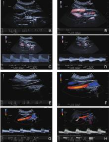
Figure 1
The ultrasound images of a 67-years old male patient with right internal carotid artery (ICA) severe (70-99%) stenosis before and after carotid artery stenting. (A). The 2-D image showed there was a heterogenous plaque at the bulb site of right ICA; (B). The CDFI image showed the local stenosis of ICA with a residual diameter was 1.2 mm; (C). The spectral waveform showed the peak systolic velocity (PSV) at the stenosis site was 514 cm/s and the end diastolic velocity (EDV) was 217 cm/s; (D). The spectral waveform of the distal ICA showed the PSV and EDV were 85.9 cm/s and 37.6 cm/s respectively; (E). The 2-D image of ICA stent with a length of stent 49.3 mm and the diameter of proximal, mid and distal of stent were 5.4 mm, 3.3 mm and 3.5 mm; (F). The CDFI image of the stent showed the blood flow within the stent was sufficient; (G). The spectral waveform showed the PSV and EDV within the stent were 92.5 cm/s and 38.8 cm/s; (H). The spectral waveform showed the PSV and EDV at the distal of ICA were 128 cm/s and 47.2 cm/s."
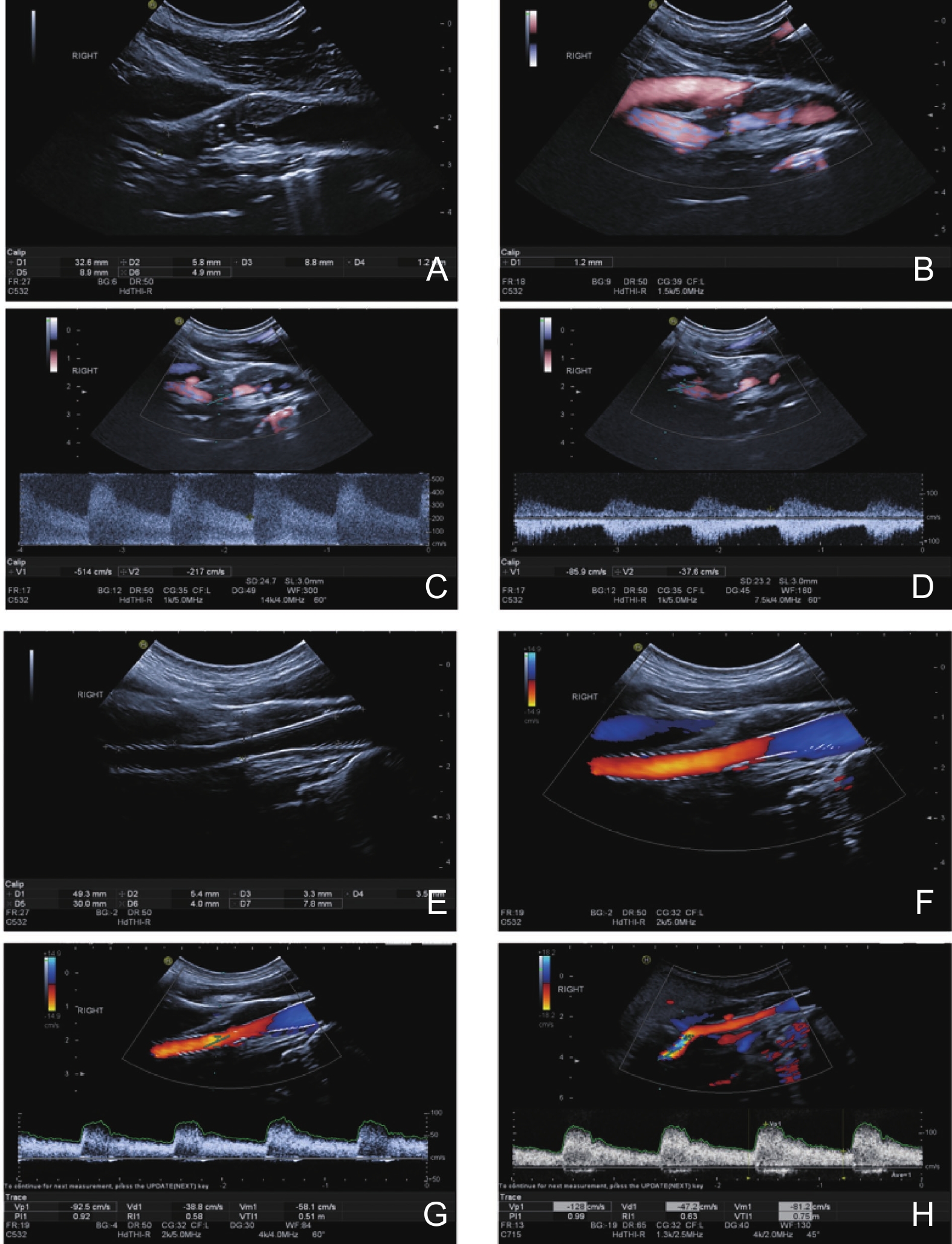
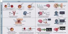
Figure 2
A schematic diagram of application of carotid ultrasound and transcranial color-coded Sonography/transcranial Doppler in carotid artery revascularization."

| [1] | Roth GA, Mensah GA, Johnson CO, Addolorato G, Ammirati E, Baddour LM, et al. GBD-NHLBI-JACC Global Burden of Cardiovascular Diseases Writing Group. Global burden of cardiovascular diseases and risk factors, 1990-2019: update from the GBD 2019 study. J Am Coll Cardiol 2020; 76: 2982-3021. |
| [2] |
Heck D, Jost A. Carotid stenosis, stroke, and carotid artery revascularization. Prog Cardiovasc Dis 2021; 65: 49-54.
doi: 10.1016/j.pcad.2021.03.005 |
| [3] | Barnett HJ, Taylor DW, Eliasziw M, Fox AJ, Ferguson GG, Haynes RB, et al. Benefit of carotid endarterectomy in patients with symptomatic moderate or severe stenosis. North American Symptomatic Carotid Endarterectomy Trial Collaborators. N Engl J Med 1998; 339: 1415-1425. |
| [4] |
European Carotid Surgery Trialists’ Collaborative Group. Randomised trial of endarterectomy for recently symptomatic carotid stenosis: final results of the MRC European Carotid Surgery Trial (ECST). Lancet 1998; 351: 1379-1387.
doi: 10.1016/S0140-6736(97)09292-1 |
| [5] | Halliday A, Mansfield A, Marro J, Peto C, Peto R, Potter J, et al. MRC Asymptomatic Carotid Surgery Trial (ACST) Collaborative Group. Prevention of disabling and fatal strokes by successful carotid endarterectomy in patients without recent neurological symptoms: randomised controlled trial. Lancet 2004; 363: 1491-1502. |
| [6] | Gurm HS, Yadav JS, Fayad P, Katzen BT, Mishkel GJ, Bajwa TK, et al. SAPPHIRE Investigators. Long-term results of carotid stenting versus endarterectomy in high-risk patients. N Engl J Med 2008; 358: 1572-1579. |
| [7] |
Hassani S, Fisher M. Management of atherosclerotic carotid artery disease: a brief overview and update. Am J Med 2022; 135: 430-434.
doi: 10.1016/j.amjmed.2021.09.027 |
| [8] |
Cuffe RL, Rothwell PM. Effect of nonoptimal imaging on the relationship between the measured degree of symptomatic carotid stenosis and risk of ischemic stroke. Stroke 2006; 37: 1785-1791.
doi: 10.1161/01.STR.0000227231.85653.ea |
| [9] |
Naylor R, Rantner B, Ancetti S, de Borst GJ, De Carlo M, Halliday A, et al. Editor's Choice - European Society for Vascular Surgery (ESVS) 2023 Clinical Practice Guidelines on the Management of Atherosclerotic Carotid and Vertebral Artery Disease. Eur J Vasc Endovasc Surg 2023; 65: 7-111.
doi: 10.1016/j.ejvs.2022.04.011 |
| [10] | Oushy SH, Essibayi MA, Savastano LE, Lanzino G. Carotid artery revascularization: endarterectomy versus endovascular therapy. J Neurosurg Sci 2021; 65: 322-326. |
| [11] | Brott TG, Howard G, Lal BK, Voeks JH, Turan TN, Roubin GS, et al. CREST-2 Investigators. Medical Management and Revascularization for Asymptomatic Carotid Stenosis. N Engl J Med 2025. |
| [12] |
Kwolek CJ, Jaff MR, Leal JI, Hopkins LN, Shah RM, Hanover TM, et al. Results of the ROADSTER multicenter trial of transcarotid stenting with dynamic flow reversal. J Vasc Surg 2015; 62: 1227-1234.
doi: 10.1016/j.jvs.2015.04.460 |
| [13] | Kleindorfer DO, Towfighi A, Chaturvedi S, Cockroft KM, Gutierrez J, Lombardi-Hill D, et al. 2021 Guideline for the prevention of stroke in patients with stroke and transient ischemic attack: a guideline from the American Heart Association/American Stroke Association. Stroke 2021; 52: e364-e467. |
| [14] |
AbuRahma AF, Avgerinos ED, Chang RW, Darling RC 3rd, Duncan AA, Forbes TL, et al. Society for vascular surgery clinical practice guidelines for management of extracranial cerebrovascular disease. J Vasc Surg 2022; 75: 4S-22S.
doi: 10.1016/j.jvs.2022.03.339 |
| [15] | Pellerito JS, Polak JF. Introduction to vascular ultrasonography. 7th edn. Elsevier Press: Amsterd 2019. |
| [16] |
Kargiotis O, Safouris A, Magoufis G, Georgala M, Roussopoulou A, Stamboulis E, et al. The role of neurosonology in the diagnosis and management of patients with carotid artery disease: a review. J Neuroimaging 2018; 28: 239-251.
doi: 10.1111/jon.12495 |
| [17] | Grant EG, Benson CB, Moneta GL, Alexandrov AV, Baker JD, Bluth EI, et al. Carotid artery stenosis: gray-scale and Doppler US diagnosis--Society of Radiologists in Ultrasound Consensus Conference. Radiology 2003; 229: 340-346. |
| [18] | Hua Y, Liu BB, Ling C, Duan C, Liu Q, Miao ZR, et al. Accurate assessment of the diagnosis between 50-69% and 70-99% carotid stenoses with ultrasonography. Chinese J Cerebrovasc Dis 2006; 3: 211-218. |
| [19] |
The Professional Committee of Vascular Ultrasound of Stroke Prevention, Treatment Expert, Committee of the National Health Commission; The Professional Committee of Superficial Organ, Peripheral Vascular Ultrasound of the Chinese Medical Ultrasound Engineering; The Professional Committee of Craniocerebral, Cervical Vascular Ultrasound of the Chinese Medical Ultrasound Engineering. Expert consensus on some issues of cerebral and carotid vascular ultrasonography. Advanced Ultrasound in Diagnosis and Therapy 2021; 5: 153-162.
doi: 10.37015/AUDT.2021.200057 |
| [20] |
Xia M, Hua Y, Jia L, Liu B, Jiao L, Ma Y. Effect of anterior communicating artery patency on the flow velocity in bilateral carotid artery stenosis after carotid endarterectomy. Vasc Med 2023; 28: 308-314.
doi: 10.1177/1358863X231171611 |
| [21] |
Brinjikji W, Huston J 3rd, Rabinstein AA, Kim GM, Lerman A, Lanzino G. Contemporary carotid imaging: from degree of stenosis to plaque vulnerability. J Neurosurg 2016; 124: 27-42.
doi: 10.3171/2015.1.JNS142452 |
| [22] |
Naghavi M, Libby P, Falk E, Casscells SW, Litovsky S, Rumberger J, et al. From vulnerable plaque to vulnerable patient: a call for new definitions and risk assessment strategies: Part I. Circulation 2003; 108: 1664-1672.
doi: 10.1161/01.CIR.0000087480.94275.97 |
| [23] | Vergallo R, Park SJ, Stone GW, Erlinge D, Porto I, Waksman R, et al. Vulnerable or high-risk plaque: a JACC: cardiovascular imaging position statement. JACC Cardiovasc Imaging 2025; 18: 709-740. |
| [24] |
Clevert DA, Sommer WH, Zengel P, Helck A, Reiser M. Imaging of carotid arterial diseases with contrast-enhanced ultrasound (CEUS). Eur J Radiol 2011; 80: 68-76.
doi: 10.1016/j.ejrad.2010.12.103 |
| [25] |
Yao J, van Sambeek MR, Dall'Agata A, van Dijk LC, Kozakova M, Koudstaal PJ, et al. Three-dimensional ultrasound study of carotid arteries before and after endarterectomy; analysis of stenotic lesions and surgical impact on the vessel. Stroke 1998; 29: 2026-2031.
doi: 10.1161/01.STR.29.10.2026 |
| [26] |
Mishra B, Pandit AK, Miyachi S, Ohshima T, Kawaguchi R, Vishnu VY, et al. Clinical utility of intravascular ultrasound (IVUS) in carotid artery interventions: a systematic review and meta-analysis. J Endovasc Ther 2022; 29: 678-691.
doi: 10.1177/15266028211064824 |
| [27] |
Zhou F, Hua Y, Ji X, Jia L, Zhang K, Li Q, et al. Ultrasound-based carotid plaque characteristics help predict new cerebral ischemic lesions after endarterectomy. Ultrasound Med Biol 2021; 47: 244-251.
doi: 10.1016/j.ultrasmedbio.2020.09.025 |
| [28] |
Zhou F, Hua Y, Ji X, Jia L. A systemic review into carotid plaque features as predictors of restenosis after carotid endarterectomy. J Vasc Surg 2021; 73: 2179-2188.
doi: 10.1016/j.jvs.2020.10.084 |
| [29] |
Cao J, Zeng Y, Zhou Y, Yao Z, Tan Z, Huo G, et al. The value of contrast-enhanced ultrasound in assessing carotid plaque vulnerability and predicting stroke risk. Sci Rep 2025; 15: 5850.
doi: 10.1038/s41598-025-90319-2 |
| [30] |
Alonso A, Artemis D, Hennerici MG. Molecular imaging of carotid plaque vulnerability. Cerebrovasc Dis 2015; 39: 5-12.
doi: 10.1159/000369123 |
| [31] |
Xu X, Hua Y, Wang L, Hou W, Xia M. Correlation between risk factors of cerebrovascular disease and calcified plaque characteristics in patients with atherosclerotic severe carotid stenosis. Neurol Res 2020; 42: 83-89.
doi: 10.1080/01616412.2019.1710403 |
| [32] |
Tao Y, Hua Y, Jia L, Jiao L, Liu B. Risk factors for residual Stenosis after carotid artery stenting. Front Neurol 2021; 11: 606924.
doi: 10.3389/fneur.2020.606924 |
| [33] |
Jia L, Hua Y, Jiao L, Ma Y, Xing Y, Wang L, et al. Effects of plaque characteristics and artery hemodynamics on the residual stenosis after carotid artery stenting. J Vasc Surg 2023; 78: 428-435.
doi: 10.1016/j.jvs.2023.03.500 |
| [34] |
Xu X, Feng Y, Bai X, Ma Y, Wang Y, Chen Y, et al. Risk factors for silent new ischemic cerebral lesions following carotid artery stenting. Neuroradiology 2020; 62: 1177-1184.
doi: 10.1007/s00234-020-02447-3 |
| [35] |
Saba L, Nardi V, Cau R, Gupta A, Kamel H, Suri JS, et al. Carotid artery plaque calcifications: lessons from histopathology to diagnostic imaging. Stroke 2022; 53: 290-297.
doi: 10.1161/STROKEAHA.121.035692 |
| [36] |
Barrett HE, Van der Heiden K, Farrell E, Gijsen FJH, Akyildiz AC. Calcifications in atherosclerotic plaques and impact on plaque biomechanics. J Biomech 2019; 87: 1-12.
doi: 10.1016/j.jbiomech.2019.03.005 |
| [37] | Saba L, Cau R, Murgia A, Nicolaides AN, Wintermark M, Castillo M, et al. Carotid Plaque-RADS: A Novel Stroke Risk Classification System. JACC Cardiovasc Imaging 2024; 17: 62-75. |
| [38] | The Stroke Prevention and Treatment Project Committee, National Health and Family Planning Commission of China. Chinese guidelines for stroke prevention and treatment. People’s Medical Publishing House. Beijing 2021. |
| [39] | Jiao LQ, Song G, Hua Y, Ma Y, Chen YF, Wang YB, et al. Recanalization of extracranial internal carotid artery occlusion: a 12-yearretrospective study. Neural Regen Res 2013; 8: 2204-2206. |
| [40] |
Liu YM, Jia LY, Liu BB, Meng XF, Yang J, Li JZ, et al. Evaluation of endarterectomy recanalization under ultrasound guidance in symptomatic patients with carotid artery occlusion. PLoS One 2015; 10: e0144381.
doi: 10.1371/journal.pone.0144381 |
| [41] |
Wang GJ, Beck AW, DeMartino RR, Goodney PP, Rockman CB, Fairman RM. Insight into the cerebral hyperperfusion syndrome following carotid endarterectomy from the national Vascular Quality Initiative. J Vasc Surg 2017; 65: 381-389.
doi: 10.1016/j.jvs.2016.07.122 |
| [42] |
Banga PV, Varga A, Csobay-Novák C, Kolossváry M, Szántó E, Oderich GS, et al. Incomplete circle of Willis is associated with a higher incidence of neurologic events during carotid eversion endarterectomy without shunting. J Vasc Surg 2018; 68: 1764-1771.
doi: 10.1016/j.jvs.2018.03.429 |
| [43] |
Hetzel A, von Reutern G, Wernz MG, Droste DW, Schumacher M. The carotid compression test for therapeutic occlusion of the internal carotid artery. Comparison of angiography with transcranial Doppler sonography. Cerebrovasc Dis 2000; 10: 194-199.
doi: 10.1159/000016056 |
| [44] |
Tang SC, Huang YW, Shieh JS, Huang SJ, Yip PK, Jeng JS. Dynamic cerebral autoregulation in carotid stenosis before and after carotid stenting. J Vasc Surg 2008; 48: 88-92.
doi: 10.1016/j.jvs.2008.02.025 |
| [45] |
Reinhard M, Roth M, Müller T, Guschlbauer B, Timmer J, Czosnyka M, et al. Effect of carotid endarterectomy or stenting on impairment of dynamic cerebral autoregulation. Stroke 2004; 35: 1381-1387.
doi: 10.1161/01.STR.0000127533.46914.31 |
| [46] | Li N, Zhou F, Lu X, Chen H, Liu R, Chen S, et al. Impaired dynamic cerebral autoregulation as a predictor for cerebral hyperperfusion after carotid endarterectomy: A prospective observational study. World Neurosurg 2023;S1878-8750. |
| [47] |
Manojlovic V, Budakov N, Budinski S, Milosevic D, Nikolic D, Manojlovic V. Cerebrovacular reserve predicts the cerebral hyperperfusion syndrome after carotid endarterectomy. J Stroke Cerebrovasc Dis 2020; 29: 105318.
doi: 10.1016/j.jstrokecerebrovasdis.2020.105318 |
| [48] | Martinelli O, Benedetti-Valentini F. Trancranial Doppler: value in clinical practice. Int Angiol 2009; 28: 249-253. |
| [49] |
Ackerstaff RGA. Cerebral circulation monitoring in carotid endarterectomy and carotid artery stenting. Front Neurol Neurosci 2006; 21: 229-238.
doi: 10.1159/000092435 |
| [50] | Basic identification criteria of Doppler microembolic signals. Consensus Committee of the Ninth International Cerebral Hemodynamic Symposium. Stroke 1995; 26: 1123 |
| [51] | Faraglia V, Palombo G, Stella N, Taurino M, Iocca ML, Romano A, et al. Cerebral embolization in patients undergoing protected carotid-artery stenting and carotid surgery. J Cardiovasc Surg (Torino) 2007; 48: 683-688. |
| [52] |
Ackerstaff RG, Suttorp MJ, van den Berg JC, Overtoom TT, Vos JA, Bal ET, ; Antonius Carotid Endarterectomy, Angioplasty Study Group, et al. Prediction of early cerebral outcome by transcranial Doppler monitoring in carotid bifurcation angioplasty and stenting. J Vasc Surg 2005; 41: 618-624.
doi: 10.1016/j.jvs.2005.01.034 |
| [53] |
Wolf O, Heider P, Heinz M, Poppert H, Sander D, Greil O, et al. Microembolic signals detected by transcranial Doppler sonography during carotid endarterectomy and correlation with serial diffusion-weighted imaging. Stroke 2004; 35: e373-375.
doi: 10.1161/01.str.0000143184.69343.ec |
| [54] |
Almekhlafi MA, Demchuk AM, Mishra S, Bal S, Menon BK, Wiebe S, et al. Malignant emboli on transcranial Doppler during carotid stenting predict postprocedure diffusion-weighted imaging lesions. Stroke 2013; 44: 1317-1322.
doi: 10.1161/STROKEAHA.111.000659 |
| [55] |
Aburahma AF, Mousa AY, Stone PA. Shunting during carotid endarterectomy. J Vasc Surg 2011; 54: 1502-1510.
doi: 10.1016/j.jvs.2011.06.020 |
| [56] |
Chuatrakoon B, Nantakool S, Rerkasem A, Orrapin S, Howard DP, Rerkasem K. Routine or selective carotid artery shunting for carotid endarterectomy (and different methods of monitoring in selective shunting). Cochrane Database Syst Rev 2022; 6: CD000190.
doi: 10.1002/14651858.cd000190.pub2 |
| [57] |
Panneton JM, Berger MW, Lewis BD, Hallett JW Jr, Bower TC, Gloviczki P, et al. Intraoperative duplex ultrasound during carotid endarterectomy. J Vasc Surg 2001; 35: 1-9.
doi: 10.1177/153857440103500102 |
| [58] |
Knappich C, Kuehnl A, Tsantilas P, Schmid S, Breitkreuz T, Kallmayer M, et al. Intraoperative completion studies, local anesthesia, and antiplatelet medication are associated with lower risk in carotid endarterectomy. Stroke 2017; 48: 955-962.
doi: 10.1161/STROKEAHA.116.014869 |
| [59] |
Normahani P, Khan B, Sounderajah V, Poushpas S, Anwar M, Jaffer U. Applications of intraoperative Duplex ultrasound in vascular surgery: a systematic review. Ultrasound J 2021; 13: 8.
doi: 10.1186/s13089-021-00208-8 |
| [60] |
Yokoya S, Furutani Y, Nishimura Y, Oka H, Hino A. String-like mobile echodensities on postoperative ultrasound after CEA: A retrospective observational study. J Clin Neurosci 2022; 101: 175-179.
doi: 10.1016/j.jocn.2022.04.025 |
| [61] | Arquizan C, Trinquart L, Touboul PJ, Long A, Feasson S, Terriat B, et al. EVA-3S Investigators. Restenosis is more frequent after carotid stenting than after endarterectomy: the EVA-3S study. Stroke 2011; 42: 1015-1020. |
| [62] |
Xin WQ, Li MQ, Li K, Li QF, Zhao Y, Wang WH, et al. Systematic and comprehensive comparison of incidence of restenosis between carotid endarterectomy and carotid artery stenting in patients with atherosclerotic carotid Stenosis. World Neurosurg 2019; 125: 74-86.
doi: 10.1016/j.wneu.2019.01.118 |
| [63] |
Vernhet H, Jean B, Lust S, Laroche JP, Bonafé A, Sénac JP, et al. Wall mechanics of the stented extracranial carotid artery. Stroke 2003; 34: e222-224.
doi: 10.1161/01.str.0000092490.21761.7c |
| [64] | Lal BK, Hobson RW 2nd, Goldstein J, Chakhtoura EY, Durán WN. Carotid artery stenting: is there a need to revise ultrasound velocity criteria?. J Vasc Surg 2004; 39: 58-66. |
| [65] |
Jia L, Hua Y, Li J, Duan C, Zhou Y, Jiao L. Optimal ultrasound criteria for defining the severity of vertebral artery in-stent restenosis. Ultrasound Med Biol 2015; 41: 775-780.
doi: 10.1016/j.ultrasmedbio.2014.10.009 |
| [66] |
Spies C, Doshi R, Spoon J, Snell RJ. Carotid artery stent type influences duplex ultrasonography derived peak systolic velocity: findings of an in-vitro model. Catheter Cardiovasc Interv 2007; 70: 309-315.
doi: 10.1002/ccd.21224 |
| [67] |
Lal BK, Hobson RW 2nd, Tofighi B, Kapadia I, Cuadra S, Jamil Z. Duplex ultrasound velocity criteria for the stented carotid artery. J Vasc Surg 2008; 47: 63-73.
doi: 10.1016/j.jvs.2007.09.038 |
| [68] |
AbuRahma AF, Abu-Halimah S, Bensenhaver J, Dean LS, Keiffer T, Emmett M, et al. Optimal carotid duplex velocity criteria for defining the severity of carotid in-stent restenosis. J Vasc Surg 2008; 48: 589-594.
doi: 10.1016/j.jvs.2008.04.004 |
| [69] |
Bitsko LJ, Ryer EJ, Penn EP, Salzler GG, Major M, Irvan J, et al. Defining duplex ultrasound criteria for in-Stent restenosis of the carotid artery using computed tomographic angiography. Cureus 2022; 14: e26700.
doi: 10.7759/cureus.26700 |
| [70] |
Pizzolato R, Hirsch JA, Romero JM. Imaging challenges of carotid artery in-stent restenosis. J Neurointerv Surg 2014; 6: 32-41.
doi: 10.1136/neurintsurg-2012-010618 |
| [71] |
AbuRahma AF, Stone P, Deem S, Dean LS, Keiffer T, Deem E. Proposed duplex velocity criteria for carotid restenosis following carotid endarterectomy with patch closure. J Vasc Surg 2009; 50: 286-291.
doi: 10.1016/j.jvs.2009.01.065 |
| [72] |
Lin YH, Liu HM. Update on cerebral hyperperfusion syndrome. J Neurointerv Surg 2020; 12: 788-793.
doi: 10.1136/neurintsurg-2019-015621 |
| [73] |
Fujimoto S, Toyoda K, Inoue T, Hirai Y, Uwatoko T, Kishikawa K, et al. Diagnostic impact of transcranial color-coded real-time sonography with echo contrast agents for hyperperfusion syndrome after carotid endarterectomy. Stroke 2004; 35: 1852-1856.
doi: 10.1161/01.STR.0000133131.93900.ff |
| [74] | Ogasawara K, Sakai N, Kuroiwa T, Hosoda K, Iihara K, Toyoda K, et al. Japanese Society for Treatment at Neck in Cerebrovascular Disease Study Group. Intracranial hemorrhage associated with cerebral hyperperfusion syndrome following carotid endarterectomy and carotid artery stenting: retrospective review of 4494 patients. J Neurosurg 2007; 107: 1130-1136. |
| [75] |
Li Q, Hua Y, Liu J, Zhou F, Du L, Li J, et al. Intraoperative transcranial Doppler monitoring predicts the risk of cerebral hyperperfusion syndrome after carotid endarterectomy. World Neurosurg 2022; 165: e571-580.
doi: 10.1016/j.wneu.2022.06.100 |
| [1] | Yumei Liu, MD, PhD, Yang Hua, MD, Yabing Wang, MD, PhD, Nan Zhang, MS, Ting Ma, MD, PhD, Yue Zhao, MS, Na Li, MS, Na Lei, MS, Ran Liu, MS. Decreased Cerebral Flow Velocities from General Anesthesia are Not Associated with Cerebral Hyperperfusion Syndrome [J]. Advanced Ultrasound in Diagnosis and Therapy, 2023, 7(3): 248-253. |
| [2] | Yanting Wang, MD, Zhenzhen Hu, MD, Yu He, MD, PhD. New Vessels or MCA Stem: A Case of Asymptomatic Chronic Middle Cerebral Artery Occlusion on TCCS [J]. Advanced Ultrasound in Diagnosis and Therapy, 2021, 5(2): 109-111. |
| Viewed | ||||||
|
Full text |
|
|||||
|
Abstract |
|
|||||
Share: WeChat
Copyright ©2018 Advanced Ultrasound in Diagnosis and Therapy
|
 Advanced Ultrasound in Diagnosis and Therapy (AUDT)
is licensed under a Creative Commons Attribution 4.0 International License.
