
Advanced Ultrasound in Diagnosis and Therapy ›› 2026, Vol. 10 ›› Issue (3): 158-168.doi: 10.26599/AUDT.2026.250112
• REVIEW ARTICLE • Previous Articles Next Articles
Jiang Xingtinga,1, Fang Wenlingb,1, Hu Panpanc,1, Yu Rongd, Ma Jinge, Wang Ninge, Wu Tianyud, Ye Lind, Ruan Wenhuie, Li Zhaojunb,c,d,e,f,*( ), Jin Lina,*()
Received:2025-11-30
Revised:2026-01-20
Accepted:2026-02-06
Online:2026-09-30
Published:2026-10-12
Contact:
Li Zhaojun, Jin Lin,
E-mail:lzj_1975@sina.com;jinlin205@163.com
About author:First author contact:1 Xingting Jiang and Wenling Fang and Panpan Hu contributed equally to this study.
Jiang Xingting, Fang Wenling, Hu Panpan, Yu Rong, Ma Jing, Wang Ning, Wu Tianyu, Ye Lin, Ruan Wenhui, Li Zhaojun, Jin Lin. Fetal Ventricular-arterial Coupling: Development, Physiology, and Ultrasonographic Advances. Advanced Ultrasound in Diagnosis and Therapy, 2026, 10(3): 158-168.
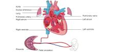
Figure 1
Fetal cardiac circulation. Deoxygenated blood returns via the superior and inferior venae cavae to the right atrium and is ejected by the right ventricle (RV) into the pulmonary artery. Owing to high pulmonary vascular resistance, most RV output bypasses the lungs through the ductus arteriosus, enters the systemic circulation, and returns to the placenta via the umbilical arteries."

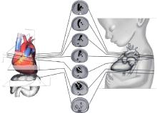
Figure 2
Anatomical structures examined in the four cardiac chambers and outflow tracts of the fetal heart [32]."
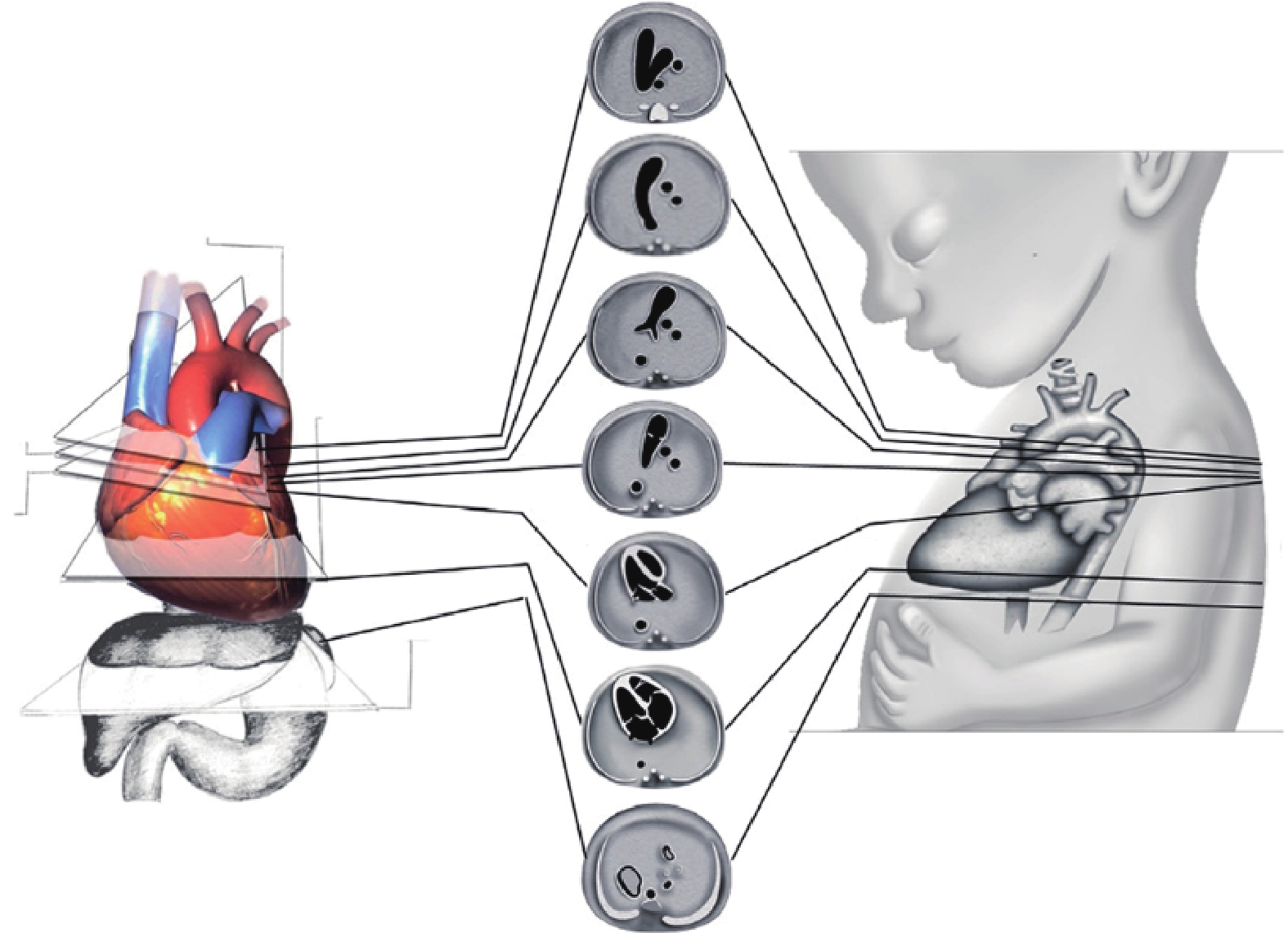

Table 1
Fetal cardiac function assessment indicators."
| Index | Main point |
| GLS, global longitudinal strain; SV, stroke volume; EDVD, end-diastolic ventricular dimension; ESVD, end-systolic ventricular dimension; MPI, myocardial performance index; ICT, isovolumetric contraction time; IRT, isovolumetric relaxation time; ET, ejection time. | |
| Cardiothoracic ratio | From a 4-chamber (4C) view, Cardiomegaly if > 0.35; Severe if > 0.50 [ |
| Global longitudinal strain (GLS) | −21.5 ± 2.0% at 28-32 weeks [ |
| Cardiac output (CO) | CO=SV x heart rate; Median combined CO: 425 mL/min/kg; Right/left CO: 1.4 [ |
| Ejection fraction (EF) | Reference values > 50-60% [ |
| Fractional shortening (FS) | (EDVD - ESVD)/EDVD; Reference values > 28% [ |
| Tei index or Myocardial performance index (MPI) | (ICT + IRT)/ET; 0.39-0.55 at 11- 41 weeks [ |
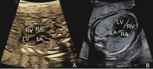
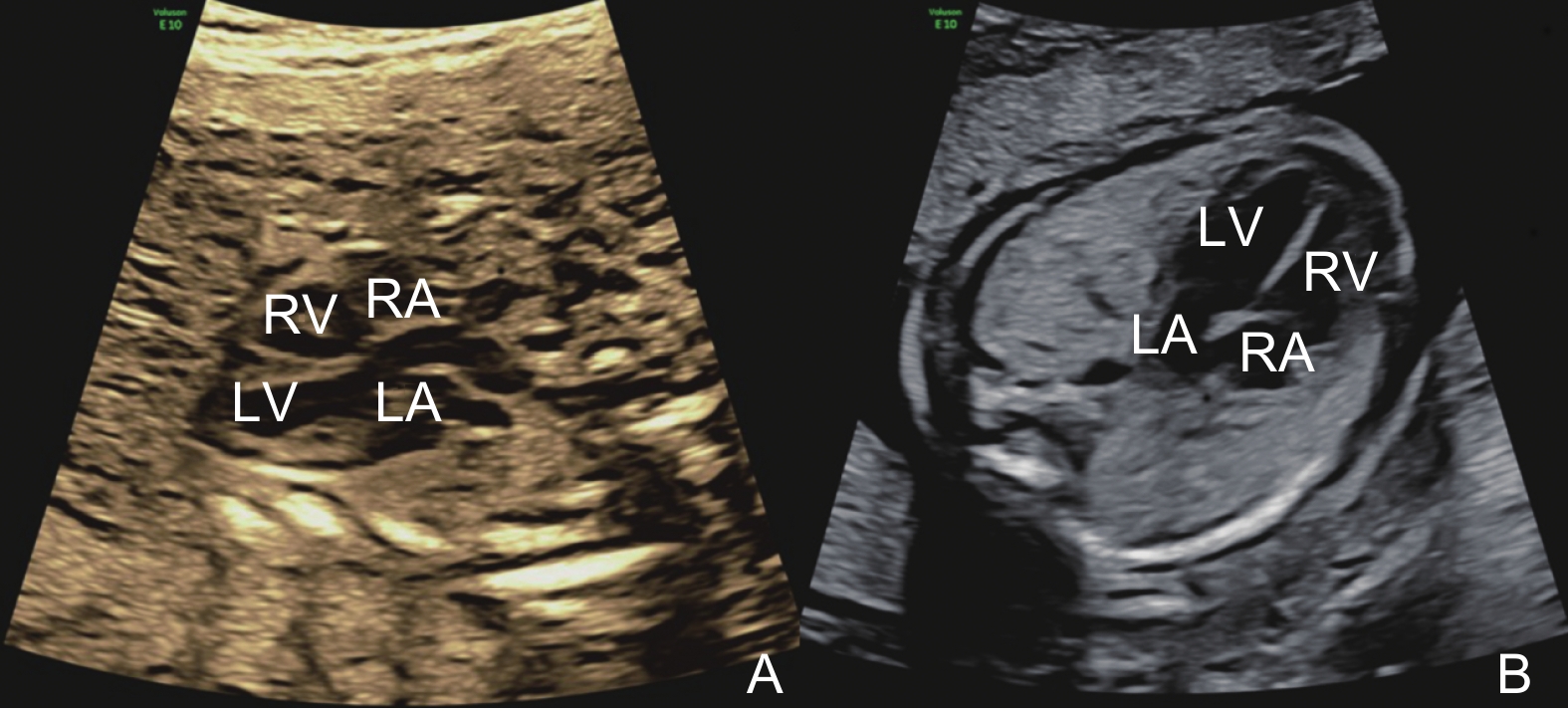
Figure 3
Ultrasound images of the fetal heart. (A) is the normal fetal heart, (B) is the fetal heart in a case of Fetal Growth Restriction (FGR). RA, right atrium; RV, right ventricle; LV, left ventricle; LA, left atrium."
| [1] |
Frenneaux M, Williams L. Ventricular-arterial and ventricular-ventricular interactions and their relevance to diastolic filling. Prog Cardiovasc Dis 2007; 49: 252-262.
doi: 10.1016/j.pcad.2006.08.004 |
| [2] |
Zhenyi Li, Ya Chen, Wang Xinqi, Yang Lan, Chen Anni, Li Zhaojun, et al. Left and right ventricular interaction: insight from echocardiography imaging. Advanced Ultrasound in Diagnosis and Therapy 2024; 8: 195-204.
doi: 10.37015/AUDT.2024.240058 |
| [3] |
Tan CMJ, Lewandowski AJ. The transitional heart: from early embryonic and fetal development to neonatal Life. Fetal Diagn Ther 2020; 47: 373-386.
doi: 10.1159/000501906 |
| [4] |
Acharya G, Gui Y, Cnota W, Huhta J, Wloch A. Human embryonic cardiovascular function. Acta Obstet Gynecol Scand 2016; 95: 621-628.
doi: 10.1111/aogs.12860 |
| [5] |
Chen CH, Fetics B, Nevo E, Rochitte CE, Chiou KR, Ding PA, et al. Noninvasive single-beat determination of left ventricular end-systolic elastance in humans. J Am Coll Cardiol 2001; 38: 2028-2034.
doi: 10.1016/S0735-1097(01)01651-5 |
| [6] |
Zhang X, Yang Y, Zhang R, Zhang L, Xie Y, Wu W, et al. Noninvasive evaluation of left ventricular-arterial coupling: methodologies and clinical relevance. Advanced Ultrasound in Diagnosis and Therapy 2024; 8: 149-158.
doi: 10.37015/AUDT.2024.240063 |
| [7] | Abdulla R, Blew GA, Holterman MJ. Cardiovascular embryology. Pediatr Cardiol 2004; 25: 191-200. |
| [8] |
Odackal NJ, Crume M, Naik T, Stiver C. Cardiac development and related clinical considerations. Neoreviews 2024; 25: 401-414.
doi: 10.1542/neo.25-7-e401 |
| [9] |
Matthews J, Rajakumar B, Carreon CK, Morton SU. Placental-heart axis: an evolutionary perspective. Int J Mol Sci 2024; 25: 11212.
doi: 10.3390/ijms252011212 |
| [10] |
Gauster M, Moser G, Wernitznig S, Kupper N, Huppertz B. Early human trophoblast development: from morphology to function. Cell Mol Life Sci 2022; 79: 345.
doi: 10.1007/s00018-022-04377-0 |
| [11] |
Mahadevan A, Tipler A, Jones H. Shared developmental pathways of the placenta and fetal heart. Placenta 2023; 141: 35-42.
doi: 10.1016/j.placenta.2022.12.006 |
| [12] |
Benagiano G, Mancuso S, Guo SW, Di Renzo GC. Events leading to the establishment of pregnancy and placental formation: the need to fine-tune the nomenclature on pregnancy and gestation. Int J Mol Sci 2023; 24: 15420.
doi: 10.3390/ijms242015420 |
| [13] |
Matthiesen NB, Henriksen TB, Agergaard P, Gaynor JW, Bach CC, Hjortdal VE, et al. Congenital heart defects and indices of placental and fetal growth in a nationwide study of 924 422 liveborn infants. Circulation 2016; 134: 1546-1556.
doi: 10.1161/CIRCULATIONAHA.116.021793 |
| [14] |
Zubrzycki M, Schramm R, Costard-Jäckle A, Grohmann J, Gummert JF, Zubrzycka M, et al. Cardiac development and factors influencing the development of congenital heart defects (CHDs): part I. Int J Mol Sci 2024; 25: 7117.
doi: 10.3390/ijms25137117 |
| [15] |
Buijtendijk MFJ, Barnett P, van den Hoff MJB. Development of the human heart. Am J Med Genet C Semin Med Genet 2020; 184: 7-22.
doi: 10.1002/ajmg.c.31778 |
| [16] | Villavicencio-Guzmán L, Sánchez-Gómez C, Jaime-Cruz R, Ramírez-Fuentes TC, Patiño-Morales CC, Salazar-García M, et al. Human heart morphogenesis: a new vision based on in vivo labeling and cell tracking. Life 2023;13.165. |
| [17] |
Bulatovic I, Månsson-Broberg A, Sylvén C, Grinnemo K-H. Human fetal cardiac progenitors: the role of stem cells and progenitors in the fetal and adult heart. Best Practice & Research Clinical Obstetrics & Gynaecology 2016; 31: 58-68.
doi: 10.1016/j.bpobgyn.2015.08.008 |
| [18] |
Combémorel N, Cavell N, Tyser RCV. Early heart development: examining the dynamics of function-form emergence. Biochemical Society Transactions 2024; 52: 1579-1589.
doi: 10.1042/BST20230546 |
| [19] |
Mielke G, Benda N. Cardiac output and central distribution of blood flow in the human fetus. Circulation 2001; 103: 1662-1668.
doi: 10.1161/01.CIR.103.12.1662 |
| [20] |
Rudolph AM, Heymann MA. The fetal circulation. Annu Rev Med 1968; 19: 195-206.
doi: 10.1146/annurev.me.19.020168.001211 |
| [21] |
Mann D, Mehta V. Cardiovascular embryology. Int Anesthesiol Clin 2004; 42: 15-28.
doi: 10.1097/00004311-200404240-00004 |
| [22] | Little WC, Cheng CP. Left ventricular-arterial coupling in conscious dogs. Am J Physiol 1991; 261: 70-76. |
| [23] | Little WC, Cheng CP. Effect of exercise on left ventricular-arterial coupling assessed in the pressure-volume plane. Am J Physiol 1993; 264: 1629-1633. |
| [24] |
Chakkarapani AA, Roehr CC, Hooper SB, Te Pas AB, Gupta S. Transitional circulation and hemodynamic monitoring in newborn infants. Pediatr Res 2024; 96: 595-603.
doi: 10.1038/s41390-022-02427-8 |
| [25] |
Gao Y, Raj JU. Regulation of the pulmonary circulation in the fetus and newborn. Physiol Rev 2010; 90: 1291-1335.
doi: 10.1152/physrev.00032.2009 |
| [26] |
Jain A, Mohamed A, Kavanagh B, Shah PS, Kuipers BCW, El-Khuffash A, et al. Cardiopulmonary adaptation during first day of life in human neonates. J Pediatr 2018; 200: 50-57.
doi: 10.1016/j.jpeds.2018.04.051 |
| [27] | Yee K, Lui F. Anatomy, Thorax, Heart foramen ovale. 2023 Jul 24. In: StatPearls [Internet]. Treasure Island (FL): StatPearls Publishing 2026. |
| [28] |
Sørensen K, Fadnes S, Myklebust T, Løvstakken L, Mertens L, Nyrnes SA, et al. Changes in left ventricular diastolic flow dynamics in the neonatal transition period and beyond. Am J Physiol Heart Circ Physiol 2025; 328: 1333-1343.
doi: 10.1152/ajpheart.00214.2025 |
| [29] |
Crispi F, Valenzuela-Alcaraz B, Cruz-Lemini M, Gratacós E. Ultrasound assessment of fetal cardiac function. Australas J Ultrasound Med 2013; 16: 158-167.
doi: 10.1002/j.2205-0140.2013.tb00242.x |
| [30] |
Moon-Grady AJ, Donofrio MT, Gelehrter S, Hornberger L, Kreeger J, Lee W, et al. Guidelines and recommendations for performance of the fetal echocardiogram: an update from the american society of echocardiography. J Am Soc Echocardiogr 2023; 36: 679-723.
doi: 10.1016/j.echo.2023.04.014 |
| [31] |
DeVore GR, Klas B, Cuneo B, Satou G, Sklansky M. Review of speckle tracking analysis to measure the size, shape, and contractility of the fetal heart in fetuses with congenital heart defects. Echocardiography 2024; 41: 15870.
doi: 10.1111/echo.15870 |
| [32] |
Dong J, Liu S, Liao Y, Wen H, Lei B, Li S, et al. A Generic Quality Control Framework for Fetal Ultrasound Cardiac Four-Chamber Planes. IEEE J Biomed Health Inform 2020; 24: 931-942.
doi: 10.1109/JBHI.2019.2948316 |
| [33] |
Muñoz H, Enríquez G, Ortega X, Pinto M, Hosiasson S, Germain A, et al. Diagnóstico de cardiopatías congénitas: ecografía de cribado, ecocardiografía fetal y medicina de precisión. Revista Médica Clínica Las Condes 2023; 34: 44-56.
doi: 10.1016/j.rmclc.2023.01.001 |
| [34] |
International Society of Ultrasound in O, Gynecology, Carvalho JS, Allan LD, Chaoui R, Copel JA, et al. ISUOG practice guidelines (updated): sonographic screening examination of the fetal heart. Ultrasound Obstet Gynecol 2013; 41: 348-359.
doi: 10.1002/uog.12403 |
| [35] | Cardiac screening examination of the fetus: guidelines for performing the 'basic' and 'extended basic' cardiac scan. Ultrasound Obstet Gynecol 2006;27:107-113. |
| [36] |
Young ID, Clarke M. Lethal malformations and perinatal mortality: a 10 year review with comparison of ethnic differences. Br Med J (Clin Res Ed) 1987; 295: 89-91.
doi: 10.1136/bmj.295.6590.89 |
| [37] |
Lee J. Hemodynamics in doppler ultrasonography. Ultrasonography 2024; 43: 413-423.
doi: 10.14366/usg.24126 |
| [38] |
Gómez-Montes E, Herraiz I, Villalain C, Galindo A. Second trimester echocardiography. Best Pract Res Clin Obstet Gynaecol 2025; 100: 102592.
doi: 10.1016/j.bpobgyn.2025.102592 |
| [39] |
DeVore GR. Assessing fetal cardiac ventricular function. Semin Fetal Neonatal Med 2005; 10: 515-541.
doi: 10.1016/j.siny.2005.08.009 |
| [40] |
Hata T, Takayoshi R, Sugihara M, Koyanagi A, Miyake T. Transvaginal slowflowHD for embryonic and fetal hearts: human cardiac development in first trimester of pregnancy. Ultrasound in Obstetrics & Gynecology 2024; 65: 662-666.
doi: 10.1002/uog.29095 |
| [41] |
Hata T, Koyanagi A, Yamanishi T, Bouno S, Takayoshi R, Miyake T, et al. Fetal abdominal blood vessels and organ microvasculature detected by slowflow HD. Ultrasound Obstet Gynecol 2020; 56: 955-957.
doi: 10.1002/uog.22043 |
| [42] |
Youssef L, Castellani R, Valenzuela-Alcaraz B, Sepulveda-Martinez Á, Crovetto F, Crispi F, et al. Cardiac remodeling from the fetus to adulthood. J Clin Ultrasound 2023; 51: 249-264.
doi: 10.1002/jcu.23336 |
| [43] |
Słodki M, Respondek-Liberska M, Pruetz JD, Donofrio MT. Fetal cardiology: changing the definition of critical heart disease in the newborn. J Perinatol 2016; 36: 575-580.
doi: 10.1038/jp.2016.20 |
| [44] |
Wang L, Fu X, Ren X, Guo Y, Zhou H, Yue H, et al. Evaluation of fetal cardiac morphology and function in hypertensive disorders of pregnancy using Fetal HQ technology combined with uterine artery ultrasound parameters. Front Cardiovasc Med 2025; 12: 1558034.
doi: 10.3389/fcvm.2025.1558034 |
| [45] |
Hata T, Koyanagi A, Yamanishi T, Bouno S, Takayoshi R, AboEllail MAM, et al. Evaluation of 24-segment sphericity index of fetal heart using FetalHQ. J Matern Fetal Neonatal Med 2022; 35: 4573-4579.
doi: 10.1080/14767058.2020.1856808 |
| [46] |
Huluta I, Wright A, Cosma LM, Hamed K, Nicolaides KH, Charakida M, et al. Fetal cardiac function at midgestation in women who subsequently develop gestational diabetes. JAMA Pediatrics 2023; 177: 718-725.
doi: 10.1001/jamapediatrics.2023.1174 |
| [47] |
Comas M, Crispi F. Assessment of fetal cardiac function using tissue doppler techniques. Fetal Diagn Ther 2012; 32: 30-38.
doi: 10.1159/000335028 |
| [48] |
Palalioglu RM, Erbiyik HI, Kaya B, Kiyak H, Gedikbasi A. Investigation of fetal cardiac function using tissue doppler imaging in fetuses compromised by growth restriction. Ginekol Pol 2021; 92: 195-204.
doi: 10.5603/gp.a2020.0175 |
| [49] |
Peixoto AB, Bravo-Valenzuela NJM, Martins WP, Słodki M, Mattar R, Moron AF, et al. Impact of type I and type II maternal diabetes mellitus on fetal cardiac function assessment parameters using spectral and tissue doppler. Int J Cardiovasc Imaging 2020; 36: 1237-1247.
doi: 10.1007/s10554-020-01821-9 |
| [50] |
Di Salvo G, Russo MG, Paladini D, Felicetti M, Castaldi B, Tartaglione A, et al. Two-dimensional strain to assess regional left and right ventricular longitudinal function in 100 normal foetuses. Eur J Echocardiogr 2008; 9: 754-756.
doi: 10.1093/ejechocard/jen134 |
| [51] |
van Oostrum NHM, de Vet CM, Clur SB, van der Woude DAA, van den Heuvel ER, Oei SG, et al. Fetal myocardial deformation measured with two-dimensional speckle-tracking echocardiography: longitudinal prospective cohort study of 124 healthy fetuses. Ultrasound Obstet Gynecol 2022; 59: 651-659.
doi: 10.1002/uog.24781 |
| [52] |
Crispi F, Sepulveda-Swatson E, Cruz-Lemini M, Rojas-Benavente J, Garcia-Posada R, Dominguez JM, et al. Feasibility and reproducibility of a standard protocol for 2D speckle tracking and tissue doppler-based strain and strain rate analysis of the fetal heart. Fetal Diagn Ther 2012; 32: 96-108.
doi: 10.1159/000337329 |
| [53] | Badano LP, Boccalini F, Muraru D, Bianco LD, Peluso D, Bellu R, et al. Current clinical applications of transthoracic three-dimensional echocardiography. J Cardiovasc Ultrasound 2012; 20: 1-22. |
| [54] | Lang RM, Badano LP, Mor-Avi V, Afilalo J, Armstrong A, Ernande L, et al. Recommendations for cardiac chamber quantification by echocardiography in adults: an update from the american society of echocardiography and the european association of cardiovascular imaging. J Am Soc Echocardiogr 2015; 28: 1-39. |
| [55] |
Shimada YJ, Shiota T. A meta-analysis and investigation for the source of bias of left ventricular volumes and function by three-dimensional echocardiography in comparison with magnetic resonance imaging. Am J Cardiol 2011; 107: 126-138.
doi: 10.1016/j.amjcard.2010.08.058 |
| [56] |
Dorosz JL, Lezotte DC, Weitzenkamp DA, Allen LA, Salcedo EE. Performance of 3-dimensional echocardiography in measuring left ventricular volumes and ejection fraction: a systematic review and meta-analysis. J Am Coll Cardiol 2012; 59: 1799-1808.
doi: 10.1016/j.jacc.2012.01.037 |
| [57] |
Muraru D, Badano LP, Ermacora D, Piccoli G, Iliceto S. Sources of variation and bias in assessing left ventricular volumes and dyssynchrony using three-dimensional echocardiography. Int J Cardiovasc Imaging 2012; 28: 1357-1368.
doi: 10.1007/s10554-011-9985-0 |
| [58] |
Viñals F, Poblete P, Giuliano A. Spatio-temporal image correlation (STIC): a new tool for the prenatal screening of congenital heart defects. Ultrasound Obstet Gynecol 2003; 22: 388-394.
doi: 10.1002/uog.883 |
| [59] |
Avnet H, Thomas S, Erenbourg A, Yagel S, Welsh A. Evaluating fetal tricuspid and mitral annular plane systolic excursion (TAPSE and MAPSE) using spatiotemporal image correlation (STIC) M-mode. J Perinat Med 2024; 52: 645-653.
doi: 10.1515/jpm-2023-0423 |
| [60] |
Ebbing C, Kessler J, Moster D, Rasmussen S. Single umbilical artery and risk of congenital malformation: population-based study in norway. Ultrasound Obstet Gynecol 2020; 55: 510-515.
doi: 10.1002/uog.20359 |
| [61] |
Zhang J, Xiao S, Zhu Y, Zhang Z, Cao H, Xie M, et al. Advances in the application of artificial intelligence in fetal echocardiography. J Am Soc Echocardiogr 2024; 37: 550-561.
doi: 10.1016/j.echo.2023.12.013 |
| [62] |
Taksoee-Vester CA, Mikolaj K, Bashir Z, Christensen AN, Petersen OB, Sundberg K, et al. AI supported fetal echocardiography with quality assessment. Sci Rep 2024; 14: 5809.
doi: 10.1038/s41598-024-56476-6 |
| [63] | Tay J, Foo L, Masini G, Bennett PR, McEniery CM, Wilkinson IB, et al. Early and late preeclampsia are characterized by high cardiac output, but in the presence of fetal growth restriction, cardiac output is low: insights from a prospective study. Am J Obstet Gynecol 2018; 218: 511-517. |
| [64] |
Crispi F, Bijnens B, Figueras F, Bartrons J, Eixarch E, Le Noble F, et al. Fetal growth restriction results in remodeled and less efficient hearts in children. Circulation 2010; 121: 2427-2436.
doi: 10.1161/CIRCULATIONAHA.110.937995 |
| [65] |
Sarvari SI, Rodriguez-Lopez M, Nuñez-Garcia M, Sitges M, Sepulveda-Martinez A, Camara O, et al. Persistence of cardiac remodeling in preadolescents with fetal growth restriction. Circ Cardiovasc Imaging 2017; 10: e005270.
doi: 10.1161/CIRCIMAGING.116.005270 |
| [66] |
Cruz-Lemini M, Crispi F, Valenzuela-Alcaraz B, Figueras F, Sitges M, Bijnens B, et al. Fetal cardiovascular remodeling persists at 6 months in infants with intrauterine growth restriction. Ultrasound Obstet Gynecol 2016; 48: 349-356.
doi: 10.1002/uog.15767 |
| [67] |
Tintu A, Rouwet E, Verlohren S, Brinkmann J, Ahmad S, Crispi F, et al. Hypoxia induces dilated cardiomyopathy in the chick embryo: mechanism, intervention, and long-term consequences. PLoS One 2009; 4: 5155.
doi: 10.1371/journal.pone.0005155 |
| [68] | Ling HZ, Guy GP, Bisquera A, Nicolaides KH, Kametas NA. Maternal cardiac adaptation and fetal growth. Am J Obstet Gynecol 2021; 224: 601. |
| [69] |
Marinari E, Rizza A, Iacobelli R, Iodice F, Favia I, Romagnoli S, et al. Ventricular-arterial coupling in children and infants with congenital heart disease after cardiopulmonary bypass surgery: observational study. Pediatr Crit Care Med 2019; 20: 753-758.
doi: 10.1097/PCC.0000000000001982 |
| [70] |
Connor JA, Thiagarajan R. Hypoplastic left heart syndrome. Orphanet J Rare Dis 2007; 2: 23.
doi: 10.1186/1750-1172-2-23 |
| [71] |
Patey O, Hornberger LK, McBrien A, Lin L, Khoo NS, Eckersley L. Perinatal cardiac functional adaptation in hypoplastic left heart syndrome: a longitudinal analysis. J Am Soc Echocardiogr 2024; 37: 1062-1072.
doi: 10.1016/j.echo.2024.06.020 |
| [72] |
Carter E, Rogers LS. Transposition of the great arteries: anatomy, physiology and surgical outcomes today. Curr Opin Pediatr 2025; 37: 419-424.
doi: 10.1097/MOP.0000000000001495 |
| [73] |
Pasqualin G, Misra A, Gauvreau K, Sanders SP, Desai AS, Alexander A, et al. Ventricular-arterial coupling is associated with clinical outcomes in patients with systemic right ventricle. Am Heart J 2025; 285: 29-38.
doi: 10.1016/j.ahj.2025.02.013 |
| [74] | Haas NA, Driscoll DJ, Rickert-Sperling S. Clinical presentation and therapy of tetralogy of fallot and double-outlet right ventricle. Adv Exp Med Biol 2024; 1441: 617-627. |
| [75] | Horenstein MS, Diaz-Frias J, Guillaume M. Tetralogy of Fallot.StatPearls. Treasure Island (FL): StatPearls Publishing Copyright © 2025, StatPearls Publishing LLC 2025. |
| [76] |
Geva T, Wald RM, Bucholz E, Cnota JF, McElhinney DB, Mercer-Rosa LM, et al. Long-term management of right ventricular outflow tract dysfunction in repaired tetralogy of fallot: a scientific statement from the american heart association. Circulation 2024; 150: 689-707.
doi: 10.1161/cir.0000000000001291 |
| [77] |
Kim SJ, Li MH, Noh CI, Kim SH, Lee CH, Yoon JK, et al. Impact of pulmonary arterial elastance on right ventricular mechanics and exercise capacity in repaired tetralogy of fallot. Korean Circ J 2023; 53: 406-417.
doi: 10.4070/kcj.2022.0228 |
| [78] |
Buddhe S, Jani V, Sarikouch S, Gaur L, Schuster A, Beerbaum P, et al. Differences in right ventricular-pulmonary vascular coupling and clinical indices between repaired standard tetralogy of Fallot and repaired tetralogy of Fallot with pulmonary atresia. Diagn Interv Imaging 2021; 102: 85-91.
doi: 10.1016/j.diii.2020.05.008 |
| [79] | Youssef L, Miranda J, Paules C, Garcia-Otero L, Vellvé K, Kalapotharakos G, et al. Fetal cardiac remodeling and dysfunction is associated with both preeclampsia and fetal growth restriction. Am J Obstet Gynecol 2020; 222: 79.71-79. |
| [80] |
Hendem DU, Oluklu D, Beser DM, Yildirim M, Turgut E, Tanacan A, et al. Evaluation of fetal cardiac functions in preeclampsia: does the severity or proteinuria affect fetal cardiac functions?. J Ultrasound Med 2023; 42: 2415-2424.
doi: 10.1002/jum.16268 |
| [81] |
Yuan LJ, Duan YY, Xue D, Cao TS, Zhou N. Ultrasound study of carotid and cardiac remodeling and cardiac-arterial coupling in normal pregnancy and preeclampsia: a case control study. BMC Pregnancy Childbirth 2014; 14: 113.
doi: 10.1186/1471-2393-14-113 |
| [82] | Huhta J. Congestive heart failure in the fetus. In: Yagel S, Silverman NH, Gembruch U, editors. Fetal cardiology. second ed. New York: Informa Healthcare USA, Inc 2009. p.561-578. |
| [83] |
Rocha LA, Rolo LC, Araujo Júnior E. How to perform a functional assessment of the fetal heart: a pictorial review. Ultrasonography 2019; 38: 365-373.
doi: 10.14366/usg.18065 |
| [84] |
De Vore GR, Siassi B, Platt LD. Use of femur length as a means of assessing m-mode ventricular dimensions during second and third trimesters of pregnancy in normal fetus. J Clin Ultrasound 1985; 13: 619-625.
doi: 10.1002/j.1097-0096.1985.tb00003.x |
| [85] |
Cruz-Martínez R, Figueras F, Bennasar M, García-Posadas R, Crispi F, Hernández-Andrade E, et al. Normal reference ranges from 11 to 41 weeks' gestation of fetal left modified myocardial performance index by conventional Doppler with the use of stringent criteria for delimitation of the time periods. Fetal Diagn Ther 2012; 32: 79-86.
doi: 10.1159/000330798 |
| [1] | Guo Fangqi, Lin Jing, Yang Qirui, Ji Qianwen, Zhao Jiaqi. Discussion on the Application of Combined Transthoracic and Transesophageal Echocardiography in the 'Four-Step Repair Method' for Treating Rheumatic Heart Valve Disease and Its Short-Term Outcomes [J]. Advanced Ultrasound in Diagnosis and Therapy, 2026, 10(3): 203-215. |
| [2] | Hu Xuelin, Zhu Ye, Zhang Zisang, Quan Yuanting, Chen Wenwen, Chen Leichong, Xu Guangyu, Qin Luning, Xie Mingxing, Zhang Li. Explainable Artificial Intelligence in Echocardiography [J]. Advanced Ultrasound in Diagnosis and Therapy, 2025, 9(4): 409-425. |
| [3] | Feng Qing, Yang Huihui, Xu Wanting, He Yu. Application of Two-Dimensional Speckle Tracking Echocardiography in Evaluation of Neonatal Pulmonary Hypertension [J]. Advanced Ultrasound in Diagnosis and Therapy, 2025, 9(3): 254-259. |
| [4] | Lohith Kumar Bittugondanahalli Prakash, Shivakumar Neeraj, Gaduputi Jahnavi, Kashif Mohammed S, K Praneethi, Reddy Manda Pranay, S Sampangi Ramaiah, Krishnamurthy Umesh, Prabhakar Suman. Comparative Analysis of Fetal Ventricular Function: AGA vs. SGA Fetuses Using 2D Speckle-Tracking [J]. Advanced Ultrasound in Diagnosis and Therapy, 2025, 9(3): 290-297. |
| [5] | Elkouahy Fatima Ezzahra, Bennis Ahmed, Merke Nicolas, Ouahid Hajar, Malali Hamid El, Taleb Lhoucine Ben, Mouhsen Azeddine. Advanced Diagnosis of Aortic Stenosis Disease Based on Ultrasound Images: A Novel Artificial Intelligence Approach [J]. Advanced Ultrasound in Diagnosis and Therapy, 2025, 9(3): 298-306. |
| [6] | Qin Shuxuan, He Qing, Wu Zhenni, Lin Yixia, Ji Mengmeng, Zhang Li, Xie Mingxing, Li Yuman. Clinical Utility of Speckle Tracking Echocardiography in Heart Transplantation [J]. Advanced Ultrasound in Diagnosis and Therapy, 2025, 9(2): 103-116. |
| [7] | Liu Jianjun, Zhang Jun, Xue Yafang, Chen Ying, Qiu Mei, Guo Yanli. Ultrasonographic Characteristics and Outcomes of Fetal Umbilical-portal-systemic Venous Shunts: A Single-center Study [J]. Advanced Ultrasound in Diagnosis and Therapy, 2025, 9(2): 181-188. |
| [8] | Yang Lan, Li Zhenyi, Chen Ya, Chen Anni, Wang Xinqi, Jin Lin, Li Zhaojun. Clinical Usefulness of Atrioventricular Coupling in Cardiovascular Disease [J]. Advanced Ultrasound in Diagnosis and Therapy, 2025, 9(1): 1-9. |
| [9] | Chen Anni, Yang Lan, Li Zhenyi, Wang Xinqi, Chen Ya, Jin Lin, Li Zhaojun. Left Ventricular-Arterial Coupling in Cardiovascular Health: Development, Assessment Methods, and Future Directions [J]. Advanced Ultrasound in Diagnosis and Therapy, 2024, 8(4): 159-171. |
| [10] | Zhang Xin, Yang Yun, Zhang Ruize, Zhang Linyue, Xie Yuji, Wu Wenqian, Zhang Jing, Lv Qing, Wang Jing, Xie Mingxing. Noninvasive Evaluation of Left Ventricular-Arterial Coupling: Methodologies and Clinical Relevance [J]. Advanced Ultrasound in Diagnosis and Therapy, 2024, 8(4): 149-158. |
| [11] | Yang Yun, Zhang Xin, Zhang Ruize, Jiang Jingrong, Xie Yuji, Fang Lingyun, Zhang Jing, Xie Mingxing, Wang Jing. Current Status and Progress in Arterial Stiffness Evaluation: A Comprehensive Review [J]. Advanced Ultrasound in Diagnosis and Therapy, 2024, 8(4): 172-182. |
| [12] | Li Zhenyi, Chen Ya, Wang Xinqi, Yang Lan, Chen Anni, Li Zhaojun, Jin Lin. Left and Right Ventricular Interaction: Insight from Echocardiography Imaging [J]. Advanced Ultrasound in Diagnosis and Therapy, 2024, 8(4): 195-204. |
| [13] | Wang Xinqi, Chen Anni, Yang Lan, Chen Ya, Li Zhenyi, Li Zhaojun, Jin Lin. Evaluation Methods and Progress of Right Ventricular-pulmonary Artery Coupling [J]. Advanced Ultrasound in Diagnosis and Therapy, 2024, 8(4): 205-216. |
| [14] | Junrong Hong, MD, Pingyang Zhang, MD, PhD, Mengyao Fei, MD, Lingling Wang, MD. A Study on Left Atrial Function in Patients with Essential Hypertension Using Four-Dimensional Echocardiography [J]. Advanced Ultrasound in Diagnosis and Therapy, 2024, 8(2): 64-73. |
| [15] | Hao Feng, MM, Yaqin Sun, MM, Jingjing Zhang, MM, Jiajia Wang, MM, Shuai Han, MM, Shumin Wang, PhD. Ultrasound Assessment of Effect of Maternal Thyroid Function During Pregnancy on Fetal and Neonatal Bone Development [J]. Advanced Ultrasound in Diagnosis and Therapy, 2024, 8(2): 41-48. |
| Viewed | ||||||
|
Full text |
|
|||||
|
Abstract |
|
|||||
Share: WeChat
Copyright ©2018 Advanced Ultrasound in Diagnosis and Therapy
|
 Advanced Ultrasound in Diagnosis and Therapy (AUDT)
is licensed under a Creative Commons Attribution 4.0 International License.
