
Advanced Ultrasound in Diagnosis and Therapy ›› 2026, Vol. 10 ›› Issue (3): 137-147.doi: 10.26599/AUDT.2026.250063
• REVIEW ARTICLE • Previous Articles Next Articles
Li Zuoyana, Du Lianfangb, Li Fanc,*( )
Received:2025-07-18
Revised:2025-11-16
Accepted:2025-11-24
Online:2026-09-30
Published:2026-10-12
Contact:
Li Fan,
E-mail:medicineli@sjtu.edu.cn
Li Zuoyan, Du Lianfang, Li Fan. Ultrasound Image Segmentation in Cardiovascular Disease: A Review of Techniques and Clinical Applications. Advanced Ultrasound in Diagnosis and Therapy, 2026, 10(3): 137-147.

Figure 1
General workflow of ultrasound image segmentation, comprising preprocessing, ROI localization, and pixel-wise segmentation, which supports clinical tasks such as defect detection and quantitative analysis."

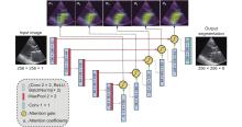
Figure 2
This picture shows the architecture of the Attention UNet with deep supervision. Attention gates generate spatial coefficient maps (α1–α5) that highlight the thin, rapidly moving mitral valve leaflets while suppressing responses from surrounding myocardium and blood cavity. This selective focusing improves boundary delineation of low-contrast, fast-moving valve tissue. Deep supervision further stabilizes the attention weights across training epochs, ensuring consistent localization throughout the cardiac cycle. Together, these mechanisms address the major challenges of echocardiographic segmentation, poor contrast, motion blur, and leaflet thinness, thereby yielding more temporally consistent and anatomically reliable results. (Reprinted with permission from copyright Elsevier) [38]"
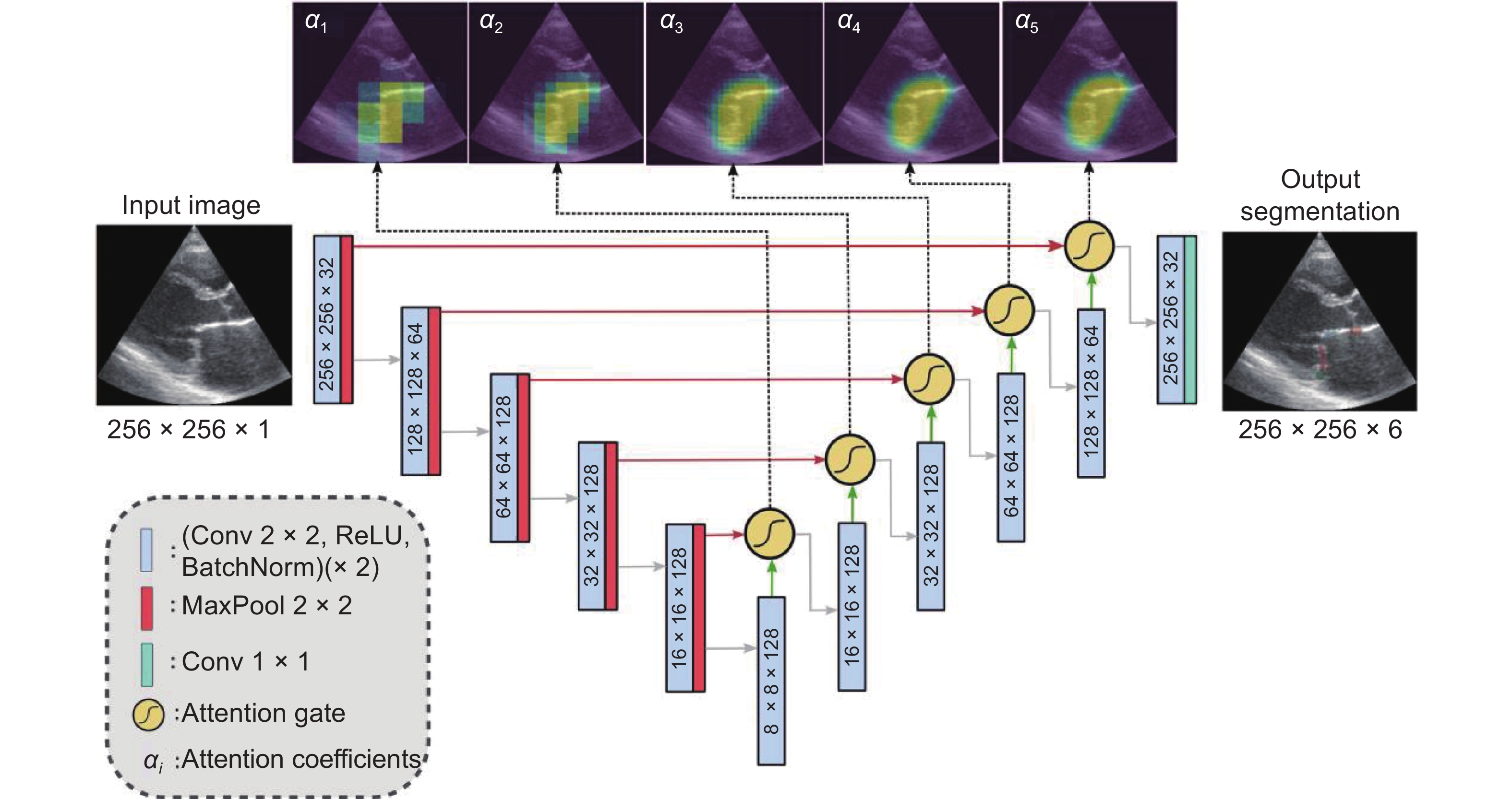
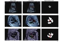
Figure 3
The sample of annotated image by maternal–fetal clinician for heart defect detection in case: (A) ASD; (B) VSD; and (C) AVSD. In the annotation, the green line is RA, the red line is LA, the purple line is RV, the blue line is LV, and the yellow line is defective. (Reprinted with permission from copyright MDPI) [47]"
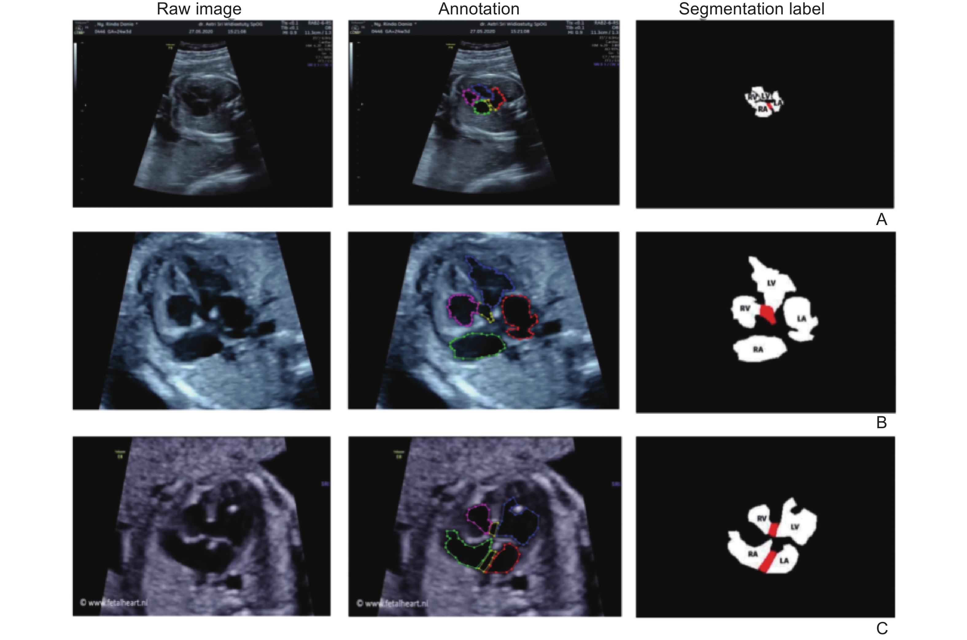
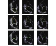
Figure 4
These nine images illustrate segmentation outcomes across three echocardiographic views. Images (A, D, G) correspond to apical two-chamber views with normal mitral valves, (B, E, H) represent apical three-chamber views with functional MR, and (C, F, I) show apical four-chamber views with mitral valve prolapse. The Dice scores for LA, LV and MV were 0.935, 0.915 and 0.757, respectively, indicating strong performance in normal and functional MR cases but reduced accuracy in prolapse, where leaflet thickening, asymmetric deformation and blurred coaptation boundaries make segmentation substantially more challenging. (Reprinted with permission from MDPI) [60]"

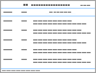
Table 1
Representative literature on heart segmentation"
| Category | Author | Model | Content | Dataset | Evaluation |
| VHD, valvular heart disease; SVM, support vector machines; CDFI, color doppler flow imaging; AV, aortic valve; MV, mitral valve; LR, logistics regression; ANN, artificial neural network; NB, naive bayes; RF, random forest; STS, the Society of Thoracic Surgeons; ACC, The American College of Cardiology; TVT, transcatheter valve therapy; TAVR, transcatheter aortic valve replacement; AUC, area under curve; DL, deep learning; CNN, convolutional neural network; LA, left atrium; AUROC, area under receiver operating characteristic curve; LSTM, long short-term memory; AS, aortic stenosis; MS, mitral stenosis; MVP, mitral valve prolapse; MR, mitral regurgitation; CHD, congenital heart disease; XGBoost, eXtreme Gradient Boosting; NLP, natural language processing; RSF, random survival forest; PMVSD, perimembranous ventricular septal defects; ML, machine learning; IoU, Intersection over Union; Echo, echocardiography; DICOM, digital imaging and communications in medicine; DMM, dynamic metric map; ACM, active contour model; FNF: false negative fraction; FPF: false positive fraction; TPF: the positive fraction; LV, left ventricular; ED, end diastolic; ES, end systole; DBN, deep belief network; PCA, principal component analysis; EF, ejection fractions; LVEF, left ventricular ejection fractions. | |||||
| VHD | Çomak et al. [ | LS-SVM (ML) | Identify abnormalities in valvular CDFI | Patients: 215 | AUC: 0.950 |
| Avci [ | GSVM (ML) | Classify the CDFI of heart valve diseases | Patients: 215 | Accuracy: 0.950 | |
| Hernandezet al. [ | LR, ANN, NB, RF (ML) | Predict in-hospital mortality after TAVR | Patients: 10883 | AUC: 0.920 | |
| Vrudhula et al. [ | 3D CNN (DL) | Detect mitral regurgitation severity | Videos: 80833 | AUC: 0.998 | |
| Holste et al. [ | CNN (DL) | Detect severe aortic stenosis | Videos: 17570 | AUROC: 0.978 | |
| Yazan et al. [ | CNN, LSTM (DL) | Classify different heart valvular diseases | Videos: 4541 | Accuracy: 0.979 | |
| CHD | Yan et al.[ | SVM, LR, RF, XGBoost (ML) | Predict postoperative arrhythmia | Patients: 1384 | AUC: 0.863 |
| Sun et al. [ | NLP, RSF (ML) | Predict spontaneous closure of PMVSD. | Patients: 29142 | AUC: 0.950 | |
| Nurmaini et al. [ | Mask-RCNN (DL) | Segment and detect congenital heart defects | Images: 1149 | Dice: 0.983 | |
| Nova et al. [ | U-Net, V-Net (DL) | Segment cardiac septal defects from 2D Echo images | Images: 2609 | Accuracy: 0.991 | |
| Tan et al. [ | CNN (DL) | Detect CHD from fetal US images | Patients: 100 | Precision: 0.890 Recall: 0.820 | |
| Jiang et al. [ | ResNet50 (DL) | Classify seven CHD views | Patients: 1411 Images: 14838 | Accuracy: 0.923 | |
| Nemri et al. [ | Swin Transformer, U-Net (DL) | Segment echocardiographic images | Videos: 10030 | Dice: 0.870 | |
| Ventricle | Huang et al. [ | DMM-ACM (Traditional Method) | Localize and segment the heart | Images: 1280 | FNF: 0.033 FPF: 0.057 TPF: 0.967 |
| LV | Khellaf et al. [ | SRF (ML) | Segment the LV in 3D echocardiograms | Videos: 45 | Dice (ED): 0.899 Dice (ES): 0.864 |
| Carneiro et al. [ | DBN, PCA (DL) | Segment LV in small dataset | Images: 480 | TPR: 0.950 FPR: 0.008 | |
| Jafari et al. [ | 3D U-Net (DL) | Estimate LV ejection fraction from Echo | Patients: 45 | Dice (ED): 0.933 Dice (ES): 0.909 | |
| LVEF | Liao et al. [ | Transformer, K-Net (DL) | Segment LV correctly and assess LVEF | Videos: 10030 | Dice: 0.929 |
| Jafari et al. [ | Deep Bayesian (ML) | Estimate ejection fraction and detect foreshortening | Patients: 2680 | Dice: 0.920 | |
| [1] |
Zhou J, Du M, Chang S, Chen ZY. Artificial intelligence in echocardiography: detection, functional evaluation, and disease diagnosis. Cardiovasc Ultrasound 2021; 19: 11.
doi: 10.1186/s12947-021-00240-7 |
| [2] |
Mielnicki W, Dyla A, Malyszek-Tumidajewicz J. A systematic approach to transoesophageal echocardiography in the intensive care unit - a practical guide for intensivists. Anaesth Intensive Ther 2021; 53: 329-335.
doi: 10.5114/ait.2021.107945 |
| [3] | Li Y. Computer-aided segmentation and quantification in contrast-enhanced echocardiography. Imperial College London 2018. |
| [4] |
Leclerc S, Smistad E, Pedrosa J, Ostvik A, Cervenansky F, Espinosa F, et al. Deep learning for segmentation using an open large-scale dataset in 2D echocardiography. IEEE Trans Med Imaging 2019; 38: 2198-2210.
doi: 10.1109/TMI.2019.2900516 |
| [5] |
Zhang J, Gajjala S, Agrawal P, Tison GH, Hallock LA, Beussink-Nelson L, et al. Fully automated echocardiogram interpretation in clinical practice: feasibility and diagnostic accuracy. Circulation 2018; 138: 1623-1635.
doi: 10.1161/CIRCULATIONAHA.118.034338 |
| [6] |
Dreyfus J, Taramasso M, Kresoja K-P, Omran H, Iliadis C, Russo G, et al. Prognostic implications of residual tricuspid regurgitation grading after transcatheter tricuspid valve repair. JACC Cardiovasc Interv 2024; 17: 1485-1495.
doi: 10.1016/j.jcin.2024.04.023 |
| [7] |
Lancellotti P, Pibarot P, Chambers J, La Canna G, Pepi M, Dulgheru R, et al. Multi-modality imaging assessment of native valvular regurgitation: an EACVI and ESC council of valvular heart disease position paper. Eur Heart J Cardiovasc Imaging 2022; 23: E171-E232.
doi: 10.1093/ehjci/jeab253 |
| [8] |
Morris DA, Belyayskiy E, Aravind-Kumar R, Kropf M, Frydas A, Braunauer K, et al. Potential usefulness and clinical relevance of adding left atrial strain to left atrial volume index in the detection of left ventricular diastolic dysfunction. JACC Cardiovasc Imaging 2018; 11: 1405-1415.
doi: 10.1016/j.jcmg.2017.07.029 |
| [9] |
Thavendiranathan P, Poulin F, Lim K-D, Plana JC, Woo A, Marwick TH. Use of myocardial strain imaging by echocardiography for the early detection of cardiotoxicity in patients during and after cancer chemotherapy: a systematic review. J Am Coll Cardiol 2014; 63: 2751-2768.
doi: 10.1016/j.jacc.2014.01.073 |
| [10] |
Satou GM, Rheuban K, Alverson D, Lewin M, Mahnke C, Marcin J, et al. Telemedicine in pediatric cardiology: a scientific statement from the American Heart Association. Circulation 2017; 135: e648-e678.
doi: 10.1161/CIRCULATIONAHA.116.025427 |
| [11] |
Trippi JA, Lee KS, Kopp G, Nelson D, Kovacs R. Emergency echocardiography telemedicine: An efficient method to provide 24-hour consultative echocardiography. J Am Coll Cardiol 1996; 27: 1748-1752.
doi: 10.1016/0735-1097(96)00042-3 |
| [12] |
Garcia-Canadilla P, Sanchez-Martinez S, Crispi F, Bijnens B. Machine learning in fetal cardiology: what to expect. Fetal Diagn Ther 2020; 47: 363-372.
doi: 10.1159/000505021 |
| [13] |
Kumar N, Verma R, Anand D, Zhou Y, Onder OF, Tsougenis E, et al. A multi-organ nucleus segmentation challenge. IEEE Trans Med Imaging 2020; 39: 1380-1391.
doi: 10.1109/TMI.2019.2947628 |
| [14] |
Carneiro G, Nascimento JC, Freitas A. The segmentation of the left ventricle of the heart from ultrasound data using deep learning architectures and derivative-based search methods. IEEE Trans Image Process 2012; 21: 968-982.
doi: 10.1109/TIP.2011.2169273 |
| [15] |
Zhang X, Dai L. Image enhancement based on rough set and fractional order differentiator. Fractal and Fractional 2022; 6: 214.
doi: 10.3390/fractalfract6040214 |
| [16] |
Liu XB, Song LP, Liu S, Zhang YD. A review of deep-learning-based medical image segmentation methods. Sustainability 2021; 13: 1224.
doi: 10.3390/su13031224 |
| [17] |
Kotaridis I, Lazaridou M. Remote sensing image segmentation advances: A meta-analysis. ISPRS-J Photogramm Remote Sens 2021; 173: 309-322.
doi: 10.1016/j.isprsjprs.2021.01.020 |
| [18] |
Zhang W, Zhuang P, Sun H-H, Li G, Kwong S, Li C. Underwater image enhancement via minimal color loss and locally adaptive contrast enhancement. IEEE Trans Image Process 2022; 31: 3997-4010.
doi: 10.1109/TIP.2022.3177129 |
| [19] |
Li Y, Zhang Y, Cui W, Lei B, Kuang X, Zhang T. Dual encoder-based dynamic-channel graph convolutional network with edge enhancement for retinal vessel segmentation. IEEE Trans Med Imaging 2022; 41: 1975-1989.
doi: 10.1109/TMI.2022.3151666 |
| [20] | Pachitariu M, Stringer C. Cellpose 2.0: how to train your own model. Nat Methods 2022; 19: 1634-1641. |
| [21] |
Liu Y, Li H, Hu C, Luo S, Luo Y, Chen CW. Learning to aggregate multi-scale context for instance segmentation in remote sensing images. IEEE Trans Neural Netw Learn Syst 2025; 36: 595-609.
doi: 10.1109/TNNLS.2023.3336563 |
| [22] |
Ragab DA, Sharkas M, Marshall S, Ren J. Breast cancer detection using deep convolutional neural networks and support vector machines. Peerj 2019; 7: e6201.
doi: 10.7717/peerj.6201 |
| [23] |
Sinha A, Dolz J. Multi-scale self-guided attention for medical image segmentation. IEEE J Biomed Health Inform 2021; 25: 121-130.
doi: 10.1109/JBHI.2020.2986926 |
| [24] |
Mou L, Zhao Y, Fu H, Liu Y, Cheng J, Zheng Y, et al. CS2-Net: Deep learning segmentation of curvilinear structures in medical imaging. Med Image Anal 2021; 67: 101874.
doi: 10.1016/j.media.2020.101874 |
| [25] |
Nieminen MS, Heikkilä J, Karjalainen J. Echocardiography in acute infectious myocarditis: relation to clinical and electrocardiographic findings. Am J Cardiol 1984; 53: 1331-1337.
doi: 10.1016/0002-9149(84)90089-4 |
| [26] |
Jin Q, Meng Z, Tuan DP, Chen Q, Wei L, Su R. DUNet: A deformable network for retinal vessel segmentation. Knowledge-Based Systems 2019; 178: 149-162.
doi: 10.1016/j.knosys.2019.04.025 |
| [27] |
Timmis A, Aboyans V, Vardas P, Townsend N, Torbica A, Kavousi M, et al. European society of cardiology: the 2023 atlas of cardiovascular disease statistics. Eur Heart J 2024; 45: 4019-4062.
doi: 10.1093/eurheartj/ehae466 |
| [28] | Chan V, Malas T, Lapierre H, Boodhwani M, Lam B-K, Rubens FD, et al. Reoperation of left heart valve bioprostheses according to age at implantation. Circulation 2011; 124: S75-S80. |
| [29] |
Timmis A, Townsend N, Gale C, Grobbee R, Maniadakis N, Flather M, et al. European society of cardiology: cardiovascular disease statistics 2017. Eur Heart J 2018; 39: 508-579.
doi: 10.1093/eurheartj/ehx628 |
| [30] | Edler I, Lindström K. The history of echocardiography. Ultrasound in medicine & biology 2004; 30: 1565-1644. |
| [31] |
Hirasawa K, Izumo M, Akashi YJ. Stress echocardiography in valvular heart disease. Front Cardiovasc Med 2023; 10: 1233924.
doi: 10.3389/fcvm.2023.1233924 |
| [32] |
Çomak E, Arslan A, Türkoğlu İ. A decision support system based on support vector machines for diagnosis of the heart valve diseases. Comput Biol Med 2007; 37: 21-27.
doi: 10.1016/j.compbiomed.2005.11.002 |
| [33] |
Avci E. A new intelligent diagnosis system for the heart valve diseases by using genetic-SVM classifier. Expert Systems with Applications 2009; 36: 10618-10626.
doi: 10.1016/j.eswa.2009.02.053 |
| [34] |
Hernandez-Suarez Dagmar F, Kim Y, Villablanca P, Gupta T, Wiley J, Nieves-Rodriguez Brenda G, et al. Machine learning prediction models for in-hospital mortality after transcatheter aortic valve replacement. JACC Cardiovasc Interv 2019; 12: 1328-1338.
doi: 10.1016/j.jcin.2019.06.013 |
| [35] | Zhang J, Liu Y, Xu W. Identification of mitral annulus hinge point based on local context feature and additive SVM classifier. Comput Math Methods Med 2015; 2015: 419826. |
| [36] |
Vrudhula A, Duffy G, Vukadinovic M, Liang D, Cheng S, Ouyang D. High-throughput deep learning detection of mitral regurgitation. Circulation 2024; 150: 923-933.
doi: 10.1161/CIRCULATIONAHA.124.069047 |
| [37] |
Long A, Haggerty CM, Finer J, Hartzel D, Jing L, Keivani A, et al. Deep learning for echo analysis, tracking, and evaluation of mitral regurgitation (DELINEATE-MR). Circulation 2024; 150: 911-922.
doi: 10.1161/CIRCULATIONAHA.124.068996 |
| [38] |
Wifstad SV, Kildahl HA, Grenne B, Holte E, Hauge SW, Saebo S, et al. Mitral valve segmentation and tracking from transthoracic echocardiography using deep learning. Ultrasound Med Biol 2024; 50: 661-670.
doi: 10.1016/j.ultrasmedbio.2023.12.023 |
| [39] |
An S, Zhu H, Wang Y, Zhou F, Zhou X, Yang X, et al. A category attention instance segmentation network for four cardiac chambers segmentation in fetal echocardiography. Comput Med Imaging Graph 2021; 93: 101983.
doi: 10.1016/j.compmedimag.2021.101983 |
| [40] |
Van der Linde D, Konings EE, Slager MA, Witsenburg M, Helbing WA, Takkenberg JJ, et al. Birth prevalence of congenital heart disease worldwide: a systematic review and meta-analysis. J Am Coll Cardiol 2011; 58: 2241-2247.
doi: 10.1016/j.jacc.2011.08.025 |
| [41] |
Zhao LJ, Chen LZ, Yang TB, Wang TT, Zhang SM, Chen LT, et al. Birth prevalence of congenital heart disease in China, 1980-2019: a systematic review and meta-analysis of 617 studies. Eur J Epidemiol 2020; 35: 631-642.
doi: 10.1007/s10654-020-00653-0 |
| [42] |
De Fauw J, Ledsam JR, Romera-Paredes B, Nikolov S, Tomasev N, Blackwell S, et al. Clinically applicable deep learning for diagnosis and referral in retinal disease. Nat Med 2018; 24: 1342-1350.
doi: 10.1038/s41591-018-0107-6 |
| [43] |
Esteva A, Kuprel B, Novoa RA, Ko J, Swetter SM, Blau HM, et al. Dermatologist-level classification of skin cancer with deep neural networks. Nature 2017; 542: 115-118.
doi: 10.1038/nature21056 |
| [44] |
Yan L, Meng Y, Sun H, Liu X, Han B. Application of machine learning in predicting postoperative arrhythmia following transcatheter closure of perimembranous ventricular septal defects. Kardiol Pol 2025; 83: 295-304.
doi: 10.33963/v.phj.103535 |
| [45] |
Sun J, Feng T, Wang B, Li F, Han B, Chu M, et al. Leveraging artificial intelligence for predicting spontaneous closure of perimembranous ventricular septal defect in children: a multicentre, retrospective study in China. Lancet Digit Health 2025; 7: e44-e53.
doi: 10.1016/S2589-7500(24)00245-0 |
| [46] |
Du X, Wang H, Wang S, He Y, Zheng J, Zhang H, et al. Machine learning model for predicting risk of in-hospital mortality after surgery in congenital heart disease patients. Rev Cardiovasc Med 2022; 23: 376.
doi: 10.31083/j.rcm2311376 |
| [47] |
Nurmaini S, Rachmatullah MN, Sapitri AI, Darmawahyuni A, Tutuko B, Firdaus F, et al. Deep learning-based computer-aided fetal echocardiography: application to heart standard view segmentation for congenital heart defects detection. Sensors (Basel) 2021; 21: 8007.
doi: 10.3390/s21238007 |
| [48] |
Nova R, Nurmaini S, Partan RU, Putra ST. Automated image segmentation for cardiac septal defects based on contour region with convolutional neural networks: A preliminary study. Informatics in Medicine Unlocked 2021; 24: 100601.
doi: 10.1016/j.imu.2021.100601 |
| [49] |
Nurmaini S, Rachmatullah MN, Sapitri AI, Darmawahyuni A, Jovandy A, Firdaus F, et al. Accurate detection of septal defects with fetal ultrasonography images using deep learning-based multiclass instance segmentation. IEEE access 2020; 8: 196160-196174.
doi: 10.1109/ACCESS.2020.3034367 |
| [50] |
Wang H, Chai K, Du M, Wang S, Cai J-P, Li Y, et al. Prevalence and incidence of heart failure among urban patients in china: a national population-based analysis. Circ Heart Fail 2021; 14: e008406.
doi: 10.1161/CIRCHEARTFAILURE.121.008406 |
| [51] |
Dunlay SM, Roger VL, Redfield MM. Epidemiology of heart failure with preserved ejection fraction. Nat Rev Cardiol 2017; 14: 591-602.
doi: 10.1038/nrcardio.2017.65 |
| [52] |
MEMBERS WC, Hunt SA, Abraham WT, Chin MH, Feldman AM, Francis GS, et al. ACC/AHA 2005 Guideline update for the diagnosis and management of chronic heart failure in the adult. Circulation 2005; 112: e154-e235.
doi: 10.1161/CIRCULATIONAHA.105.553370 |
| [53] | Bansod P, B. DU, N. MS, and Burkule N. Segmentation of left ventricle in short-axis echocardiographic sequences by weighted radial edge filtering and adaptive recovery of dropout regions. Comput Methods Biomech Biomed Engin 2011; 14: 603-613. |
| [54] | Huang JZ, Huang XL, Metaxas D, Axel L. Dynamic texture based heart localization and segmentation in 4-D cardiac images. I S Biomed Imaging 2007:852-855. |
| [55] | Mühlenbruch G, Das M, Hohl C, Wildberger JE, Rinck D, Flohr TG, et al. Global left ventricular function in cardiac CT. Evaluation of an automated 3D region-growing segmentation algorithm. Eur Radiol 2006; 16: 1117-1123. |
| [56] | Khellaf F, Leclerc S, Voorneveld JD, Bandaru RS, Bosch JG, Bernard O, editors. Left ventricle segmentation in 3D ultrasound by combining structured random forests with active shape models. Medical Imaging 2018: Image Processing. SPIE 2018;10574:120-129. |
| [57] | Yu L, Guo Y, Wang Y, Yu J, Chen P. Segmentation of fetal left ventricle in echocardiographic sequences based on dynamic convolutional neural networks. IEEE Trans Biomed Eng 2016; 64: 1886-1895. |
| [58] | Amer A, Ye XJ, Janan F. ResDUnet: A deep learning-based left ventricle segmentation method for echocardiography. IEEE access 2021; 9: 159755-159763. |
| [59] |
Moradi S, Oghli MG, Alizadehasl A, Shiri I, Oveisi N, Oveisi M, et al. MFP-Unet: A novel deep learning based approach for left ventricle segmentation in echocardiography. Physica Medica 2019; 67: 58-69.
doi: 10.1016/j.ejmp.2019.10.001 |
| [60] |
Huang H, Ge Z, Wang H, Wu J, Hu C, Li N, et al. Segmentation of echocardiography based on deep learning model. Electronics 2022; 11: 1714.
doi: 10.3390/electronics11111714 |
| [61] |
Liao M, Lian Y, Yao Y, Chen L, Gao F, Xu L, et al. Left ventricle segmentation in echocardiography with transformer. Diagnostics 2023; 13: 2365.
doi: 10.3390/diagnostics13142365 |
| [62] |
Holste G, Oikonomou EK, Mortazavi BJ, Coppi A, Faridi KF, Miller EJ, et al. Severe aortic stenosis detection by deep learning applied to echocardiography. Eur Heart J 2023; 44: 4592-4604.
doi: 10.1093/eurheartj/ehad456 |
| [63] |
Al-Issa Y, Alqudah AM. A lightweight hybrid deep learning system for cardiac valvular disease classification. Sci Rep 2022; 12: 14297.
doi: 10.1038/s41598-022-18293-7 |
| [64] | Tan J, Au A, Meng QJ, FinesilverSmith S, Simpson J, Rueckert D, et al. Automated detection of congenital heart disease in fetal ultrasound screening. International Workshop on Advances in Simplifying Medical Ultrasound. Cham: Springer International Publishing 2020: 243-252. |
| [65] |
Jiang X, Yu J, Ye J, Jia W, Xu W, Shu Q. A deep learning-based method for pediatric congenital heart disease detection with seven standard views in echocardiography. World J Pediatr Surg 2023; 6: e000580.
doi: 10.1136/wjps-2023-000580 |
| [66] |
Nemri S, Duong L. Automatic segmentation of echocardiographic images using a shifted windows vision transformer architecture. Biomed Phys Eng Express 2024; 10: 065017.
doi: 10.1088/2057-1976/ad7594 |
| [67] |
Smistad E, Ostvik A, Salte IM, Melichova D, Nguyen TM, Haugaa K, et al. Real-time automatic ejection fraction and foreshortening detection using deep learning. IEEE Trans Ultrason Ferroelectr Freq Control 2020; 67: 2595-2604.
doi: 10.1109/TUFFC.2020.2981037 |
| [68] | Jafari MH, Van Woudenberg N, Luong C, Abolmaesumi P, Tsang T. Deep bayesian image segmentation for a more robust ejection fraction estimation. 2021 Ieee 18th International Symposium on Biomedical Imaging (Isbi) 2021:1264-1268. |
| [1] | Ruby A.Usha, Chandran.J George Chellin. Efficient Fetal Ultrasound Image Segmentation with MFP-Unet: A Deep Learning Approach [J]. Advanced Ultrasound in Diagnosis and Therapy, 2026, 10(3): 216-222. |
| [2] | Sun Siguo, Huang Jiahui, Ma Bin, Liu Zemin, Liu Xi. Intelligent Sonographic Identification of Closed Fractures Using Deep Learning [J]. Advanced Ultrasound in Diagnosis and Therapy, 2026, 10(3): 223-231. |
| [3] | Cui Yuanjie, Guo Cuixia, Li Zhen, Zhang Juan, Wu Yuting, Wu Qingqing, Sun Lijuan. Use of Artificial Intelligence in Fetal Brain Ultrasound Examination: A review [J]. Advanced Ultrasound in Diagnosis and Therapy, 2026, 10(2): 90-97. |
| [4] | Gao Yuanjing, Niu Zihan, Luo Yanwen, Zhou Mengyuan, Xiao Mengsu, Jiang Yuxin, Zhu Qingli. Research Progress on Ultrasound Radiomics in Preoperative Prediction of Axillary Lymph Node Metastasis in Breast Cancer [J]. Advanced Ultrasound in Diagnosis and Therapy, 2026, 10(2): 79-89. |
| [5] | Jin Tong, Yu Xiaohu, Ai Zheng, Guo Hongcheng. Artificial Intelligence in Ultrasound Imaging: A Review of Progress from Machine Learning to Large Language Model [J]. Advanced Ultrasound in Diagnosis and Therapy, 2025, 9(4): 483-496. |
| [6] | Zhong Xian, Xie Xiaoyan. Multimodal Ultrasound Radiomics in Liver Disease: Current Status and Future Directions [J]. Advanced Ultrasound in Diagnosis and Therapy, 2025, 9(4): 388-408. |
| [7] | Li Yanran, Cui Yuanjie, Wu Qingqing, Zhang Na. Current Applications of Artificial Intelligence in Obstetric Ultrasound [J]. Advanced Ultrasound in Diagnosis and Therapy, 2025, 9(4): 449-456. |
| [8] | Hu Xuelin, Zhu Ye, Zhang Zisang, Quan Yuanting, Chen Wenwen, Chen Leichong, Xu Guangyu, Qin Luning, Xie Mingxing, Zhang Li. Explainable Artificial Intelligence in Echocardiography [J]. Advanced Ultrasound in Diagnosis and Therapy, 2025, 9(4): 409-425. |
| [9] | Raymond Sutjiadi, MS, Siti Sendari, PhD, Heru Wahyu Herwanto, PhD, Yosi Kristian, PhD. Deep Learning for Segmentation and Classification in Mammograms for Breast Cancer Detection: A Systematic Literature Review [J]. Advanced Ultrasound in Diagnosis and Therapy, 2024, 8(3): 94-105. |
| [10] | Yuhang Zheng, BS, Jianqiao Zhou, MD. Deep Learning in Ultrasound Localization Microscopy [J]. Advanced Ultrasound in Diagnosis and Therapy, 2024, 8(3): 86-92. |
| [11] | V. Merin Shobi, MCA, MPhil , ME, F. Ramesh Dhanaseelan, MSc, MTech , PhD. Review on Image Inpainting using Intelligence Mining Techniques [J]. Advanced Ultrasound in Diagnosis and Therapy, 2023, 7(4): 366-372. |
| [12] | Changyan Wang, BS, Haobo Chen, MS, Jieyi Liu, BS, Changchun Li, BS, Weiwei Jiao, BS, Qihui Guo, BS, Qi Zhang, PhD. Deep Learning on Ultrasound Imaging for Breast Cancer Diagnosis and Treatment: Current Applications and Future Perspectives [J]. Advanced Ultrasound in Diagnosis and Therapy, 2023, 7(2): 91-113. |
| [13] | Rendong Chen, PhD, Xiaoqian Wang, BS, Ping Liang, MD, Xiaoping Ouyang, PhD, Dexing Kong, PhD. Intelligent Ultrasonic Diagnosis and Clinical Application: Technical Development and Prospectives [J]. Advanced Ultrasound in Diagnosis and Therapy, 2023, 7(2): 73-81. |
| [14] | Siyi Xun, MA, Wei Ke, PhD, Mingfu Jiang, MA, Huachao Chen, BA, Haoming Chen, BA, Chantong Lam, PhD, Ligang Cui, MD, Tao Tan, PhD. Current Status, Prospect and Bottleneck of Ultrasound AI Development: A Systemic Review [J]. Advanced Ultrasound in Diagnosis and Therapy, 2023, 7(2): 61-72. |
| [15] | Won-Chul Bang, PhD, Vice President, Yeong Kyeong Seong, PhD, Jinyong Lee. The Impact of Deep Learning on Ultrasound in Diagnosis and Therapy: Enhancing Clinical Decision Support, Workflow Efficiency, Quantification, Image Registration, and Real-time Assistance [J]. Advanced Ultrasound in Diagnosis and Therapy, 2023, 7(2): 204-216. |
| Viewed | ||||||
|
Full text |
|
|||||
|
Abstract |
|
|||||
Share: WeChat
Copyright ©2018 Advanced Ultrasound in Diagnosis and Therapy
|
 Advanced Ultrasound in Diagnosis and Therapy (AUDT)
is licensed under a Creative Commons Attribution 4.0 International License.
