Advanced Ultrasound in Diagnosis and Therapy ›› 2026, Vol. 10 ›› Issue (4): 360-375.doi: 10.26599/AUDT.2026.260039
• REVIEW ARTICLE • Previous Articles Next Articles
Han Xiaoa, Ma Xiaa, Yu Fengxiaa, Cai Linkunb, Ren Penglingb,*(
), Shi Xian-Quana,*(
), Wang Zhenchangb,*(
)
Received:2026-09-15
Revised:2026-09-30
Accepted:2026-11-17
Online:2026-12-30
Published:2026-11-02
Contact:
Ren Pengling, Shi Xian-Quan, Wang Zhenchang,
E-mail:yihan3469@126.com;sonoshixq@ccmu.edu.cn;cjr.wzhch@vip.163.com
Han Xiao, Ma Xia, Yu Fengxia, Cai Linkun, Ren Pengling, Shi Xian-Quan, Wang Zhenchang. Wearable Ultrasound in Cardiovascular Monitoring: Clinical Evidence and Translational Challenges. Advanced Ultrasound in Diagnosis and Therapy, 2026, 10(4): 360-375.
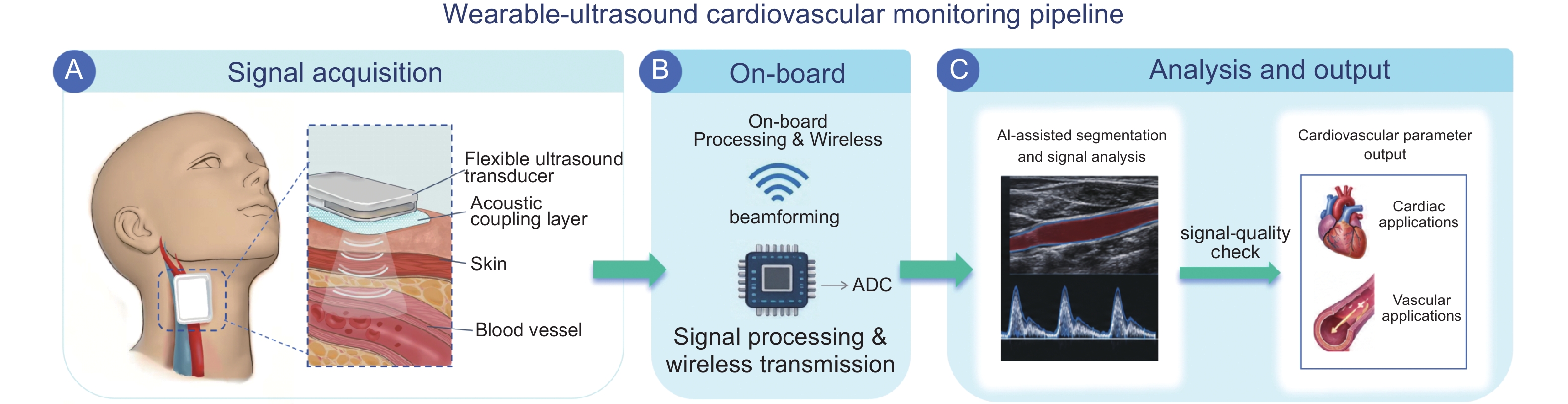
Figure 1
Conceptual pipeline for wearable ultrasound cardiovascular monitoring. (A) Signal acquisition using a flexible ultrasound transducer and an acoustic coupling layer at the skin interface; (B) On-board electronics support signal acquisition, processing, and wireless transmission; (C) AI-assisted segmentation and signal analysis, together with signal-quality assessment, enable parameter extraction for cardiac and vascular applications. The schematic illustrates a general workflow; the implementation and location of processing steps vary across devices. ADC, analog-to-digital converter; AI, artificial intelligence."
Table 1
Representative studies of wearable ultrasound for continuous cardiac function monitoring"
| Author/year | Population | Device/mode | Integration level | Freq./depth | Cardiac target | Monitoring | Comparator | Key findings | Limitation/ evidence stage |
| US, ultrasound; CBF, cerebral blood flow; LV, left ventricle; DL, deep learning; PLAX, parasternal long-axis; PSAX, parasternal short-axis; A4C, apical four-chamber; A2C, apical two-chamber; EDV, end-diastolic volume; ESV, end-systolic volume; SV, stroke volume; CO, cardiac output; LVEF, left ventricular ejection fraction; PFO, patent foramen ovale; AV, aortic valve; RV, right ventricular; DSA, digital subtraction angiography; TEE, transesophageal echocardiography; GI, gastrointestinal; PDA, patent ductus arteriosus; NICU, neonatal intensive care unit; echo, echocardiography; f/u, follow-up. Evidence stage is graded as follows: Technical proof-of-concept, demonstration of device functionality in phantoms or very small samples; Physiological validation, evaluation of measurements or physiological responses in healthy volunteers; Early clinical feasibility, preliminary evaluation in patient populations via case series or small-scale clinical studies; Clinical reference-standard validation, systematic evaluation of measurement agreement or diagnostic performance against an established clinical reference method in a clinical cohort. | |||||||||
| Wang/2021[ | Healthy volunteers | Stretchable US phased array (Doppler) | Wired probe | ~2 MHz; ≤ 14 cm | Cardiac tissues; central vessels; CBF | Real-time monitoring | Commercial rigid phased array (Verasonics P4-2v) | Active beam focusing/steering (−20° to +20°); deep-tissue Doppler performance comparable to commercial device | Healthy only; no LV functional quantification Physiological validation |
| Hu/2023[ | Healthy volunteers | Orthogonal wearable cardiac imager (B/M-mode + DL) | Wired wearable patch | 3 MHz | LV; standard echo views (PLAX, PSAX, A4C, A2C) | Rest/exercise/ recovery | Commercial echocardiography | Automated EDV, ESV, SV, CO, LVEF from continuous LV volume curves; good agreement with commercial echocardiography for LV volume and derived functional indices | Healthy only; limited clinical validation Physiological validation |
| Zhang/2025[ | 1 adult PFO patient (49 y/o, bicuspid AV) | WISMED wearable US (W30PC) | Wireless wearable patch | Not reported (proprietary) | RV inflow/outflow tract; PFO occluder position | PFO closure (intraprocedural guidance + postop monitoring) | DSA + TEE (conventional PFO guidance) | Continuous hands-free imaging during intervention; real-time device guidance; avoids esophageal intubation | Single case/ Early clinical feasibility |
| Wang/2025[ | 1 neonate (3.75 kg, GI bleeding, PFO/PDA) | Wearable phased-array echo (P2-5-64, color Doppler) | Wireless wearable patch | 3.6 MHz | LV (volume, contractility); standard neonatal views | Perioperative anesthesia + NICU monitoring | Preoperative clinical echocardiography | Continuous contractility and volume assessment; guides fluid management in neonatal setting | Single case/ Early clinical feasibility |
Table 2
Representative studies of wearable ultrasound for continuous non-invasive blood pressure monitoring"
| Author/year | Population | Device/method | Target vessel | Calibration | Monitoring/duration | Comparator | Key findings | Integration | Evidence stage |
| US, ultrasound; BP, blood pressure; DBP, diastolic BP; PMUT, piezoelectric micromachined ultrasound transducer; PWV, pulse wave velocity; PPG, photoplethysmography; SBP, systolic BP; MAE, mean absolute error; SD, standard deviation; HR, heart rate; USoP, ultrasonic-system-on-patch; ML, machine learning; HIIT, high-intensity interval training; CO, cardiac output; ICU, intensive care unit; MAP, mean arterial pressure; ISO, International Organization for Standardization; MAD, mean absolute difference; SAFU, skin-adaptive focused ultrasound; BMI, body mass index; cath, cardiac catheterization. Evidence stage is graded as follows: Technical proof-of-concept, demonstration of device functionality in phantoms or very small samples; Physiological validation, evaluation of measurements or physiological responses in healthy volunteers; Early clinical feasibility, preliminary evaluation in patient populations via case series or small-scale clinical studies; Clinical reference-standard validation, systematic evaluation of measurement agreement or diagnostic performance against an established clinical reference method in a clinical cohort. | |||||||||
| Wang/2018 [ | 1 healthy male volunteer | Conformal US patch; pulse-echo arterial wall tracking | Carotid artery + jugular vein | Yes; BP derived from vessel diameter–pressure relationship (brachial cuff for DBP reference) | Rest and exercise; continuous waveform monitoring; up to 2 h skin integration | Commercial tonometry | Continuous central and peripheral arterial pulse/BP waveforms from vessels up to ~4 cm deep; good agreement with tonometry | Wired probe | Technical proof-of-concept |
| Sempionatto/2021 [ | Healthy volunteers | Multimodal epidermal patch (US + electrochemical sensors) | Carotid artery | Yes; commercial cuff BP calibration | Food intake, exercise, daily stimuli; continuous simultaneous monitoring | Commercial cuff BP monitor | Simultaneous BP/HR + glucose/lactate/caffeine/alcohol monitoring; detected expected BP/HR responses to exercise and metabolic changes | Wired wearable patch | Physiological validation |
| Zhou/2025(IEEE) [ | 17 healthy subjects (12M/5F, 22-36 y) | Wearable PMUT; pulse-echo arterial diameter + local PWV | Two sites along a single artery (local PWV) | Yes; initial BP cuff calibration required | Rest and dynamic BP changes; wearable/mobile monitoring | Cuff sphygmomanometer; PPG finger clip (waveform) | SBP/DBP MAE <4 mmHg, SD < 3 mmHg; HR error < 3 bpm; waveform r = 0.951-0.998 | Wireless wearable (PMUT) | Physiological validation |
| Lin/2024 [ | Healthy adult volunteers | Fully integrated USoP (A/B/M-mode + ML target tracking) | Carotid artery/central arteries | Yes; tonometer calibration for BP | Moving subjects; exercise (cycling, HIIT); ≤ 12 h continuous | Tonometer; reference BP devices | ML-based autonomous vascular target tracking (96.13% accuracy with domain adaptation); central BP error < 1.58 mmHg post-calibration; continuous BP/HR/CO | Fully integrated wireless system | Physiological validation |
| Zhou/2025(NatBE) [ | 117 participants (7 daily-activity, 85 outpatient, 21 catheterization, and 4 ICU participants) | Wearable US BP sensor; closely spaced sonographic windows + backing layer; arterial wall-motion sensing | Brachial and radial arteries (main) | Yes; reference calibration used | Daily activities; supine–standing challenge; cardiac catheterization; ICU 10–12 h | Cuff BP; invasive arterial line (cath lab & ICU) | Cath lab: Bland–Altman SBP 0.34 ± 3.90, MAP −0.18 ± 2.20, DBP −0.43 ± 2.66 mmHg; met ISO 81060-2 criteria; outpatient MAD ≤ 4.90 mmHg | Wireless wearable patch | Clinical reference-standard validation |
| Yuan/2025 [ | Healthy volunteers; multiple small BP-validation cohorts | SAFU flexible US array (2.4 MHz, self-adhesive hydrogel); arterial wall tracking | Fingertip, radial, brachial, carotid (multi-artery) | Yes; vessel mechanics linked to reference BP | Multi-artery testing, exercise, 24-h BP monitoring | Mercury sphygmomanometer | SBP error 1.8 ± 2.98, DBP error 1.1 ± 2.43 mmHg (representative); 24-h monitoring ±2.48/±1.99 mmHg; robust to moderate misalignment | Flexible wearable patch | Physiological validation |
Table 3
Representative studies of wearable ultrasound for vascular hemodynamics and arterial stiffness assessment"
| Study | Population | Device | Target | Key parameter | Comparator | Key quantitative finding | Integration | Evidence stage |
| PLR, passive leg raising; CW, continuous-wave; ccFT, corrected carotid flow time; SV, stroke volume; AUROC, area under the receiver operating characteristic curve; CABG, coronary artery bypass grafting; TEE, transesophageal echocardiography; CCA, common carotid artery; CPB, cardiopulmonary bypass; IJ, internal jugular; VTI, velocity-time integral; LVET, left ventricular ejection time; CSA, cross-sectional area; US, ultrasound; HT, hypertension; SAFU, skin-adaptive focused ultrasound; BP, blood pressure; PWV, pulse wave velocity; PW, pulsed-wave; MAE, mean absolute error; MAPE, mean absolute percentage error; ccVTI, carotid velocity-time integral; Δ, change. Evidence stage is graded as follows: Technical proof-of-concept, demonstration of device functionality in phantoms or very small samples; Physiological validation, evaluation of measurements or physiological responses in healthy volunteers; Early clinical feasibility, preliminary evaluation in patient populations via case series or small-scale clinical studies; Clinical reference-standard validation, systematic evaluation of measurement agreement or diagnostic performance against an established clinical reference method in a clinical cohort. | ||||||||
| Kenny/2023 Front Biosci Elite [ | 19 volunteers; 36 PLR | Wireless CW Doppler patch | Carotid | ccFT | Non-invasive pulse-contour SV | 8,856 cycles; ccFT tracks SV change (AUROC 0.77 for +5% ΔSV) | Wireless patch | Physiological validation |
| Kenny/2021 Bioengineering [ | 2 CABG patients (proof-of-principle) | Wireless CW Doppler patch (4 MHz) | Carotid | ccVTIΔ | TEE | 10-beat averaged ccVTIΔ: AUROC 0.95 for +10% ΔSV | Wireless patch | Early clinical feasibility |
| Kenny/2025 CHEST Crit Care [ | CABG patients (post-op) | Wireless CW Doppler patch | Carotid | ΔccFT | TEE ΔSV | Validated ΔccFT for detecting ≥10% SV change | Wireless patch | Clinical reference-standard validation |
| Munding/2023 [ | 14 on-pump CABG patients | Hands-free CW Doppler patch | Carotid | Max CCA velocity | CPB pump flow | 13.6% ΔCCA velocity ↔ 10% ΔCPB flow | Hands-free patch | Early clinical feasibility |
| Kenny/2023 Ultrasound J [ | 2 illustrative cases (1 healthy + 1 clinical) | Wireless CW Doppler patch (4 MHz) | Carotid + IJ vein | ccFT, VTI, venous Doppler | Non-invasive SV (case 1); clinical context (case 2) | "Doppler Starling curve" framework; differential preload-output responses | Wireless patch | Early clinical feasibility |
| Hofer/2025 [ | 347 participants; 137,521 cycles | Wireless CW Doppler patch | Carotid | ccFT/LVET (4 correction equations) | — (reference distribution) | Mean ccFT 295–311 ms across correction formulas; 99.1% signal validity across 137,521 cycles | Wireless patch | Physiological validation |
| Kong/2026 [ | In vitro + human volunteers | Wearable T-shaped B-mode + Doppler patch (8.5 MHz, 1.3 mm) | Carotid | CSA + VTI → volumetric flow | Commercial US system | In vitro max deviation 6.3%; simultaneous dual-axis imaging | T-shaped array patch | Physiological validation |
| Yuan/2025 [ | Paired subgroups (n = 2 per pair): HT/normo, elderly/young, athlete/sedentary | SAFU flexible pulse-echo array (2.4 MHz) | Radial (stiffness); multiple sites (BP) | β-stiffness index, local PWV | Arteriosclerosis detector (VBP-9) | HT β = 13.1 vs normo β = 9.9; PWV relative error < 25% | Flexible self-adhesive array | Early clinical feasibility |
| Li/2026 [ | In vitro phantom only | Stretchable PW Doppler patch (5.05 MHz, 6 transducers, ~30% stretch) | Flow phantom | Flow velocity | Reference pump velocity | MAE 3.56 cm/s, MAPE 5.99% (20-100 cm/s) | Stretchable patch | Technical proof-of-concept |
| [1] |
Lindstrom M, DeCleene N, Dorsey H, Fuster V, Johnson CO, LeGrand KE, et al. Global burden of cardiovascular diseases and risks collaboration, 1990-2021. J Am Coll Cardiol 2022; 80: 2372-2425.
doi: 10.1016/j.jacc.2022.11.001 |
| [2] |
Xie H, Yang L, Jiang B, Huang Z, Lin Y. State-of-the-art wearable sensors for cardiovascular health: a review. npj Cardiovascular Health 2025; 2: 53.
doi: 10.1038/s44325-025-00090-6 |
| [3] |
Buetti N, MimoZ O, Schwebel C, Ruckly S, Castry M, Dupuis C, et al. Insertion site and infection risk among peripheral arterial catheters. Am J Respir Crit Care Med 2021; 203: 630-633.
doi: 10.1164/rccm.202007-3008LE |
| [4] |
O'Grady NP, Alexander M, Burns LA, Dellinger EP, Garland J, Heard SO, et al. Guidelines for the prevention of intravascular catheter-related infections. Clin Infect Dis 2011; 52: e162-e193.
doi: 10.1093/cid/cir257 |
| [5] |
Lin M, Zhang Z, Gao X, Bian Y, Wu RS, Park G, et al. A fully integrated wearable ultrasound system to monitor deep tissues in moving subjects. Nat Biotechnol 2024; 42: 448-457.
doi: 10.1038/s41587-023-01800-0 |
| [6] | Song P, Andre M, Chitnis P, Xu S, Croy T, Wear K, et al. Clinical, safety, and engineering perspectives on wearable ultrasound technology: a review. IEEE Trans Ultrason Ferroelectr Freq Control 2024; 71: 730-744. |
| [7] |
Yan M, Wang Z, Li Z, Wang P, Wu P, Chen X, et al. Wearable ultrasound: a review of core technologies and clinical applications in cardiovascular monitoring. Microsyst Nanoeng 2026; 12: 246.
doi: 10.1038/s41378-026-01158-8 |
| [8] |
Fang M, Yu R, Meng F, Liu X, Tang N, Yuan M, et al. Wearable ultrasound technology in health monitoring and therapy: technological innovations, differentiated applications, and future directions. ACS Sens 2025; 10: 5433-5455.
doi: 10.1021/acssensors.5c01767 |
| [9] |
Hu H, Huang H, Li M, Gao X, Yin L, Qi R, et al. A wearable cardiac ultrasound imager. Nature 2023; 613: 667-675.
doi: 10.1038/s41586-022-05498-z |
| [10] | Zhou S, Park G, Longardner K, Lin M, Qi B, Yang X, et al. Clinical validation of a wearable ultrasound sensor of blood pressure. Nat Biomed Eng 2025; 9: 865-881. |
| [11] | Kenny JES, Horner C, Elfarnawany M, Eibl AM, Eibl JK. Carotid artery corrected flow time measured by wearable Doppler ultrasound accurately detects changing stroke volume during the passive leg raise in ambulatory volunteers. Front Biosci (Elite Ed) 2023; 15: 12 |
| [12] | Kong F, Tang H, Liu P, Ruan R, Lou K, Liu M, et al. Continuous monitoring of carotid artery flow volume using a wearable T-shaped ultrasound patch. IEEE Trans Biomed Eng 2026:1-9. |
| [13] |
Zhou S, Gao X, Park G, Yang X, Qi B, Lin M, et al. Transcranial volumetric imaging using a conformal ultrasound patch. Nature 2024; 629: 810-818.
doi: 10.1038/s41586-024-07381-5 |
| [14] |
Dong L, Zhang X, Li M, Li Y, Guo J, Lu S, et al. AI-enabled wearable ultrasound for noninvasive central venous pressure monitoring. Cyborg Bionic Syst 2026; 7: 0653.
doi: 10.34133/cbsystems.0653 |
| [15] | Huang H, Wu RS, Lin M, Xu S. Emerging wearable ultrasound technology. IEEE Trans Ultrason Ferroelectr Freq Control 2024; 71: 713-729. |
| [16] |
Joshi SV, Sadeghpour S, Kuznetsova N, Wang C, Kraft M. Flexible micromachined ultrasound transducers (MUTs) for biomedical applications. Microsyst Nanoeng 2025; 11: 9.
doi: 10.1038/s41378-024-00783-5 |
| [17] |
Wang C, Qi B, Lin M, Zhang Z, Makihata M, Liu B, et al. Continuous monitoring of deep-tissue haemodynamics with stretchable ultrasonic phased arrays. Nat Biomed Eng 2021; 5: 749-758.
doi: 10.1038/s41551-021-00763-4 |
| [18] |
Wang C, Chen X, Wang L, Makihata M, Liu HC, Zhou T, et al. Bioadhesive ultrasound for long-term continuous imaging of diverse organs. Science 2022; 377: 517-523.
doi: 10.1126/science.abo2542 |
| [19] |
Lin S, Xu H, Li Y, Ge X, Chen K, Chen L, et al. Synchronous wearable ultrasound for early detection of coronary and carotid artery comorbidity. Sci Adv 2026; 12: eaeed2114.
doi: 10.1126/sciadv.aed2114 |
| [20] |
Ferreira DL, Lau C, Salaymang Z, Arnaout R. Self-supervised learning for label-free segmentation in cardiac ultrasound. Nat Commun 2025; 16: 4070.
doi: 10.1038/s41467-025-59451-5 |
| [21] |
Chao CJ, Gu YR, Kumar W, Xiang T, Appari L, Wu J, et al. Foundation versus domain-specific models for left ventricular segmentation on cardiac ultrasound. NPJ Digit Med 2025; 8: 341.
doi: 10.1038/s41746-025-01730-y |
| [22] |
Zhang P, Luo X, Shao Z, Liang G, Fu J, Song H. Wearable ultrasound monitoring in minimally invasive cardiac surgery: a case report. JCA Adv 2025; 2: 100168.
doi: 10.1016/j.jcadva.2025.100168 |
| [23] |
Wang Y, Wu G, Song J, Song H, Chu Q. Innovative wearable technology for continuous echocardiographic monitoring: first-in-neonate case report. Front Pediatr 2025; 13: 1567386.
doi: 10.3389/fped.2025.1567386 |
| [24] |
Scholte NTB, van Ravensberg AE, Shakoor A, Boersma E, Ronner E, de Boer RA, et al. A scoping review on advancements in noninvasive wearable technology for heart failure management. NPJ Digit Med 2024; 7: 279.
doi: 10.1038/s41746-024-01268-5 |
| [25] |
Wang C, Li X, Hu H, Zhang L, Huang Z, Lin M, et al. Monitoring of the central blood pressure waveform via a conformal ultrasonic device. Nat Biomed Eng 2018; 2: 687-695.
doi: 10.1038/s41551-018-0287-x |
| [26] |
Sempionatto JR, Lin M, Yin L, De la Paz E, Pei K, Sonsa-Ard T, et al. An epidermal patch for the simultaneous monitoring of haemodynamic and metabolic biomarkers. Nat Biomed Eng 2021; 5: 737-748.
doi: 10.1038/s41551-021-00685-1 |
| [27] |
Zhou J, Qu M, Liu W, Wang Y, Zhang G, Zheng Y, et al. Continuous monitoring of blood pressure by measuring local pulse wave velocity using wearable micromachined ultrasonic probes. IEEE Trans Biomed Eng 2025; 72: 1615-1624.
doi: 10.1109/TBME.2024.3514878 |
| [28] |
Yuan J, Li Z, Zhao Y, Luo R, Qin S, Li J, et al. Skin-adaptive focused flexible micromachined ultrasound transducers for wearable cardiovascular health monitoring. Sci Adv 2025; 11: eadw7632.
doi: 10.1126/sciadv.adw7632 |
| [29] | Cohen JB, Byfield RL, Hardy ST, Juraschek SP, Houston Miller N, Mukkamala R, et al. Cuffless devices for the measurement of blood pressure: a scientific statement from the American Heart Association. Hypertension 2026; 83: e00254 |
| [30] |
Stergiou GS, Avolio AP, Palatini P, Kyriakoulis KG, Schutte AE, Mieke S, et al. European Society of Hypertension recommendations for the validation of cuffless blood pressure measuring devices: European Society of Hypertension Working Group on Blood Pressure Monitoring and Cardiovascular Variability. J Hypertens 2023; 41: 2074-2087.
doi: 10.1097/HJH.0000000000003483 |
| [31] |
Kenny JES, Clarke G, Myers M, Elfarnaway M, Eibl AM, Eibl JK, et al. A wireless wearable Doppler ultrasound detects changing stroke volume: proof-of-principle comparison with trans-esophageal echocardiography during coronary bypass surgery. Bioengineering (Basel) 2021; 8: 203.
doi: 10.3390/bioengineering8120203 |
| [32] |
Kenny JES, Clarke G, Atwi S, Kerrebijn I, Savery T, Knott M, et al. Carotid artery corrected flow time measured by wearable Doppler ultrasound detects stroke volume change measured by transesophageal echocardiography after coronary artery bypass grafting. CHEST Crit Care 2025; 3: 100138.
doi: 10.1016/j.chstcc.2025.100138 |
| [33] | Munding CE, Kenny JES, Yang Z, Clarke G, Elfarnaway M, Eibl AM, et al. Detecting the change in total circulatory flow with a wireless, wearable Doppler ultrasound patch: a pilot study. Crit Care Explor 2023; 5: e0914 |
| [34] |
Kenny JES. Functional hemodynamic monitoring with a wireless ultrasound patch. J Cardiothorac Vasc Anesth 2021; 35: 1509-1515.
doi: 10.1053/j.jvca.2021.01.040 |
| [35] | Hofer LM, Kenny JES, Munding CE, Kerrebijn I, Atwi S, Younan A, et al. Defining the physiological bounds of left ventricular ejection time with a wireless, wearable ultrasound: an analysis of over 137,000 cardiac cycles. Digit Health 2025; 11: 20552076251323838 |
| [36] |
Kenny JES, Gibbs SO, Eibl JK, Eibl AM, Yang Z, Johnston D, et al. Simultaneous veno-arterial Doppler during preload augmentation: illustrating the Doppler Starling curve. Ultrasound J 2023; 15: 32.
doi: 10.1186/s13089-023-00330-9 |
| [37] |
Li Y, Li Z, Peng C. A stretchable wearable Doppler ultrasound patch for continuous vascular monitoring. Measurement 2026; 266: 120490.
doi: 10.1016/j.measurement.2026.120490 |
| [38] |
Rola P, Haycock K, Spiegel R, Beaubien-Souligny W, Denault A. VExUS: common misconceptions, clinical use and future directions. Ultrasound J 2024; 16: 49.
doi: 10.1186/s13089-024-00395-0 |
| [39] |
Kenny JES, Gibbs SO, Johnston D, Yang Z, Hofer LM, Elfarnaway M, et al. The time cost of physiologically ineffective intravenous fluids in the emergency department: an observational pilot study employing wearable Doppler ultrasound. J Intensive Care 2023; 11: 7.
doi: 10.1186/s40560-023-00655-6 |
| [40] |
Da Luz L, Atwi S, Notario L, Irvine R, Farah D, Johnston D, et al. The feasibility of monitoring trauma patients with a wireless, wearable Doppler ultrasound. Transfusion 2025; 65: S123-S130.
doi: 10.1111/trf.18241 |
| [41] |
Krüger GM, Birkeli SG, Hansen ØM, Faldaa B, Norvik A, Lund HJ, et al. Real-time detection of spontaneous circulation in humans during cardiopulmonary resuscitation using a continuous hands-free carotid Doppler: a pilot study. Resusc Plus 2025; 26: 101080.
doi: 10.1016/j.resplu.2025.101080 |
| [42] |
Kaufman CS, Wu D, Eibl JK, Kenny JES. Ultrasonographic measures of safe and fluid effective resuscitation (SaFER): incorporating the VExUS and VTI into protocolized care. SN Compr Clin Med 2026; 8: 73.
doi: 10.1007/s42399-026-02318-7 |
| [43] | Gong C, Lu G, Liu B, Che ZP, Ji J, Wei F, et al. A wearable non-invasive sonogenetic pacemaker. Nat Biomed Eng 2026. |
| [44] | Park G, Bian Y, Huang H, Zhou S, Qin S, Lin M, et al. Fetal monitoring for high-risk pregnancies using a wearable ultrasound patch. Nat Biotechnol 2026. |
| [45] | Stevenson LW, Ross HJ, Rathman LD, Boehmer JP. Remote monitoring for heart failure management at home. J Am Coll Cardiol 2023; 81: 2272-2291. |
| [46] |
Bioeffects Committee of the American Institute of Ultrasound in M. American Institute of Ultrasound in Medicine consensus report on potential bioeffects of diagnostic ultrasound: executive summary. J Diagn Med Sonogr 2011; 27: 3-13.
doi: 10.1177/8756479310394986 |
| [47] |
Amado-Rey AB, Gonçalves Seabra AC, Stieglitz T. Towards ultrasound wearable technology for cardiovascular monitoring: from device development to clinical validation. IEEE Rev Biomed Eng 2025; 18: 93-112.
doi: 10.1109/RBME.2024.3410399 |

| No related articles found! |
| Viewed | ||||||
|
Full text |
|
|||||
|
Abstract |
|
|||||
Share: WeChat
Copyright ©2018 Advanced Ultrasound in Diagnosis and Therapy
|
Advanced Ultrasound in Diagnosis and Therapy (AUDT) a>
is licensed under a Creative Commons Attribution 4.0 International License a>.