
Advanced Ultrasound in Diagnosis and Therapy ›› 2026, Vol. 10 ›› Issue (3): 203-215.doi: 10.26599/AUDT.2026.250085
• ORIGINAL RESEARCH • Previous Articles Next Articles
Guo Fangqia, Lin Jinga, Yang Qiruia, Ji Qianwena, Zhao Jiaqia,*( )
Received:2025-08-09
Revised:2025-11-14
Accepted:2026-05-26
Online:2026-09-30
Published:2026-10-12
Contact:
Zhao Jiaqi,
E-mail:ultrasoundczzjq@163.com
Guo Fangqi, Lin Jing, Yang Qirui, Ji Qianwen, Zhao Jiaqi. Discussion on the Application of Combined Transthoracic and Transesophageal Echocardiography in the 'Four-Step Repair Method' for Treating Rheumatic Heart Valve Disease and Its Short-Term Outcomes. Advanced Ultrasound in Diagnosis and Therapy, 2026, 10(3): 203-215.

Table 1
Baseline data of 105 patients with rheumatic mitral valve disease [$\bar{x}±s$, n (%)]"
| Parameter | Mean |
| NYHA: New York Heart Association (NYHA) Functional Classification | |
| Age | 45.19 ± 10.30 |
| Male | 15 |
| Female | 90 |
| Height | 161.65 ± 6.41 |
| Weight | 59.56 ± 10.17 |
| Body surface area | 1.62 ± 0.15 |
| Systolic blood pressure (mmHg) | 120.55 ± 14.05 |
| Diastolic blood pressure (mmHg) | 75.27 ± 11.05 |
| Heart rate | 81.10 ± 12.12 |
| NYHA Heart failure classification | |
| II | 19 |
| III | 86 |
| LVEF (%) | 63.70 ± 4.81 |
| Atrial fibrillation | 44 |
| Left atrial thrombus | 3 |
| Pulmonary hypertension | 24 |
| Mitral valve annuloplasty ring | 33.45 ± 1.64 |
| Extracorporeal circulation time | 162.05 ± 42.01 |
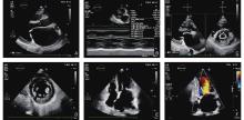
Figure 1
(A) Long-axis view of the left ventricle showing thickening and enlargement of the mitral valve leaflets, with the anterior leaflet tip open and bulging; (B) Long-axis M-mode measurement of the left ventricle showing "wall-like" changes; (C) Parasternal short-axis view of the left ventricle using the biplane method, showing "fish-mouth" changes in the anterior and posterior mitral valve leaflets, with a valve orifice area of 0.8 cm2; (D) Fusion of the papillary muscles; (E) Thickening and shortening of the chordae tendineae; (F) Apical two-chamber heart color Doppler flow imaging (CDFI) showing a "flower-like" blood flow pattern at the valve orifice."
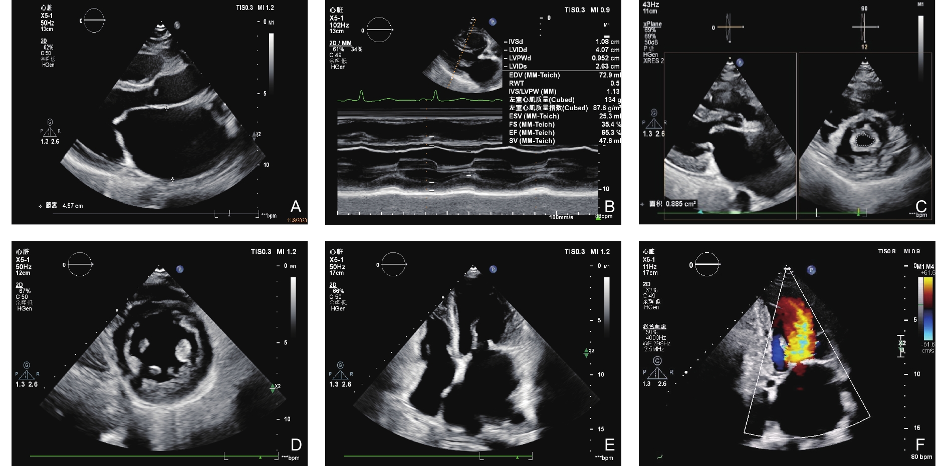
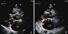
Figure 2
(A) Diastole is defined as α1 and β1, respectively; (B) Systole is defined as α2 and β2, respectively. AO, Aorta; LA, Left Atrium; LV, Left Ventricle; RV, Right Ventricle."
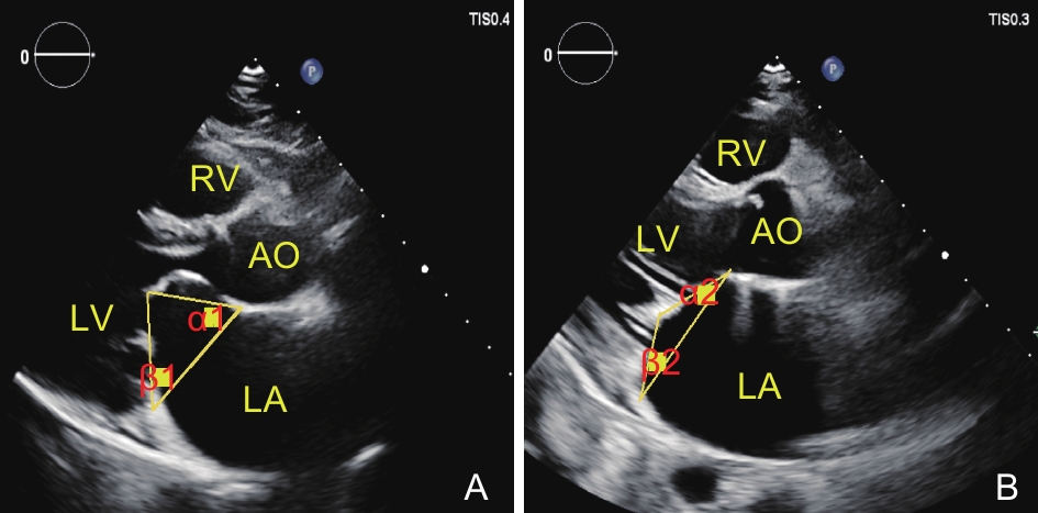
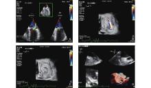
Figure 3
(A) TEE-3D software volumetric imaging shows significant mitral regurgitation at the mitral valve orifice; (B) TEE-3D bird's-eye view shows that the mitral regurgitation jet originates from the anterolateral commissure; (C) TEE-3D bird's-eye view with Color Doppler Flow Imaging (CDFI) removed shows a gap in the closure of the anterior mitral leaflet; (D) TEE-3D volumetric imaging shows a thrombus in the left atrial appendage."
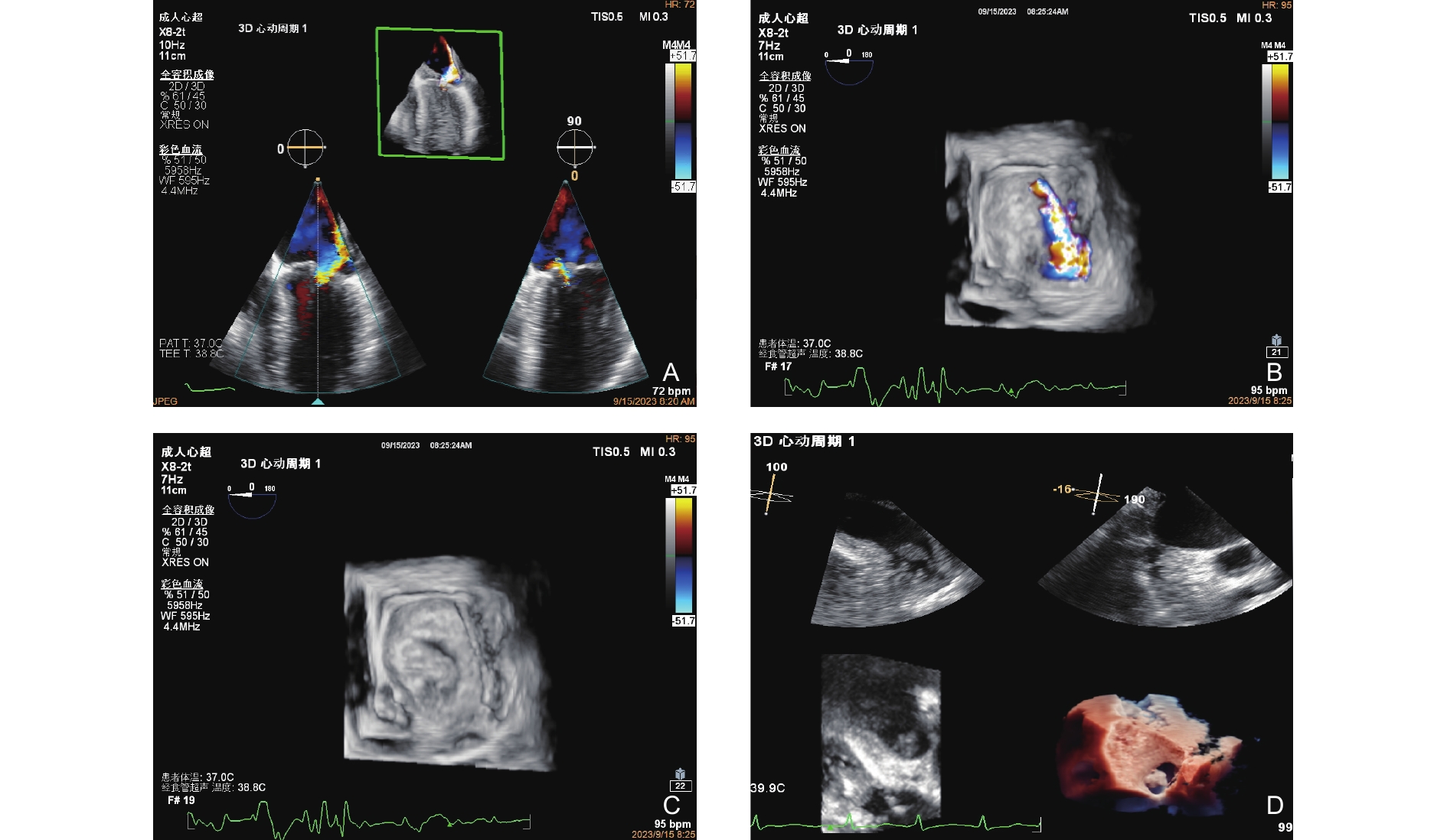
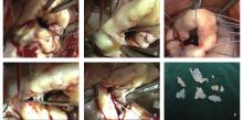
Figure 4
“Four-step method” standard for rheumatic mitral valve repair technique. (A) Exposure of the mitral valve orifice: Commissural adhesions in rheumatic mitral valve disease; (B) Step 1: Removal of commissural fibrous plaques; (C) Step 2: Observation and confirmation of the natural state of the leaflet coaptation; (D) Step 3: Incision of the adhesive commissure; (E) Step 4: Separation of fused papillary muscles; (F) Intraoperative removal of thickened leaflet tissue specimens."

Table 2
Preoperative and postoperative relevant parameters of mitral valve repair with retained mitral valve structural devices evaluated by TTE"
| Parameter | Preoperative | Postoperative | t-value | P-value |
| P-values are presented as uncorrected. After Bonferroni correction for 14 comparisons (adjusted α = 0.0036), parameters marked with an asterisk (*) remained statistically significant. Specifically, the changes in MVA, MV-Vmax, MPG, MRA, LA, LVDd, EDV, LVEF, TAPSE, and S' retained significance (all P < 0.001), whereas the change in LVDs (*P* = 0.013) did not meet the corrected threshold. LA, Left Atrium (Left-Right Diameter); LV, Left Ventricle (Anterior-Posterior Diameter); LVDd, Left ventricular end-diastolic diameter; LVDs, Left Ventricular Systolic End Diastolic Diameter; MV, Mitral Valve; MV-Vmax, Peak Forward Flow Velocity of the Mitral Valve; MRA, Mitral regurgitation area; MPG, Mean pressure gradient; EDV, Left Ventricular End-Diastolic Volume; RA, right atrial diameter; FAC, Right Ventricular Area Change Fraction; S’, Right Ventricular Systolic Velocity; TAPSE, Tricuspid Annular Plane Systolic Excursion. | ||||
| MVA (cm2) | 1.394 ± 0.0864 | 2.6313 ± 0.03453 | −13.843 | 0.000 |
| MV-Vmax (m/s) | 1.684 ± 0.0583 | 1.1612 ± 0.04075 | 9.068 | 0.000 |
| MPG (mmHg) | 6.319 ± 0.3241 | 2.252 ± 0.0960 | 11.745 | 0.000 |
| MRA (cm2) | 4.996 ± 0.4725 | 1.978 ± 0.1905 | 6.836 | 0.000 |
| LA (mm) | 45.97 ± 0.896 | 38.30 ± 0.821 | 9.086 | 0.000 |
| LVDd (mm) | 47.82 ± 0.544 | 43.53 ± 0.581 | 6.086 | 0.000 |
| LVDs (mm) | 30.86 ± 0.395 | 29.73 ± 0.421 | 2.532 | 0.0130 |
| EDV (mL) | 100.64 ± 3.444 | 82.53 ± 2.551 | 5.121 | 0.000 |
| ESV (mL) | 33.00 ± 1.121 | 31.95 ± 0.996 | .848 | 0.398 |
| LVEF (%) | 63.53 ± 0.550 | 59.10 ± 0.487 | 5.867 | 0.000 |
| RA (mm) | 35.32 ± 0.926 | 33.52 ± 0.519 | 1.846 | 0.068 |
| TAPSE (mm) | 18.168 ± 0.3512 | 10.67 ± 0.191 | 19.493 | 0.000 |
| S’ (cm/s) | 10.692 ± 0.1998 | 8.23 ± 0.143 | 10.561 | 0.000 |
| FAC (%) | 46.43 ± 0.676 | 42.37 ± 0.683 | 4.156 | 0.747 |
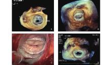
Figure 5
TEE-3D ultrasound image of rheumatic mitral stenosis. (A) Preoperative TEE-3D showing restricted opening of the mitral valve orifice with a "fish-mouth" appearance. (B) Preoperative TEE-3D showing significant thickening of the mitral valve leaflets at closure. (C) Intraoperative mitral valve repair and annuloplasty ring placement completed. (D) Postoperative TEE-3D showing an annuloplasty ring-like echo, with unrestricted leaflet opening."

Table 3
Diagnostic performance of transthoracic echocardiography (TTE) versus intraoperative findings for mitral valve lesions"
| Lesion | TTE (n = 105) | Intraoperative (n = 105) | χ2 | P-value |
| Leaflet thickening | 99 (94.3%) | 101 (96.2%) | 0.081 | 0.776 |
| Commissural adhesion | 94 (89.5%) | 105 (100%) | - | < 0.001 |
| Chordal shortening | 73 (69.5%) | 93 (88.6%) | - | < 0.001 |
| Papillary muscle fusion | 87 (82.9%) | 97 (92.4%) | - | < 0.001 |
Table 4
Diagnostic performance of transesophageal echocardiography (TEE) versus intraoperative findings for mitral valve lesions"
| Lesion | TEE (n = 105) | Intraoperative (n = 105) | χ2 | P-value |
| Leaflet thickening | 98 (93.3%) | 101 (96.2%) | 0.122 | 0.727 |
| Commissural adhesion | ||||
| C1 only | 6 (5.7%) | 10 (9.5%) | - | - |
| C2 only | 1 (1.0%) | 1 (1.0%) | - | - |
| C1 + C2 | 94 (89.5%) | 94 (89.5%) | - | - |
| Total | 101 (96.2%) | 105 (100%) | 165.429 | 0.943 |
| Chordal shortening | 93 (88.6%) | 93 (88.6%) | 76.289 | 0.283 |
| Papillary muscle fusion | 96 (91.4%) | 97 (92.4%) | 78.507 | 0.209 |
Table 5
Diagnostic agreement and performance of preoperative transthoracic and transesophageal echocardiography versus intraoperative findings for mitral valve lesions"
| Lesion | Modality | McNemar Test (P value) | Cohen‘s κ (95% CI) | Sensitivity (%) | Specificity (%) |
| P-values were derived from McNemar's test; both κ values and sensitivity/specificity were calculated using intraoperative findings as the gold standard. | |||||
| Leaflet thickening | TTE | 0.125 | 0.88 (0.80-0.96) | 98.0 | 100.0 |
| TEE | 0.065 | 0.86 (0.79-0.93) | 97.0 | 100.0 | |
| Commissural adhesion | TTE | < 0.001 | 0.45 (0.30-0.60) | 85.7 | 100.0 |
| TEE | 1.000 | 0.90 (0.82-0.98) | 100.0 | 100.0 | |
| Chordal shortening | TTE | < 0.001 | 0.78 (0.68-0.88) | 78.5 | 100.0 |
| TTE | 1.000 | 0.82 (0.72-0.92) | 100.0 | 100.0 | |
| Papillary muscle fusion | TTE | 0.002 | 0.76 (0.65-0.87) | 89.7 | 100.0 |
| TEE | 1.000 | 0.85 (0.75-0.95) | 99.0 | 100.0 | |
Table 6
Transthoracic echocardiographic assessment of mitral valve anterior and posterior leaflet motion angles perioperatively"
| Items | Preoperative average value (SD) | Postoperative average value (SD) | P-value |
| α1° | 50.03 (7.89) | 65.50 (9.71) | < 0.001 |
| β1° | 40.05 (8.80) | 50.20 (10.79) | < 0.001 |
| α2° | 26.33 (5.95) | 29.00 (7.63) | = 0.058 |
| β2° | 28.43 (7.82) | 30.55 (7.85) | = 0.106 |
| [1] |
Vieira MLC, Branco CEB, Gazola ASL, Vieira P, Benvenuti LA, Demarchi L, et al. 3D Echocardiography for Rheumatic Heart Disease Analysis: Ready for Prime Time. Front Cardiovasc Med 2021; 8: 676938.
doi: 10.3389/fcvm.2021.676938 |
| [2] |
Arifputera A, Loo G, Chang P, Kojodjojo P. An unusual case of dysphonia and dysphagia. Singapore Med J 2014; 55: e31-33.
doi: 10.11622/smedj.2013212 |
| [3] |
Harb SC, Griffin BP. Mitral Valve Disease: a Comprehensive Review. Curr Cardiol Rep 2017; 19: 73.
doi: 10.1007/s11886-017-0883-5 |
| [4] |
Tiange L, Xu M. repair strategies based on pathological characteristics of the rheumatic mitral valve in Chinese patients. Heart Lung Circ 2018; 27: 856-863.
doi: 10.1016/j.hlc.2017.05.146 |
| [5] |
Ma LY, Chen WW, Gao RL, Liu LS, Zhu ML, Wang YJ, et al. China cardiovascular diseases report 2018: an updated summary. J Geriatr Cardiol 2020; 17: 1-8.
doi: 10.26599/1671-5411.2023.06.001 |
| [6] |
Watkins DA, Johnson CO, Colquhoun SM, Karthikeyan G, Beaton A, Bukhman G, et al. Global, Regional, and National Burden of Rheumatic Heart Disease, 1990-2015. N Engl J Med 2017; 377: 713-722.
doi: 10.1056/NEJMoa1603693 |
| [7] | Rémond M, Atkinson D, White A, Brown A, Carapetis J, Remenyi B, et al. Are minor echocardiographic changes associated with an increased risk of acute rheumatic fever or progression to rheumatic heart disease?. Int J Cardiol 2015; 198: 117-122. |
| [8] | Ojha U, Marshall DC, Salciccioli JD, Al-Khayatt BM, Hammond-Haley M, Goodall R, et al. Temporal trend analysis of rheumatic heart disease burden in high-income countries between 1990 and 2019. Eur Heart J Qual Care Clin Outcomes 2022. |
| [9] |
Takahashi A, Iesaka Y, Takahashi Y, Takahashi R, Kobayashi K, Takagi K, et al. Electrical connections between pulmonary veins: implication for ostial ablation of pulmonary veins in patients with paroxysmal atrial fibrillation. Circulation 2002; 105: 2998-3003.
doi: 10.1161/01.CIR.0000019585.91146.AB |
| [10] |
Fu J, Li Y, Zhang H, Han J, Jiao Y, Du J, et al. Outcomes of mitral valve repair compared with replacement for patients with rheumatic heart disease. J Thorac Cardiovasc Surg 2021; 162: 72-82.e7.
doi: 10.1016/j.jtcvs.2020.01.053 |
| [11] |
Gaur P, Kaneko T, McGurk S, Rawn JD, Maloney A, Cohn LH. Mitral valve repair versus replacement in the elderly: short-term and long-term outcomes. J Thorac Cardiovasc Surg 2014; 148: 1400-1406.
doi: 10.1016/j.jtcvs.2014.01.032 |
| [12] |
Waikittipong S. Mitral valve repair for rheumatic mitral regurgitation: Mid-term results. Asian Cardiovasc Thorac Ann 2015; 23: 658-664.
doi: 10.1177/0218492315576282 |
| [13] |
Lazam S, Vanoverschelde JL, Tribouilloy C, Grigioni F, Suri RM, Avierinos JF, et al. Twenty-year outcome after mitral repair versus replacement for severe degenerative mitral regurgitation: Analysis of a parge, Prospective, multicenter, international registry. Circulation 2017; 135: 410-422.
doi: 10.1161/CIRCULATIONAHA.116.023340 |
| [14] |
Wang YC, Tsai FC, Chu JJ, Lin PJ. Midterm outcomes of rheumatic mitral repair versus replacement. Int Heart J 2008; 49: 565-576.
doi: 10.1536/ihj.49.565 |
| [15] |
Talwar S, Mathur A, Choudhary SK, Singh R, Kumar AS. Aortic valve replacement with mitral valve repair compared with combined aortic and mitral valve replacement. Ann Thorac Surg 2007; 84: 1219-1225.
doi: 10.1016/j.athoracsur.2007.04.115 |
| [16] |
Maganti K, Rigolin VH, Sarano ME, Bonow RO. Valvular heart disease: diagnosis and management. Mayo Clin Proc 2010; 85: 483-500.
doi: 10.4065/mcp.2009.0706 |
| [17] |
Alessandrini H, Wohlmuth P, Meincke F, Hakmi S, Ubben T, Bohnen S, Kuck KH, Willems S, Tigges E. Comprehensive echocardiographic prediction of postprocedural transmitral pressure gradient following transcatheter mitral valve repair. Int J Cardiovasc Imaging 2021; 37: 2947-2955.
doi: 10.1007/s10554-021-02290-4 |
| [18] |
Chompoosan C, Schrøder AS, Bach MBT, Møgelvang R, Willesen JL, Langhorn R, Koch J. Transthoracic two-dimensional and three-dimensional echocardiography for the measurement of mitral valve area planimetry in English Bull Terriers with and without heart disease. J Vet Cardiol 2021; 36: 169-179.
doi: 10.1016/j.jvc.2021.06.002 |
| [19] | Wunderlich NC, Beigel R, Siegel RJ. Management of mitral stenosis using 2D and 3D echo-Doppler imaging. JACC Cardiovasc Imaging 2013; 6: 1191-205. |
| [20] | Wang Yin, Tang Yangfeng, Xu Fei, et al. Chinese Expert Consensus on Surgical Indications for Rheumatic Mitral Valve Disease. Chinese Journal of Thoracic and Cardiovascular Surgery 2022; 38: 132-137. |
| [21] | Milojevic M, Milosevic G, Nikolic A, Petrovic M, Petrovic I, Bojic M, Jagodic S. Mastering the Best Practices: A Comprehensive Look at the European Guidelines for Cardiopulmonary Bypass in Adult Cardiac Surgery. J Cardiovasc Dev Dis 2023; 10: 296. |
| [22] |
von Stumm M, Dudde F, Holst T, et al. Predicting clinical outcome by indexed mitral valve tenting in functional mitral valve regurgitation. Open Heart 2021; 8: e001483.
doi: 10.1136/openhrt-2020-001483 |
| [23] |
Reshmi JL, Gopan G, Varma PK, Thushara M, Sudheer VB, Madavathazhathil RG, et al. Transesophageal echocardiographic assessment of the repaired mitral valve: A proposed decision pathway. Semin Cardiothorac Vasc Anesth 2022; 26: 68-82.
doi: 10.1177/10892532211036655 |
| [24] |
Silbiger JJ. Advances in rheumatic mitral stenosis: Echocardiographic, pathophysiologic, and hemodynamic considerations. J Am Soc Echocardiogr 2021; 34: 709-722.e1.
doi: 10.1016/j.echo.2021.02.015 |
| [25] |
Luo T, Meng X. Rheumatic mitral valve repair: the Score procedure. Asian Cardiovasc Thorac Ann 2020; 28: 377-380.
doi: 10.1177/0218492320935251 |
| [26] | Zühlke, L. J., & Steer, A. C. The global burden of rheumatic heart disease. Heart 2013; 99: 1711-1718. |
| [27] | Klein, S. L., & Flanagan, K. L. Sex differences in immune responses. Nature Reviews Immunology 2016; 16: 626-638. |
| [28] |
Rezk M, Elkilani O, Shaheen A, Gamal A, Badr H. Maternal hemodynamic changes and predictors of poor obstetric outcome in women with rheumatic heart disease: a five-year observational study. J Matern Fetal Neonatal Med 2018; 31: 1542-1547.
doi: 10.1080/14767058.2023.2235778 |
| [29] |
Chan RK, Costello BT, Paratz ED. Echoing concerns: Tackling rheumatic heart disease in pregnant first nations women. Heart Lung Circ 2024; 33: 1239-1241.
doi: 10.1016/j.hlc.2024.08.007 |
| [30] |
Muhamed B, Parks T, Sliwa K. Genetics of rheumatic fever and rheumatic heart disease. Nat Rev Cardiol 2020; 17: 145-154.
doi: 10.1038/s41569-019-0258-2 |
| [31] |
Luo T, Meng X. Clinico-pathological classification of rheumatic mitral valve damage and surgical strategy. J Thorac Dis 2021; 13: 2933-2941.
doi: 10.21037/jtd-20-3456 |
| [32] |
Iung B, Leenhardt A, Extramiana F. Management of atrial fibrillation in patients with rheumatic mitral stenosis. Heart 2018; 104: 1062-1068.
doi: 10.1136/heartjnl-2017-311425 |
| [33] |
Okuno T, Tomii D, Buffle E, Lanz J, Ryffel C, Demirel C, et al. Transcatheter aortic valve implantation in patients with rheumatic aortic stenosis. Heart 2022; 108: 1225-1233.
doi: 10.1136/heartjnl-2021-320531 |
| [34] |
Afifi A, Shehata N, Nagi M, Sultan AR, Yacoub M. Expanding valve repair in rheumatic heart disease. Front Cardiovasc Med. 2022; 8: 799652.
doi: 10.3389/fcvm.2021.799652 |
| [35] |
Fu JT, Popal MS, Zhang HB, Han W, Hu QM, Meng X, Ma CY. A meta-analysis of late outcomes of mitral valve repair in patients with rheumatic heart disease. J Thorac Dis 2017; 9: 4366-4375.
doi: 10.21037/jtd.2017.10.97 |
| [36] |
Chen SW, Chen CY, Chien-Chia Wu V, Chou AH, Cheng YT, Chang SH, Chu PH. Mitral valve repair versus replacement in patients with rheumatic heart disease. J Thorac Cardiovasc Surg 2022; 164: 57-67.e11.
doi: 10.1016/j.jtcvs.2020.07.117 |
| [37] |
Overtchouk P, Ben-Ali W, Prendergast B, Conradi L, Hahn R, Granada J, et al. Comparison of mitral valve replacement and repair for degenerative mitral regurgitation: a meta-analysis and implications for transcatheter mitral procedures. Curr Cardiol Rep 2020; 22: 79.
doi: 10.1007/s11886-020-01333-x |
| [38] |
Jiang Y, Wang C, Li G, Chen S. Clinical outcomes following surgical mitral valve repair or replacement in patients with rheumatic heart disease: a meta-analysis. Ann Transl Med 2021; 9: 204.
doi: 10.21037/atm-20-3542 |
| [39] |
Tian B, Wu F, Han J, Meng X, Jiao Y, Luo T, Pang S, Xu J. Short-term results on leaflet mobility in patients undergoing rheumatic mitral valve repair with an efficient 4-Step commissuroplasty technique. Tex Heart Inst J 2022; 49: e217801.
doi: 10.14503/thij-21-7801 |
| [40] |
Abdouni AA, Brandão CMA, Rochitte CE, Pomerantzeff PMA, Veronese ET, Pacheco AB, et al. Cardiac magnetic resonance analysis of mitral annular dynamics after mitral valve repair. Clinics (Sao Paulo) 2020; 75: e2428.
doi: 10.6061/clinics/2020/e2428 |
| [41] |
Pandian NG, Kim JK, Arias-Godinez JA, Marx GR, Michelena HI, Chander Mohan J, et al. Recommendations for the use of echocardiography in the evaluation of rheumatic heart disease: A report from the American Society of Echocardiography. J Am Soc Echocardiogr 2023; 36: 3-28.
doi: 10.1016/j.echo.2022.10.009 |
| [42] |
Ashikhmina E, Schaff HV, Daly RC, Stulak JM, Greason KL, Michelena HI, et al. Risk factors and progression of systolic anterior motion after mitral valve repair. J Thorac Cardiovasc Surg 2021; 162: 567-577.
doi: 10.1016/j.jtcvs.2019.12.106 |
| [43] | Paiva PP, Leite FM, Antunes PE, Antunes MJ. Risk-Prediction model for transfusion of erythrocyte concentrate during extracorporeal circulation in coronary surgery. Braz J Cardiovasc Surg 2021; 36: 323-330. |
| [44] | Zamper R, Prempeh A, Iglesias I, Fayad A. Intraoperative transesophageal echocardiography following mitral valve repair: a systematic review. Braz J Anesthesiol 2022; 72: 379-397. |
| [45] |
Bartoszko J, Karkouti K. Managing the coagulopathy associated with cardiopulmonary bypass. J Thromb Haemost 2021; 19: 617-632.
doi: 10.1111/jth.15195 |
| [1] | Jiang Xingting, Fang Wenling, Hu Panpan, Yu Rong, Ma Jing, Wang Ning, Wu Tianyu, Ye Lin, Ruan Wenhui, Li Zhaojun, Jin Lin. Fetal Ventricular-arterial Coupling: Development, Physiology, and Ultrasonographic Advances [J]. Advanced Ultrasound in Diagnosis and Therapy, 2026, 10(3): 158-168. |
| [2] | Hu Xuelin, Zhu Ye, Zhang Zisang, Quan Yuanting, Chen Wenwen, Chen Leichong, Xu Guangyu, Qin Luning, Xie Mingxing, Zhang Li. Explainable Artificial Intelligence in Echocardiography [J]. Advanced Ultrasound in Diagnosis and Therapy, 2025, 9(4): 409-425. |
| [3] | Feng Qing, Yang Huihui, Xu Wanting, He Yu. Application of Two-Dimensional Speckle Tracking Echocardiography in Evaluation of Neonatal Pulmonary Hypertension [J]. Advanced Ultrasound in Diagnosis and Therapy, 2025, 9(3): 254-259. |
| [4] | Lohith Kumar Bittugondanahalli Prakash, Shivakumar Neeraj, Gaduputi Jahnavi, Kashif Mohammed S, K Praneethi, Reddy Manda Pranay, S Sampangi Ramaiah, Krishnamurthy Umesh, Prabhakar Suman. Comparative Analysis of Fetal Ventricular Function: AGA vs. SGA Fetuses Using 2D Speckle-Tracking [J]. Advanced Ultrasound in Diagnosis and Therapy, 2025, 9(3): 290-297. |
| [5] | Elkouahy Fatima Ezzahra, Bennis Ahmed, Merke Nicolas, Ouahid Hajar, Malali Hamid El, Taleb Lhoucine Ben, Mouhsen Azeddine. Advanced Diagnosis of Aortic Stenosis Disease Based on Ultrasound Images: A Novel Artificial Intelligence Approach [J]. Advanced Ultrasound in Diagnosis and Therapy, 2025, 9(3): 298-306. |
| [6] | Qin Shuxuan, He Qing, Wu Zhenni, Lin Yixia, Ji Mengmeng, Zhang Li, Xie Mingxing, Li Yuman. Clinical Utility of Speckle Tracking Echocardiography in Heart Transplantation [J]. Advanced Ultrasound in Diagnosis and Therapy, 2025, 9(2): 103-116. |
| [7] | Yang Lan, Li Zhenyi, Chen Ya, Chen Anni, Wang Xinqi, Jin Lin, Li Zhaojun. Clinical Usefulness of Atrioventricular Coupling in Cardiovascular Disease [J]. Advanced Ultrasound in Diagnosis and Therapy, 2025, 9(1): 1-9. |
| [8] | Yang Yun, Zhang Xin, Zhang Ruize, Jiang Jingrong, Xie Yuji, Fang Lingyun, Zhang Jing, Xie Mingxing, Wang Jing. Current Status and Progress in Arterial Stiffness Evaluation: A Comprehensive Review [J]. Advanced Ultrasound in Diagnosis and Therapy, 2024, 8(4): 172-182. |
| [9] | Chen Anni, Yang Lan, Li Zhenyi, Wang Xinqi, Chen Ya, Jin Lin, Li Zhaojun. Left Ventricular-Arterial Coupling in Cardiovascular Health: Development, Assessment Methods, and Future Directions [J]. Advanced Ultrasound in Diagnosis and Therapy, 2024, 8(4): 159-171. |
| [10] | Zhang Xin, Yang Yun, Zhang Ruize, Zhang Linyue, Xie Yuji, Wu Wenqian, Zhang Jing, Lv Qing, Wang Jing, Xie Mingxing. Noninvasive Evaluation of Left Ventricular-Arterial Coupling: Methodologies and Clinical Relevance [J]. Advanced Ultrasound in Diagnosis and Therapy, 2024, 8(4): 149-158. |
| [11] | Li Zhenyi, Chen Ya, Wang Xinqi, Yang Lan, Chen Anni, Li Zhaojun, Jin Lin. Left and Right Ventricular Interaction: Insight from Echocardiography Imaging [J]. Advanced Ultrasound in Diagnosis and Therapy, 2024, 8(4): 195-204. |
| [12] | Wang Xinqi, Chen Anni, Yang Lan, Chen Ya, Li Zhenyi, Li Zhaojun, Jin Lin. Evaluation Methods and Progress of Right Ventricular-pulmonary Artery Coupling [J]. Advanced Ultrasound in Diagnosis and Therapy, 2024, 8(4): 205-216. |
| [13] | Junrong Hong, MD, Pingyang Zhang, MD, PhD, Mengyao Fei, MD, Lingling Wang, MD. A Study on Left Atrial Function in Patients with Essential Hypertension Using Four-Dimensional Echocardiography [J]. Advanced Ultrasound in Diagnosis and Therapy, 2024, 8(2): 64-73. |
| [14] | Rifei Li, MM, Yuanmei Zhang, MM, Chengkai Zhang, MM, Xuenian Huang, MM, Shangwei Ding, MD. Contrast Echocardiography Evaluation of Microcirculation of Myocardial Infarction Caused by Takotsubo Syndrome: Case Report and Literature Review [J]. Advanced Ultrasound in Diagnosis and Therapy, 2021, 5(3): 258-261. |
| [15] | Jianping Xu, MS, Faping Cui, MS, Shuixiu Dou, MS, Jiafu Ou, MD. Echocardiography of Marfan's Syndrome Patient with New Gene Mutation of FBN1 with 13-year Follow-up [J]. Advanced Ultrasound in Diagnosis and Therapy, 2021, 5(3): 249-253. |
| Viewed | ||||||
|
Full text |
|
|||||
|
Abstract |
|
|||||
Share: WeChat
Copyright ©2018 Advanced Ultrasound in Diagnosis and Therapy
|
 Advanced Ultrasound in Diagnosis and Therapy (AUDT)
is licensed under a Creative Commons Attribution 4.0 International License.
