
Advanced Ultrasound in Diagnosis and Therapy ›› 2026, Vol. 10 ›› Issue (4): 298-316.doi: 10.26599/AUDT.2026.260040
• REVIEW ARTICLE • Previous Articles Next Articles
Han Xuedia, Cui Liganga,*( )
Received:2026-09-16
Revised:2026-09-30
Accepted:2026-11-24
Online:2026-12-30
Published:2026-11-02
Contact:
Cui Ligang,
E-mail:cuiligang_bysy@126.com
Han Xuedi, Cui Ligang. Research Progress and Clinical Translational Application of Ultrasound Localization Microscopy in Microcirculation Imaging. Advanced Ultrasound in Diagnosis and Therapy, 2026, 10(4): 298-316.
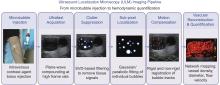
Figure 1
Conceptual schematic of the ULM imaging pipeline. ULM involves intravenous microbubble injection, ultrafast ultrasound data acquisition, clutter suppression, sub-pixel microbubble localization, motion compensation, and final vascular reconstruction with hemodynamic quantification."

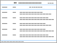
Table 1
Comparison of representative microbubble signal extraction and localization algorithms for ULM."
| Algorithm | Paper | Computational complexity | Localization accuracy | Capacity for high-density overlapping microbubbles | Dependence on SNR | Real-time performance | Main merits and limitations |
| note: “-” indicates this metric is not applicable the corresponding algorithm. | |||||||
| Clutter-suppression algorithms | |||||||
| SVD filtering | Brown 2019 | High | − | Poor | High | Poor | Delivers effective tissue clutter suppression. Suffers heavy matrix-decomposition computational cost; fails for stationary microbubbles and may introduce hundreds-of-microns localization errors under 0-2 mm/s flow; mostly limited to offline processing. |
| Pulse inversion | Brown 2019 | Moderate | − | Moderate | Moderate | Moderate | Insensitive to flow direction and velocity, suitable for slow-flow and tortuous microvasculature. Requires multiple transmit pulses and cannot separate overlapping microbubbles inherently. |
| Differential imaging (DI) | Brown 2019 | Low-Moderate | − | Moderate | High | Good | Low computational overhead. Only functional for flow rates > 0.5 mm/s under axial-dominated blood flow; performance deteriorates for slow or lateral-oriented perfusion. |
| AF-UNet | Zhang 2026 | Low | − | Moderate | Moderate | Good (GPU- dependent) | Lightweight multi-angle-fusion network achieving > 20-fold speedup relative to SVD with favourable cross-organ generalization. Better preserves microbubble point-spread function (PSF) than 3D U-Net variants but cannot intrinsically resolve spatially-overlapping microbubbles; faint microbubble signals may be attenuated. Real-time capability depends on GPU hardware, without native scanner-side implementation. relies on high-quality training datasets |
| Localization algorithms | |||||||
| Gaussian fit (GF) | Sobolewski 2024, Heiles 2022 | High | High | Limited-test | Moderate-high | Poor | Iterative nonlinear least-squares fitting. Reaches precision close to the Cramér-Rao lower bound with properly selected patch size and sampling parameters and yields the lowest standard deviation among classical approaches. Performance degrades under Rician noise and oversized patches; bimodal error occurs with undersampling; computationally expensive. Only single-microbubble simulations in cited literature; overlapping-bubble scenarios remain unevaluated. |
| YOLO-modified object-detection-based CNN | Liu 2023 | Moderate-High | High | Limited-test | Moderate | Moderate | Modified YOLO-style convolutional-neural-network detector outputting microbubble coordinates and confidence scores. Achieves 77% Jaccard index and 20 μm localization error with 7-fold speed-up against Gaussian fitting on ULTRASR-Challenge simulated datasets; confidence filtering suppresses false detections. Performance heavily relies on training-data distribution; lacks systematic overlapping-microbubble and in-vivo validations and requires GPU acceleration. Only simulated flow-like dataset in original publication; dedicated overlapping-microbubble tests are missing. |
| Tracking-association algorithms | |||||||
| Modified MCMCDA | Ackermann 2016 | High | − | Good | Moderate | Poor | Designed for high-density microbubble track linking, accounting for false alarms and track birth-death events; validated via phantom and in-vivo mouse-tumor measurements. Heavy computational overhead prevents real-time deployment. |
| Hierarchical Kalman tracker (HK) | Taghavi 2022 | High | − | Good | Moderate | Poor | Hierarchical Kalman framework adopting velocity-stratified initialization parameters. Delivers superior reconstruction accuracy under high microbubble density and large localization uncertainty. Multi-level iteration imposes high computational cost and supports only offline workflows (3 million MB positions, 5-level HK, with 35.79 min runtime). |
Table 2
Summary of preclinical and clinical application studies of ULM."
| Paper | Study design | Cohort size | Application field | Research model | Research focus | Disease background | Reference standard | Core research findings | Evidence maturity |
| Lowerison 2020 | Simulation assisted pre-clinical observational | 5 | chicken embryos | renal cell carcinoma xenograft model grown in the chorioallantoic membrane of chicken embryos | To provide a quantitative measures of tissue hypoxic status of ULM | Tumor | fluorescent histology | ULM derived intervessel distance strongly correlates with histological microvascular density, and vascular tortuosity reliably reflects tissue hypoxia levels | Pre-clinical animal |
| Lowerison 2022 | Pre-clinical observational | 99 | chicken embryos | human colorectal carcinoma tumor xenograft model engrafted into the CAM of chicken embryos | To indicate the functional microvascular status of chemotherapy sensitivity | Tumor | histology | Elevated vascular fractal dimension on ULM images is closely associated with tumor therapeutic resistance | Pre-clinical animal |
| Huang 2020 | phantom + in vivo chicken embryo proof of concept study | biological replicate number not reported, representative datasets shown | chick chorioallantoic membrane, embryo tumour xenograft | chicken embryo tumor xenograft model and chicken brain | To resolve the inherent trade-off between microbubbles concentration and acquisition duration | Tumor | optical imaging | This novel algorithm separates overlapping microbubbles based on flow velocity and direction differences, enabling robust high-resolution microvascular imaging within seconds under high microbubble dosage | Pre-clinical animal |
| Chen 2020 | Pre-clinical observational | 15 | mice | mouse acute kidney injury model | To evaluate early acute kidney injury | Kidney disease | histology and immunohistochemistry (CD31) | ULM identifies early renal microcirculatory disorders prior to serum creatinine elevation, supporting early warning of acute kidney injury | Pre-clinical animal |
| Lowerison 2022 | Pre-clinical observational | 27 | mice | aging mouse model | To varify differences of aged brain and normal brain | Aging | not appliable | Aged brains exhibit reduced blood velocity, increased vascular tortuosity, and decreased cortical blood volume across whole-brain regions on ULM | Pre-clinical animal |
| Ma 2022 | Pre-clinical observational | 8 | rat | stroke events and Alzheimer's disease | To assess microvascular features of cerebral small vessel disease | Cerebral small vessel disease | H&E staining, PTAH staining, TB staining, MRI | A controllable cerebral small vessel disease model exhibits reduced microvascular number, decreased flow velocity and diminished blood volume in sonicated lesions | Pre-clinical animal |
| Andersen 2020 | Pre-clinical interventional | 10 | rat | Sprague-Dawley rat | To identify early ischemic damage to the renal microcirculation | Kidney disease | Hematoxylin and eosin staining, Ex-vivo MRI | ULM reliably resolves 15–20 μm ascending vasa recta and distinguishes microvascular alterations after renal ischemia-reperfusion injury | Pre-clinical animal |
| Andersen 2022 | Pre-clinical interventional | 7 | rat | normal rat | To assess renal vascular velocity responses to pharmacological intervention | Kidney disease | not appliable | Pharmacological intervention with prazosin induces velocity reduction specifically in cortical arterioles and outer-medullary vasa recta, enabling compartment-specific renal hemodynamic evaluation by ULM | Pre-clinical animal |
| Beliard 2022 | Pre-clinical interventional | 10, 5 per group (4 week post contusion SCI, 8 week post contusion SCI) | rat | chronic spinal cord injury | to explore changes in spinal cord blood vessels | Spinal cord injury | immunohistochemistry (laminin and for SMI-71), BBB locomotor behavioural scoring | Spinal cord injury presents reduced perfusion velocity and asymmetric hypoperfusion, with ULM-derived vascular biomarkers closely correlating with injury severity and supporting longitudinal vascular function assessment | Pre-clinical animal |
| Qiu 2022 | Pre-clinical observational | 11 | rat | spontaneously hypertensive rats and normal Wistar-Kyoto rats | To detect features of CKD in early stage | Kidney disease | phantom experiments, Pathology (H&E staining) | ULM detects altered interlobular arterial flow velocity, allowing early diagnosis of hypertensive nephrosclerosis. | Pre-clinical animal |
| Søgaard 2022 | Pre-clinical observational | 8 male Zucker rats: 4 obese Zucker rats + 4 age matched lean Zucker rats | rat | obese Zucker rats | To quantify renal vascular density and tortuosity in vivo | Kidney disease | renal functional markers,,Histopathology (H&E, periodic acid-Schiff, Sirius Red, and Jones Silver staining) | Obese rats show increased renal vascular density and subclinical renal dysfunction without apparent histological damage, which can be non-invasively identified by ULM | Pre-clinical animal |
| Demeulenaere 2022 | Pre-clinical observational ex-vivo + in-vivo animal study | 9 Sprague Dawley male rats: 6 isolated Langendorff-perfused beating hearts (ex-vivo), 3 closed chest in-vivo anaesthetized rats | rat | isolated beating rat hearts | To visualize whole heart coronary microvasculature beyond the acoustic diffraction limit (sub-20 μm resolution) | Heart | Ex-vivo micro computed tomography of fixed hearts; aortic perfusion flow measurements from Langendorff perfusion system | ULM enables sub-20 μm three-dimensional visualization of beating-heart coronary microvasculature and accurately quantifies adenosine induced coronary flow augmentation. | Pre-clinical animal |
| Qian 2022 | Pre-clinical observational:in-vitro phantom + single animal in-vivo proof of concept study | Custom built flow channel phantoms; 1 New Zealand white rabbit imaged under three IOP levels (10 mmHg, 30 mmHg, 50 mmHg) | rat | pre-designed flow phantoms in vitro; New Zealand white rabbit eye in vivo | To monitor alterations of ocular blood flow of the deep eye vasculature | Retinal and optic nerve diseases | Phantom: preset pump driven ground truth flow speeds; in-vivo: calibrated manometer controlled intraocular pressure; literature reported OCT-A performance as comparative benchmark | Elevated intraocular pressure reduces ocular vascular density and flow velocity, and ULM provides high-resolution deep ocular imaging comparable to optical coherence tomography angiography | Pre-clinical animal |
| Zhang 2021 | Pre-clinical interventional longitudinal in-vivo animal feasibility study | 8 male New Zealand white rabbits; 6 rabbits with successfully implanted VX2 liver tumors for longitudinal baseline/post sorafenib therapy imaging | Rabbit | liver tumor model | To present a novel anesthesia protocol to enable the long image acquisition times | Tumor | Post mortem histopathology including H&E staining, CD31 immunohistochemistry for microvascular endothelium, Hypoxyprobe pimonidazole staining for intratumoral hypoxia; in-vivo physiological vital sign monitoring | Optimized apneic imaging protocols eliminate motion artifacts, enabling clear visualization of hypovascular tumor cores and intact tumoral microvascular architecture | Pre-clinical animal |
| Lei 2022 | Pre-clinical observational longitudinal in-vivo animal feasibility study (endoscopic ULM) | 6 female New Zealand white rabbits; longitudinal imaging at baseline, 7, 14 and 21 days after rectal VX2 tumour implantation | rabbit | gastrointestinal tract model | Feasibility assessment of endoscopic ULM for gastrointestinal tumour microvascular evaluation | Tumor | In-vitro flow phantom ground truth tube dimensions; ex-vivo micro-CT angiography of resected rectal tumour tissue for vascular pattern validation | Endoscopic ULM reveals significant microvascular pattern and density differences across distinct tumor growth stages | Pre-clinical animal |
| Yu 2018 | Pre-clinical observational proof of concept in-vivo animal study | 1 New Zealand white rabbit; balloon injured femoral artery atherosclerosis model, with contralateral non-injured artery as internal control | New Zealand white rabbit | atherosclerosis model | To develop deconvolution combined ULM with STIC synchronization to tolerate cardiac related physiological motion | Atherosclerosis | In-vitro synthetic phantom for algorithm validation; post mortem H&E histology and vWF immunofluorescence staining for vasa vasorum assessment | A customized deconvolution and spatiotemporal correlation workflow achieves 41 μm microvascular visualization in rabbit atherosclerotic models under cardiac motion interference | Pre-clinical animal |
| Zhu 2019 | Pre-clinical observational feasibility in-vivo animal study | 3, 2 male New Zealand white rabbits for in-vivo popliteal LN imaging; one extra rabbit for ex-vivo micro-CT vascular corrosion cast reference | rabbit | lymph node | Feasibility of 3D ULM for in-vivo lymph node microvascular and flow assessment | Lymph node disease | Ex-vivo micro-CT vascular corrosion cast (separate animal) for vessel size distribution benchmark; in-vivo ASAP enhanced power doppler as imaging comparator | 3D-ULM resolves vessels separated by 30 μm (~15-fold resolution improvement vs Doppler); most ULM detected vessels < 80 μm (PD mostly > 100-250 μm); predominant intranodal blood-flow velocity < 5 mm/s | Pre-clinical animal |
| Zhu 2022 | Prospective human feasibility pilot clinical study | 10 lymph nodes from 10 patients (4 reactive benign LNs, 6 metastatic LNs); screened from 44 patients | human | metastatic and reactive LNs | To detect and distinguish metastatic lymph nodes from benign lymphadenopathy | LNs disease | Ultrasound guided core needle biopsy histopathology | LFDI, a metric reflecting local blood flow direction disorder, is a promising ULM derived biomarker for identifying metastatic lymph nodes. This pilot study highlights major practical challenges including patient motion and scanner frame rate constraints | Human feasibility |
| Bodard 2023 | Prospective human feasibility pilot study including method optimization and proof of concept | 49 kidney transplant recipients; 35 for imaging protocol optimization; final analysis on 7 kidney allograft patients | human | human kidney allografts | Evaluate feasibility of ULM implemented on conventional low frame rate clinical ultrasound scanner for renal microvascular assessment | Kidney disease | Comparative imaging benchmark versus SMI, advanced dynamic flow (ADF) and CDFI | ULM resolved vessels 2-4-fold thinner than Doppler modes, with the minimum analyzable vessel cross-section averaging 0.3 ± 0.2 mm, and enabled quantitative cortical blood velocity measurement | Human feasibility |
| Huang 2021 | Prospective multi-organ human feasibility proof of concept study | Multiple human subjects; imaging of healthy liver, healthy kidney, pancreatic tumour and DCIS breast tumour; exact individual subject count not explicitly reported | human | a healthy liver and a diseased liver with acute-on-chronic liver failure, a kidney, a pancreatic tumor | To study a robust in human ULM on a high frame-rate clinical ultrasound scanner to achieve short acquisition time | Human multiple-organ in-vivo model (Liver disease, kidney disease, and tumor) | Contrast enhanced power doppler reconstructed from identical microbubble dataset for imaging performance comparison | Clinical high frame rate ULM enables ultrafast human microvascular imaging across multiple tissues and shows broad clinical potential, though rigorous gold-standard validation for tiny vascular structures remains necessary | Human feasibility |
| Zhang 2022 | Prospective human clinical cohort study | 46 breast masses from 46 female patients (20 benign, 26 malignant) | human | different breast masses | To evaluate its feasibility and value for the differentiation of breast masses | Breast lesions | Ultrasound guided core needle biopsy or surgical resection | ULM provides supplementary morphological and functional microvascular information for breast lesions, and MVD serves as a reliable biomarker for differentiating breast malignancy | Human feasibility |
| Li 2025 | Prospective human clinical cohort study | 31 breast lesions (14 benign,17 malignant), confirmed by pathology | human | Human breast lesions | Evaluate qualitative and quantitative ULM parameters for differentiating benign and malignant breast lesions; assess intra and inter operator reproducibility of ULM quantitative indices | Benign and malignant breast lesions | Post biopsy surgical histopathology for global lesion diagnosis | Qualitative vascular morphology patterns and quantitative ULM metrics show high diagnostic performance to discriminate benign and malignant breast lesions; ULM-derived quantitative parameters exhibit excellent intra and inter operator reproducibility, supporting its diagnostic potential for breast lesion evaluation | clinical cohort |
| Zhang 2022 | Prospective human feasibility pilot clinical study | 24 thyroid nodules from 24 patients (12 benign, 12 malignant) | human | different thyroid nodules | Pilot evaluation of ULM derived vascular metrics for distinguishing benign and malignant thyroid nodules | Thyroid lesions | FNA or surgical resection histopathology for global nodule diagnosis | Benign thyroid nodules exhibit significantly higher MVD and MFR values than malignant thyroid lesions on ULM | Human feasibility |
| Zhang 2025 | Prospective human clinical cohort study | 135 cervical LNs from 135 patients (61 reactive hyperplastic LNs, 74 metastatic LNs) | human | metastatic and reactive lymph nodes | To evaluate ULM for discriminating reactive hyperplastic versus metastatic cervical lymph nodes | Lymph nodes | Ultrasound guided core needle biopsy histopathology | integrating ULM with routine ultrasound improves diagnostic accuracy for metastatic cervical lymph nodes | Human feasibility |
| Li 2024 | Prospective human clinical cohort study | 58 Patients with obstructive azoospermia (OA) and 36 non-obstructive azoospermia (NOA) | Human | Obstructive and non-obstructive azoospermia | To evaluate testicular microcirculation via ULM; to explore correlations between ULM haemodynamic metrics and spermatogenic function; construct combined diagnostic model for differentiating NOA and OA | Human testis of azoospermia patients | Clinical diagnosis of OA/NOA; no pixel wise spatial registration between ULM vascular indices and testicular immunohistochemistry | ULM enables non-invasive detailed assessment of human testicular microvascular haemodynamics. ULM-quantified vascular parameters reflect links between testicular microcirculation and spermatogenic function. Combining serum FSH with ULM derived mean vascular diameter greatly improves diagnostic performance for discriminating NOA from OA | clinical cohort |
| Papanikolaou 2026 | Prospective case control study | Human cohorts: 10 healthy men, 9 HH patients; 11 HH patients receiving testosterone, 9 gonadotrophins, 12 untreated; 7 HH paients for 12 month fertility response follow-up; 5 rodent pubertal blockade model | human | HH and male infertility related testicular disorders | To assess ULM as a treatment responsive biomarker for testicular function in hypogonadotrophic HH; to evaluate its capability for early detection of therapeutic response | Human testis and rodent pubertal blockade testis patients | Clinical endocrinological laboratory markers | ULM yields biologically meaningful, treatment responsive vascular biomarkers for testicular function. It enables patient stratification and earlier identification of therapeutic response in HH compared with conventional clinical markers, with supporting pre-clinical evidence from rodent pubertal blockade experiments. | clinical cohort |
| Li 2026 | Prospective observational human clinical study | 20 traumatic brain injury patients after decompressive craniectomy | human | Human brain of traumatic brain injury patients after decompressive craniectomy | To evaluate bedside ULM cerebral microvascular imaging performance using a multihypothesis multiframe global optimization algorithm; investigate post-injury cerebral microvascular remodelling and prognostic value of ULM composite metrics | Traumatic brain injury following decompressive craniectomy | Invasive intracranial pressure(ICP) monitoring and 6 month clinical functional outcome assessment | A multihypothesis multiframe global optimization algorithm enables bedside cerebral ULM on clinical grade systems without strict ultrafast sequence prerequisites. Bedside ULM can depict post traumatic cerebral microvascular remodelling, correlate with invasive ICP, and stratify long term clinical outcomes for neurocritical care patients |
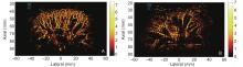
Figure 2
Comparison between normal kidney and diabetes nephropathy. (A) shows the normal kidney image of a 38 year old male; (B) shows a diabetes nephropathy of a 58 year old female which were diagnosed by histopathology (unpublished research results)."
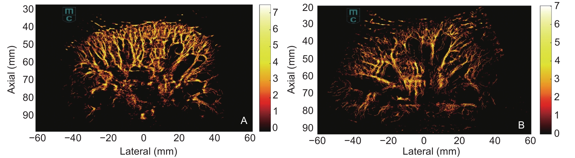
Figure 3
CEUS quantitative parameters of malignant breast lesions and benign lesions. The malignant breast lesions diagnosed as exhibited significantly higher AUC, peak intensity, but lower TTP than benign lesions (unpublished research results). (A-C) shows a poorly differentiated invasive breast cancer of 50 year old female; (D-F) shows a fibroadenoma lesion of 38 year old female."
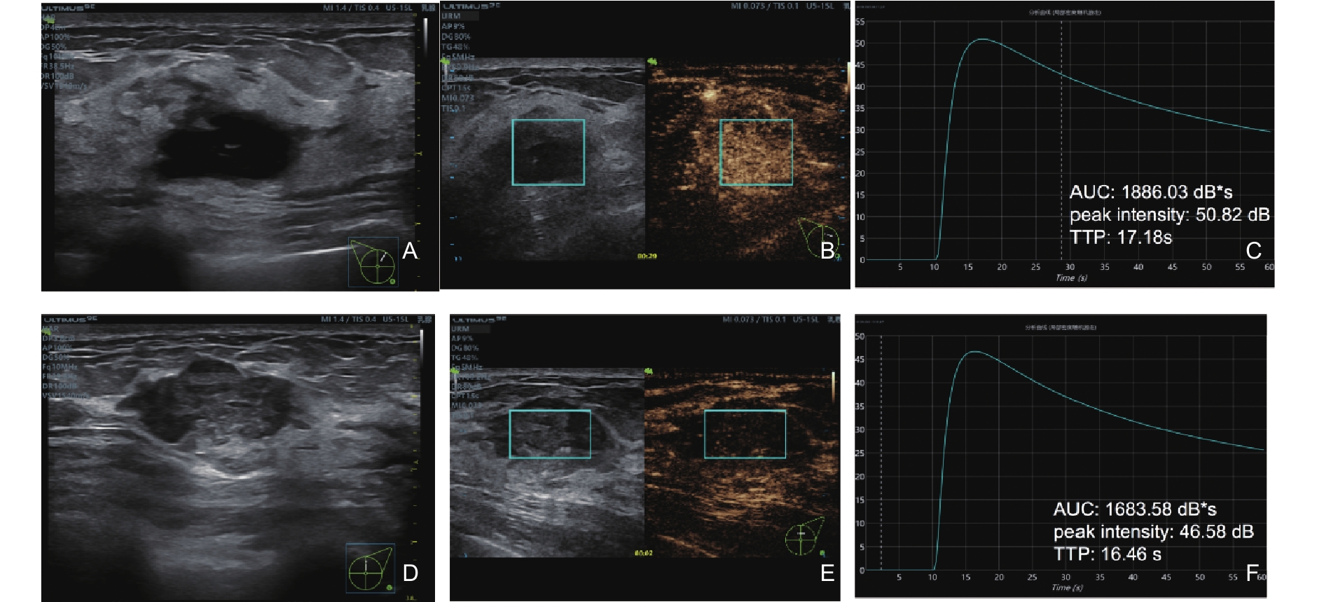
Figure 4
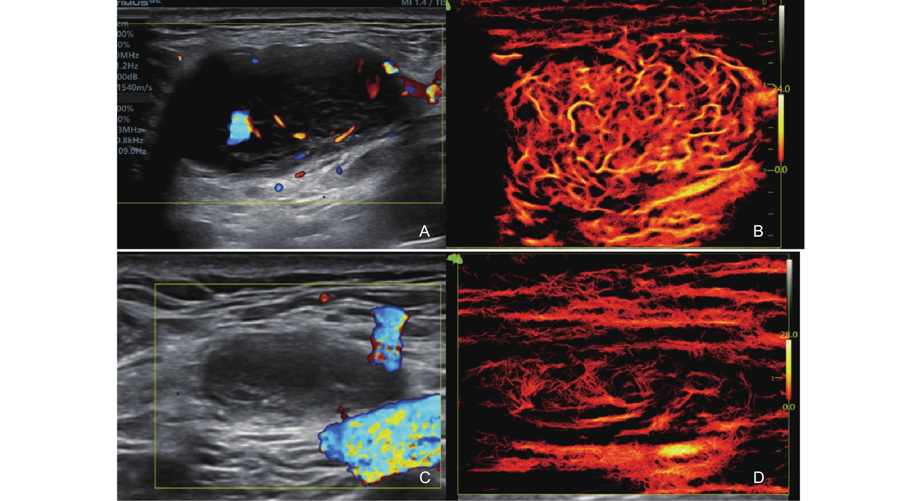
Comparison of ULM imaging before and after follicular lymphoma treatment of a LN. (A,B) showed the color images and microvascular density of LNs before treatment; (C,D) showed the corresponding results after treatment. The MVD decreased from 60.8% to 27.2% after treatment (unpublished research results)."
| [1] |
Viessmann OM, Eckersley RJ, Christensen-Jeffries K, Tang MX, Dunsby C. Acoustic super-resolution with ultrasound and microbubbles. Phys Med Biol 2013; 58: 6447-6458.
doi: 10.1088/0031-9155/58/18/6447 |
| [2] |
Ikeda O, Sato T, Suzuki K. Super-resolution imaging system using waves with a limited frequency bandwidth. JASA 1979; 65: 75-81.
doi: 10.1121/1.382270 |
| [3] | Couture O, Besson B, Montaldo G, Fink M, Tanter M. Microbubble ultrasound super-localization imaging (MUSLI). IEEE international ultrasonics symposium 2011[Conference abstract]. |
| [4] |
Foiret J, Zhang H, Ilovitsh T, Mahakian L, Tam S, Ferrara KW. Ultrasound localization microscopy to image and assess microvasculature in a rat kidney. Sci Rep 2017; 7: 13662.
doi: 10.1038/s41598-017-13676-7 |
| [5] |
Hingot V, Errico C, Tanter M, Couture O. Subwavelength motion-correction for ultrafast ultrasound localization microscopy. Ultrasonics 2017; 77: 17-21.
doi: 10.1016/j.ultras.2017.01.008 |
| [6] |
Bar-Zion A, Solomon O, Tremblay-Darveau C, Adam D, Eldar YC. SUSHI: sparsity-based ultrasound super-resolution hemodynamic imaging. IEEE Trans Ultrason Ferroelectr Freq Control 2018; 65: 2365-2380.
doi: 10.1109/TUFFC.2018.2873380 |
| [7] |
Espindola D, Lin F, Soulioti DE, Dayton PA, Pinton GF. Adaptive multifocus beamforming for contrast-enhanced-super-resolution ultrasound imaging in deep tissue. IEEE Trans Ultrason Ferroelectr Freq Control 2018; 65: 2255-2263.
doi: 10.1109/TUFFC.2018.2865903 |
| [8] |
Wang R, Lee WN. Automated microbubble discrimination in ultrasound localization microscopy by vision transformer. IEEE Trans Ultrason Ferroelectr Freq Control 2025; 72: 1134-1146.
doi: 10.1109/TUFFC.2025.3570496 |
| [9] |
Errico C, Pierre J, Pezet S, Desailly Y, Lenkei Z, Couture O, et al. Ultrafast ultrasound localization microscopy for deep super-resolution vascular imaging. Nature 2015; 527: 499-502.
doi: 10.1038/nature16066 |
| [10] |
Desailly Y, Couture O, Fink M, Tanter M. Sono-activated ultrasound localization microscopy. Applied Physics Letters 2013; 103: 174107.
doi: 10.1063/1.4826597 |
| [11] |
Tanter M, Fink M. Ultrafast imaging in biomedical ultrasound. IEEE Trans Ultrason Ferroelectr Freq Control 2014; 61: 102-19.
doi: 10.1109/TUFFC.2014.2882 |
| [12] |
Desailly Y, Pierre J, Couture O, Tanter M. Resolution limits of ultrafast ultrasound localization microscopy. Phys Med Biol 2015; 60: 8723-8740.
doi: 10.1088/0031-9155/60/22/8723 |
| [13] |
Zhang G, Yu J, Lei YM, Hu JR, Hu HM, Harput S, et al. Ultrasound super-resolution imaging for the differential diagnosis of thyroid nodules: a pilot study. Front Oncol 2022; 12: 978164.
doi: 10.3389/fonc.2022.978164 |
| [14] |
Ackermann D, Schmitz G. Detection and tracking of multiple microbubbles in ultrasound B-mode images. IEEE Trans Ultrason Ferroelectr Freq Control 2016; 63: 72-82.
doi: 10.1109/TUFFC.2015.2500266 |
| [15] |
Christensen-Jeffries K, Browning RJ, Tang MX, Dunsby C, Eckersley RJ. In vivo acoustic super-resolution and super-resolved velocity mapping using microbubbles. IEEE Trans Med Imaging 2015; 34: 433-440.
doi: 10.1109/TMI.2014.2359650 |
| [16] | Hou C, Wang Y, Wang L, Yang L, Li S, Nie F, et al. Super-resolution ultrasound imaging indirectly reveals neurovascular uncoupling in substantia nigra of a Parkinson's disease model. NPJ Parkinsons Dis 2026; 12 1 130 |
| [17] |
Denis L, Bodard S, Hingot V, Chavignon A, Battaglia J, Renault G, et al. Sensing ultrasound localization microscopy for the visualization of glomeruli in living rats and humans. EBioMedicine 2023; 91: 104578.
doi: 10.1016/j.ebiom.2023.104578 |
| [18] |
Brown J, Christensen-Jeffries K, Harput S, Zhang G, Zhu J, Dunsby C, et al. Investigation of microbubble detection methods for super-resolution imaging of microvasculature. IEEE Trans Ultrason Ferroelectr Freq Control 2019; 66: 676-691.
doi: 10.1109/TUFFC.2019.2894755 |
| [19] |
Zhang X, Liu P, Qian X. A cost-efficient multi-angle fusion deep learning for ultrasound localization microscopy. IEEE Trans Biomed Eng 2026; 73: 2123-2132.
doi: 10.1109/TBME.2025.3623140 |
| [20] |
Christensen-Jeffries K, Harput S, Brown J, Wells PNT, Aljabar P, Dunsby C. Microbubble axial localization errors in ultrasound super-resolution imaging. IEEE Trans Ultrason Ferroelectr Freq Control 2017; 64: 1644-1654.
doi: 10.1109/TUFFC.2017.2741067 |
| [21] |
Taghavi I, Andersen SB, Hoyos CAV, Schou M, Gran F, Hansen KL, et al. Ultrasound super-resolution imaging with a hierarchical Kalman tracker. Ultrasonics 2022; 122: 106695.
doi: 10.1016/j.ultras.2022.106695 |
| [22] |
Ghosh D, Xiong F, Sirsi SR, Shaul PW, Mattrey RF, Hoyt K. Toward optimization of in vivo super-resolution ultrasound imaging using size-selected microbubble contrast agents. Med Phys 2017; 44: 6304-6313.
doi: 10.1002/mp.12606 |
| [23] | Lerendegui M, Yan J, Stride E, Dunsby C, Tang MX. Understanding the effects of microbubble concentration on localization accuracy in super-resolution ultrasound imaging. Phys Med Biol 2024;69. |
| [24] |
Zhang G, Hu X, Ren X, Zhou B, Li B, Li Y, et al. In vivo ultrasound localization microscopy for high-density microbubbles. Ultrasonics 2024; 143: 107410.
doi: 10.1016/j.ultras.2024.107410 |
| [25] |
Lin F, Tsuruta JK, Rojas JD, Dayton PA. Optimizing sensitivity of ultrasound contrast-enhanced super-resolution imaging by tailoring size distribution of microbubble contrast agent. Ultrasound Med Biol 2017; 43: 2488-2493.
doi: 10.1016/j.ultrasmedbio.2017.05.014 |
| [26] | Tafsast A, Benoudjit N, Bouakaz A. Comparative analysis of semi-adaptive beamforming algorithms for ultrasound localization microscopy. ICAECCS 2025[Conference abstract]. |
| [27] |
Sobolewski J, Dencks S, Schmitz G. Influence of image discretization and patch size on microbubble localization precision. IEEE Trans Ultrason Ferroelectr Freq Control 2024; 71: 1823-1832.
doi: 10.1109/TUFFC.2024.3479710 |
| [28] |
Heiles B, Chavignon A, Hingot V, Lopez P, Teston E, Couture O. Performance benchmarking of microbubble-localization algorithms for ultrasound localization microscopy. Nat Biomed Eng 2022; 6: 605-616.
doi: 10.1038/s41551-021-00824-8 |
| [29] | Liu X, Almekkawy M. Ultrasound microbubbles localization using object detection model. IEEE IUS 2023;1-4. |
| [30] | Ramilo U, Garay G, Barolin J, Sorriba V, Bube M, Damián JP, et al. Comparison of microbubble tracking algorithms for ultrasound localization microscopy. GMEPE/PAHCE 2026[Conference abstract]. |
| [31] |
Andersen SB, Taghavi I, Hoyos CAV, Søgaard SB, Gran F, Lönn L, et al. Super-resolution imaging with ultrasound for visualization of the renal microvasculature in rats before and after renal ischemia: a pilot sudy. Diagnostics (Basel) 2020; 10: 862.
doi: 10.3390/diagnostics10110862 |
| [32] |
Harput S, Christensen-Jeffries K, Brown J, Li Y, Williams KJ, Davies AH, et al. Two-stage motion correction for super-resolution ultrasound imaging in human lower limb. IEEE Trans Ultrason Ferroelectr Freq Control 2018; 65: 803-814.
doi: 10.1109/TUFFC.2018.2824846 |
| [33] |
Opacic T, Dencks S, Theek B, Piepenbrock M, Ackermann D, Rix A, et al. Motion model ultrasound localization microscopy for preclinical and clinical multiparametric tumor characterization. Nat Commun 2018; 9: 1527.
doi: 10.1038/s41467-018-03973-8 |
| [34] |
Yu J, Lavery L, Kim K. Super-resolution ultrasound imaging method for microvasculature in vivo with a high temporal accuracy. Sci Rep 2018; 8: 13918.
doi: 10.1038/s41598-018-32235-2 |
| [35] |
Dencks S, Piepenbrock M, Opacic T, Krauspe B, Stickeler E, Kiessling F, et al. Clinical pilot application of super-resolution US imaging in breast cancer. IEEE Trans Ultrason Ferroelectr Freq Control 2019; 66: 517-526.
doi: 10.1109/TUFFC.2018.2872067 |
| [36] |
Kanoulas E, Butler M, Rowley C, Voulgaridou V, Diamantis K, Duncan WC, et al. Super-resolution contrast-enhanced ultrasound methodology for the identification of in vivo vascular dynamics in 2D. Invest Radiol 2019; 54: 500-516.
doi: 10.1097/RLI.0000000000000565 |
| [37] |
Schneider M. Characteristics of SonoVuetrade mark. Echocardiography 1999; 16: 743-746.
doi: 10.1111/j.1540-8175.1999.tb00144.x |
| [38] |
Bianconi E, Piovesan A, Facchin F, Beraudi A, Casadei R, Frabetti F. An estimation of the number of cells in the human body. Ann Hum Biol 2013; 40: 463-471.
doi: 10.3109/03014460.2013.807878 |
| [39] |
Ivanov KP, Kalinina MK, Levkovich YuI. Blood flow velocity in capillaries of brain and muscles and its physiological significance. Microvasc Res 1981; 22: 143-155.
doi: 10.1016/0026-2862(81)90084-4 |
| [40] |
Hingot V, Errico C, Heiles B, Rahal L, Tanter M, Couture O. Microvascular flow dictates the compromise between spatial resolution and acquisition time in Ultrasound Localization Microscopy. Sci Rep 2019; 9: 2456.
doi: 10.1038/s41598-018-38349-x |
| [41] |
Christensen-Jeffries K, Brown J, Harput S, Zhang G, Zhu J, Tang MX, Dunsby C, Eckersley RJ. Poisson statistical model of ultrasound super-resolution imaging acquisition time. IEEE Trans Ultrason Ferroelectr Freq Control 2019; 66: 1246-1254.
doi: 10.1109/TUFFC.2019.2916603 |
| [42] |
Huang C, Lowerison MR, Trzasko JD, Manduca A, Bresler Y, Tang S. Short acquisition time super-resolution ultrasound microvessel imaging via microbubble separation. Sci Rep 2020; 10: 6007.
doi: 10.1038/s41598-020-62898-9 |
| [43] |
Dencks S, Piepenbrock M, Schmitz G. Assessing vessel reconstruction in ultrasound localization microscopy by maximum likelihood estimation of a zero-inflated poisson model. IEEE Trans Ultrason Ferroelectr Freq Control 2020; 67: 1603-1612.
doi: 10.1109/TUFFC.2020.2980063 |
| [44] |
Solomon O, van Sloun RJG, Wijkstra H, Mischi M, Eldar YC. Exploiting flow dynamics for superresolution in contrast-enhanced ultrasound. IEEE Trans Ultrason Ferroelectr Freq Control 2019; 66: 1573-1586.
doi: 10.1109/TUFFC.2019.2926062 |
| [45] | Huang C, Zhang W, Gong P, Lok UW, Tang S, Yin T, et al. Super-resolution ultrasound localization microscopy based on a high frame-rate clinical ultrasound scanner: an in-human feasibility study. Phys Med Biol 2021; 66: 10.1088/1361-6560/abef45. |
| [46] |
Lowerison MR, Sekaran NVC, Zhang W, Dong Z, Chen X, Llano DA, et al. Aging-related cerebral microvascular changes visualized using ultrasound localization microscopy in the living mouse. Sci Rep 2022; 12: 619.
doi: 10.1038/s41598-021-04712-8 |
| [47] | Zhang Z, Hwang M, Kilbaugh TJ, Sridharan A, Katz J. Cerebral microcirculation mapped by echo particle tracking velocimetry quantifies the intracranial pressure and detects ischemia. Nat Commun 2022; 13 1 666 |
| [48] |
Shin Y, Lowerison MR, Wang Y, Chen X, You Q, Dong Z, et al. Context-aware deep learning enables high-efficacy localization of high concentration microbubbles for super-resolution ultrasound localization microscopy. Nat Commun 2024; 15: 2932.
doi: 10.1038/s41467-024-47154-2 |
| [49] |
Beliard B, Ahmanna C, Tiran E, Kanté K, Deffieux T, Tanter M, et al. Ultrafast Doppler imaging and ultrasound localization microscopy reveal the complexity of vascular rearrangement in chronic spinal lesion. Sci Rep 2022; 12: 6574.
doi: 10.1038/s41598-022-10250-8 |
| [50] |
Ma H, Yang Y, Gao M, He Q, Zhao D, Luo J, et al. A novel rat model of cerebral small vessel disease and evaluation by super-resolution ultrasound imaging. J Neurosci Methods 2022; 379: 109673.
doi: 10.1016/j.jneumeth.2022.109673 |
| [51] | Li M, Wang W, Su Z, Wu J, Yang Y, Wang L, et al. Clinically translatable ultrasound localization microscopy reveals cerebrovascular remodelling and prognosis in patients with traumatic brain injury. Nat Biomed Eng 2026. |
| [52] |
Kwiatkowska E, Kwiatkowski S, Dziedziejko V, Tomasiewicz I, Domański L. Renal microcirculation injury as the main cause of ischemic acute kidney injury development. Biology (Basel) 2023; 12: 327.
doi: 10.3390/biology12020327 |
| [53] |
Bodard S, Denis L, Hingot V, Chavignon A, Hélénon O, Anglicheau D, et al. Ultrasound localization microscopy of the human kidney allograft on a clinical ultrasound scanner. Kidney Int 2023; 103: 930-935.
doi: 10.1016/j.kint.2023.01.027 |
| [54] |
Lowerison MR, Huang C, Lucien F, Chen S, Song P. Ultrasound localization microscopy of renal tumor xenografts in chicken embryo is correlated to hypoxia. Sci Rep 2020; 10: 2478.
doi: 10.1038/s41598-020-59338-z |
| [55] |
Chen Q, Yu J, Rush BM, Stocker SD, Tan RJ, Kim K. Ultrasound super-resolution imaging provides a noninvasive assessment of renal microvasculature changes during mouse acute kidney injury. Kidney Int 2020; 98: 355-365.
doi: 10.1016/j.kint.2020.02.011 |
| [56] |
Zhang H, Huang L, Yang Y, Qiu L, He Q, Liu J, et al. Evaluation of early diabetic kidney disease using ultrasound localization microscopy: a feasibility study. J Ultrasound Med 2023; 42: 2277-2292.
doi: 10.1002/jum.16249 |
| [57] |
Søgaard SB, Andersen SB, Taghavi I, Hoyos CAV, Christoffersen C, Hansen KL, et al. Super-resolution ultrasound imaging provides quantification of the renal cortical and medullary vasculature in obese zucker rats: a pilot study. Diagnostics (Basel) 2022; 12: 1626.
doi: 10.3390/diagnostics12071626 |
| [58] |
Qiu L, Zhang J, Yang Y, Zhang H, Lee FF, He Q. In vivo assessment of hypertensive nephrosclerosis using ultrasound localization microscopy. Med Phys 2022; 49: 2295-2308.
doi: 10.1002/mp.15583 |
| [59] |
Andersen SB, Taghavi I, Søgaard SB, Hoyos CAV, Nielsen MB, Jensen JA, et al. Super-resolution ultrasound imaging can quantify alterations in microbubble velocities in the renal vasculature of rats. Diagnostics (Basel) 2022; 12: 1111.
doi: 10.3390/diagnostics12051111 |
| [60] |
Zhang W, Lowerison MR, Dong Z, Miller RJ, Keller KA, Song P. Super-resolution ultrasound localization microscopy on a rabbit liver VX2 tumor model: an initial feasibility study. Ultrasound Med Biol 2021; 47: 2416-2429.
doi: 10.1016/j.ultrasmedbio.2021.04.012 |
| [61] | Zhang L, Tian J, Chen Y, Hua Q, Yuan C, Zhang Y, et al. Super-resolution ultrasound combined with contrast-enhanced ultrasound: an exploratory approach for differentiating benign from malignant renal tumors based on microvascular characteristics. Acad Radiol 2026:S1076-6332-00550-7. |
| [62] |
Li J, Wei C, Ying T, Liu Y, Wang R, Li M, et al. Differentiation of benign and malignant breast lesions by ultrasound localization microscopy. Insights Imaging 2025; 16: 128.
doi: 10.1186/s13244-025-02013-6 |
| [63] |
Lei S, Ma T, Gao Z, Zhang Q, Wang S, Hu D, et al. Endoscopic ultrasound localization microscopy for the evaluation of the microvasculature of gastrointestinal tract tumors in rabbits. IEEE Trans Biomed Eng 2022; 69: 3438-3448.
doi: 10.1109/TBME.2022.3170557 |
| [64] |
Zhang G, Lei YM, Li N, Yu J, Jiang XY, Yu MH, et al. Ultrasound super-resolution imaging for differential diagnosis of breast masses. Front Oncol 2022; 12: 1049991.
doi: 10.3389/fonc.2022.1049991 |
| [65] |
Xia S, Hua Q, Song Y, Yuan C, Zheng Y, Tao R, et al. Super-resolution ultrasound imaging of intranodal lymphatic sinuses for predicting sentinel lymph node metastasis in breast cancer: a preliminary study. Eur Radiol 2025; 35: 6079-6088.
doi: 10.1007/s00330-025-11520-5 |
| [66] |
Lowerison M, Zhang W, Chen X, Fan T, Song P. Characterization of anti-angiogenic chemo-sensitization via longitudinal ultrasound localization microscopy in colorectal carcinoma tumor xenografts. IEEE Trans Biomed Eng 2022; 69: 1449-1460.
doi: 10.1109/TBME.2021.3119280 |
| [67] |
Demeulenaere O, Sandoval Z, Mateo P, Dizeux A, Villemain O, Gallet R, et al. Coronary flow assessment using 3-dimensional ultrafast ultrasound localization microscopy. JACC Cardiovasc Imaging 2022; 15: 1193-1208.
doi: 10.1016/j.jcmg.2022.02.008 |
| [68] |
Wang T, Zhang Y, Chen J, Chen P, Kandi F, Yao J, et al. Ultrasound localization microscopy in identifying high-risk carotid plaque using low-frame-rate ultrasound imaging. Ultrasound Med Biol 2026; 52: 123-132.
doi: 10.1016/j.ultrasmedbio.2025.08.030 |
| [69] |
Qian X, Huang C, Li R, Song BJ, Tchelepi H, Shung KK, et al. Super-resolution ultrasound localization microscopy for visualization of the ocular blood flow. IEEE Trans Biomed Eng 2022; 69: 1585-1594.
doi: 10.1109/TBME.2021.3120368 |
| [70] |
Zhu J, Rowland EM, Harput S, Riemer K, Leow CH, Clark B, et al. 3D super-resolution US imaging of rabbit lymph node vasculature in vivo by using microbubbles. Radiology 2019; 291: 642-650.
doi: 10.1148/radiol.2019182593 |
| [71] |
Zhu J, Zhang C, Christensen-Jeffries K, Zhang G, Harput S, Dunsby C, et al. Super-resolution ultrasound localization microscopy of microvascular structure and flow for distinguishing metastatic lymph nodes - an initial human study. Ultraschall Med 2022; 43: 592-598.
doi: 10.1055/a-1917-0016 |
| [72] |
He J, Yi H, Tang D, Zhang X, Cui X, Zhang W, et al. Differentiating benign and malignant superficial lymph nodes using super-resolution contrast enhanced ultrasound - a pilot study. BMC Med Imaging 2025; 25: 170.
doi: 10.1186/s12880-025-01714-0 |
| [73] |
Zhang J, Hu Y, Huang X, Huang X, Deng Q, Zhou Q. Ultrafast ultrasound localization microscopy for differential diagnosis of reactive hyperplasia and metastatic cervical lymph nodes: a pilot study. Quant Imaging Med Surg 2025; 15: 10204-10214.
doi: 10.21037/qims-2024-2876 |
| [74] |
Li M, Zhang X, Yan J, Shu H, Li Z, Ye C, et al. Non-invasive ultrasound localization microscopy (ULM) in azoospermia: connecting testicular microcirculation to spermatogenic functions. Theranostics 2024; 14: 4967-4982.
doi: 10.7150/thno.99668 |
| [75] |
Papanikolaou N, Yan J, Huang B, Davies R, Dunsterville C, De Silva NL, et al. Ultrasound localisation microscopy tracks testicular microvascular adaptations to endocrine function in male infertility. EBioMedicine 2026; 129: 106333.
doi: 10.1016/j.ebiom.2026.106333 |
| [76] |
Smith CAB, Wilson H, Yan J, Tang MX. Quantitative image markers of super-resolution ultrasound. EBioMedicine 2026; 124: 106108.
doi: 10.1016/j.ebiom.2025.106108 |
| [77] | Xia S, Zheng Y, Hua Q, Wen J, Luo X, Yan J, et al. Chinese artificial intelligence alliance for thyroid and breast ultrasound. Super-resolution ultrasound and microvasculomics: a consensus statement. Eur Radiol 2024; 34: 7503-7513. |
| [78] |
Piscaglia F, Bolondi L; Italian Society for Ultrasound in Medicine, Biology (SIUMB) Study Group on Ultrasound Contrast Agents. The safety of Sonovue in abdominal applications: retrospective analysis of 23188 investigations. Ultrasound Med Biol 2006; 32: 1369-1375.
doi: 10.1016/j.ultrasmedbio.2006.05.031 |
| [79] |
Hu C, Feng Y, Huang P, Jin J. Adverse reactions after the use of SonoVue contrast agent: characteristics and nursing care experience. Medicine (Baltimore) 2019; 98: e17745.
doi: 10.1097/MD.0000000000017745 |
| [1] | Wu Mingtai, Zhang Shijun, Xu Li, Jiang Lixin. Advances in the Application of Ultrasound Localization Microscopy in the Diagnosis of Andrological Diseases [J]. Advanced Ultrasound in Diagnosis and Therapy, 2026, 10(4): 376-384. |
| [2] | Xie Liting, Zhang Chengyue, Lou Wenjing, Xu Fan, Ma Wenyuan, Jiang Tian’an. Pulse Field Ablation in Oncology: Current Progress and Future Directions [J]. Advanced Ultrasound in Diagnosis and Therapy, 2025, 9(4): 426-436. |
| [3] | Hou Wenfei, Chen Wanting, Liu Huazhen, Tang Jiajia, Yang Meng. Applications of Ultrasound Localization Microscopy in Abdominal Imaging [J]. Advanced Ultrasound in Diagnosis and Therapy, 2025, 9(4): 347-356. |
| [4] | Xia ShuJun, Zheng YuHang, Hua Qing, Wei MinYan, Wen Jing, Luo XiaoMao, Yan JiPing, Bai BaoYan, Liu Fang, Dong YiJie, Zhou JianQiao, behalf of The Chinese Artificial Intelligence Alliance for Thyroid on, Ultrasound Breast. Super-Resolution Ultrasound-Based Habitat Imaging: A Consensus Statement [J]. Advanced Ultrasound in Diagnosis and Therapy, 2025, 9(2): 97-102. |
| [5] | Yuhang Zheng, BS, Jianqiao Zhou, MD. Deep Learning in Ultrasound Localization Microscopy [J]. Advanced Ultrasound in Diagnosis and Therapy, 2024, 8(3): 86-92. |
| [6] | Guoying Zhang, MD, Ying Tang, BS, Mingyang Wang, MD, Weina Kong, MD. Diagnostic Utility of Superb Microvascular Imaging of ultrasound Examinations to Evaluate Hepatic Ischemia-reperfusion Injury [J]. Advanced Ultrasound in Diagnosis and Therapy, 2024, 8(1): 15-21. |
| [7] | Rendong Chen, PhD, Xiaoqian Wang, BS, Ping Liang, MD, Xiaoping Ouyang, PhD, Dexing Kong, PhD. Intelligent Ultrasonic Diagnosis and Clinical Application: Technical Development and Prospectives [J]. Advanced Ultrasound in Diagnosis and Therapy, 2023, 7(2): 73-81. |
| [8] | Zehui Fu, MD, Chuxue Sun, MD, Haixia Zhou, MD, Xiaowen Lv, MD, Siqi Wang, MD, Hui Chen, MD. Multimodal Vascular Ultrasound Findings in A Young Female with Internal Carotid Artery Dissection [J]. Advanced Ultrasound in Diagnosis and Therapy, 2023, 7(1): 28-31. |
| [9] | Zehui Fu, MD, Siqi Wang, MD, Jian Zhang, MD, Yejun Lu, MD, Xiaomin Mo, MD, Ye He, MD, Chong Wang, MD, Hui Chen, MD. Diagnostic Performance of Superb Microvascular Imaging for Breast Masses: A Systematic Review and Meta-analysis [J]. Advanced Ultrasound in Diagnosis and Therapy, 2022, 6(3): 95-103. |
| Viewed | ||||||
|
Full text |
|
|||||
|
Abstract |
|
|||||
Share: WeChat
Copyright ©2018 Advanced Ultrasound in Diagnosis and Therapy
|
 Advanced Ultrasound in Diagnosis and Therapy (AUDT)
is licensed under a Creative Commons Attribution 4.0 International License.
