
Advanced Ultrasound in Diagnosis and Therapy ›› 2026, Vol. 10 ›› Issue (4): 264-276.doi: 10.26599/AUDT.2026.260027
• REVIEW ARTICLE • Previous Articles Next Articles
Khatami Seyedeh Mohaddeseha, Rahmani Arashb, Liu Ji-Bina, Eisenbrey Johna,*( )
Received:2026-07-31
Revised:2026-08-31
Accepted:2026-10-15
Online:2026-12-30
Published:2026-11-02
Contact:
Eisenbrey John,
E-mail:John.Eisenbrey@jefferson.edu
Khatami Seyedeh Mohaddeseh, Rahmani Arash, Liu Ji-Bin, Eisenbrey John. Ultrasound-Stimulated Microbubbles in Cancer Therapy: A Review of Clinical and Translational Evidence. Advanced Ultrasound in Diagnosis and Therapy, 2026, 10(4): 264-276.
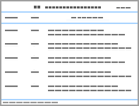
Table 1
Clinically available second-generation microbubble contrast agents and their approved diagnostic indications [7,13,14]."
| Agent | Manufacturer | Shell | Gas Core | Mean diameter (μm) | Approved Indications | Regions of approval |
| Definity (Luminity in EU) | Lantheus | Lipid | Octafluoropropane (C3F8) | 1.1-3.3 | Echocardiography | US, Canada, EU, Australia |
| Lumason/SonoVue | Bracco | Phospholipid | Sulfur hexafluoride (SF6) | 1.5-2.5 | Echocardiography; focal liver lesions; vesicoureteral reflux | US, EU, China, |
| Optison | GE Healthcare | Albumin | Octafluoropropane (C3F8) | 3.0-4.5 | Echocardiography | US, EU |
| Sonazoid | GE Healthcare | Phospholipid | Perfluorobutane (C4F10) | 2-3 | Focal liver lesions; focal breast lesions (Japan) | Japan, South Korea, Taiwan, China, Norway |
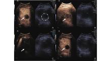
Figure 1
UTMD performed during Y90-TARE treatment of hepatocellular carcinoma in a 70-year-old man with a 3.1 cm segment 4A HCC (arrows), who subsequently achieved complete radiologic response on long-term follow-up imaging. Dual-screen acquisition is shown in each panel, with nonlinear contrast-mode imaging on the left and corresponding B-mode imaging on the right for anatomic reference. (A) Pre-contrast baseline; (B) Peak intratumoral and parenchymal enhancement following intravenous microbubble infusion; (C) Application of the high-mechanical-index destructive pulse; (D) Immediate post-pulse image demonstrating absence of contrast within the tumor, confirming microbubble destruction."

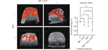
Figure 2
In vivo assessment of tumour perfusion using 3D Power Doppler (PD) ultrasound imaging. Left panel: Representative volumetric reconstructions of tumour vasculature (orange) are shown for each treatment group. Vascularity is visualized in a 3D overlay on B-mode grayscale anatomical images. Scale bar: 2 mm. Right panel: Average Vascular index (VI) across control (C), microbubble-enhanced focused ultrasound (MB + FUS), single-dose radiation therapy (XRT, 8 Gy), and MB + FUS + XRT treatment groups. Data are presented as mean ± SD. p-values ranging from P < 0.05 (*), P < 0.01 (**), P < 0.001 (***). (Reproduced from Petchiny et al32 under the Creative Commons CC-BY 4.0 license)."
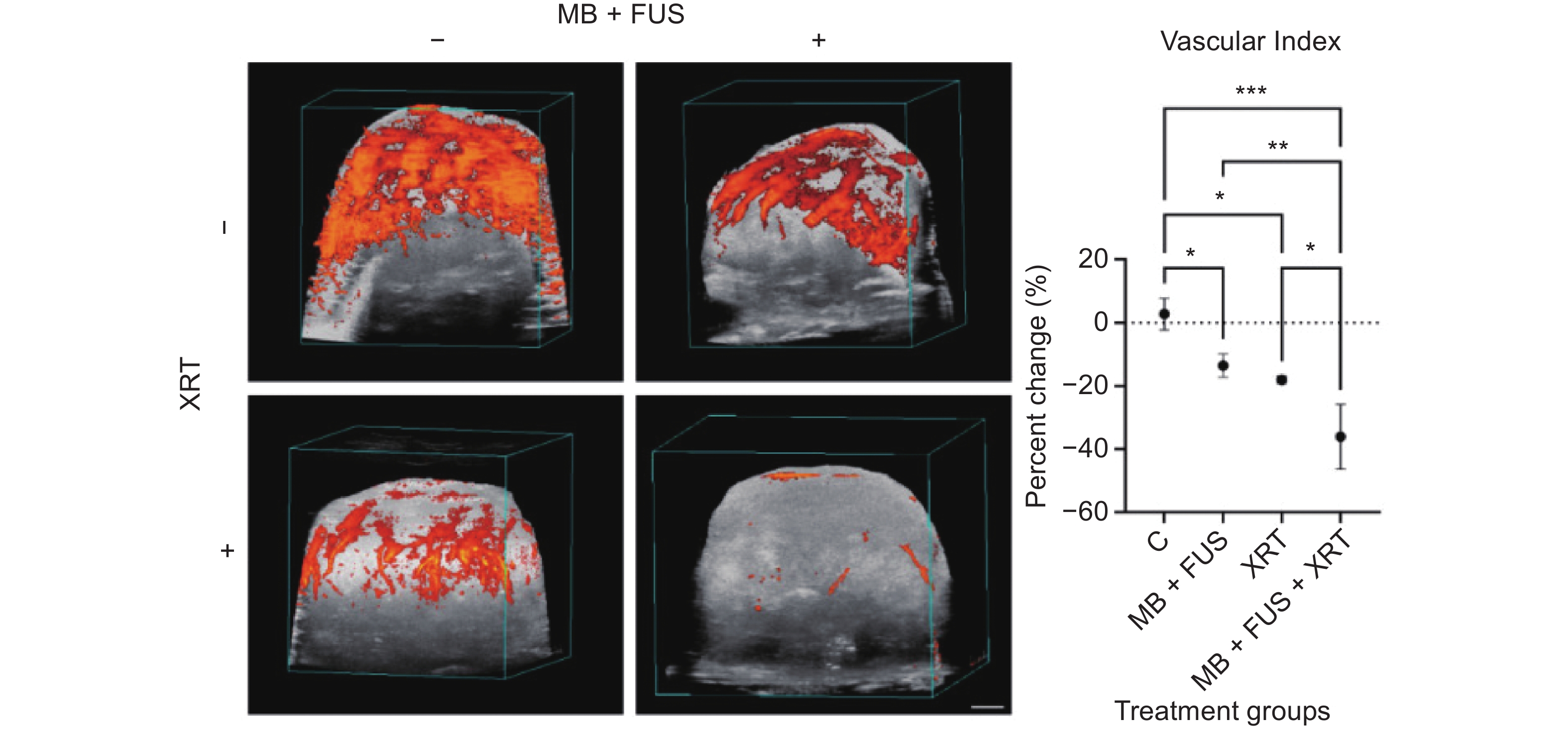
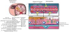
Figure 3
Schematic of intraoperative LIPU/MB procedure and time course of peritumoral brain biopsy. Photo from surgical microscope shows hyperfluorescent areas of dysfunctional BBB shortly after LIPU/MB and infusion of chemotherapy with fluorescein bolus as previously described (24). Blue arrows indicate sonicated peritumoral brain and pink arrows indicate nonsonicated peritumoral brain. Sonicated peritumoral brain biopsies were collected at early time points after LIPU/MB (within 15 minutes) and at later time points (at least 45 minutes after LIPU/MB). (Reproduced from Gould, A. et al.40 under the Creative Commons CC-BY 4.0 license)."

Table 2
Clinical trials of ultrasound-stimulated microbubbles in oncology (by year)."
| First author, year | Application | Tumor | Phase/design | n | Microbubble agent | US delivery | Ref |
| Reference numbers correspond to the bibliography of the current manuscript. Abbreviations: BBB, blood-brain barrier; CRC, colorectal cancer; DMG, diffuse midline glioma; FUS, focused ultrasound; GBM, glioblastoma; HCC, hepatocellular carcinoma; HIFU, high-intensity focused ultrasound; HNSCC, head and neck squamous cell carcinoma; ICC, intrahepatic cholangiocarcinoma; MB, microbubble; MDL, metastatic disease to liver; PDAC, pancreatic ductal adenocarcinoma; PK, pharmacokinetics; RT, radiotherapy; TARE, transarterial radioembolization; US, ultrasound; USMB, ultrasound-stimulated microbubbles; UTMD, ultrasound-triggered microbubble destruction. | |||||||
| Carpentier, 2016 | Drug delivery | Recurrent GBM | Phase 1/2a, single-arm | 15 | SonoVue | SonoCloud-1 implantable + carboplatin | [ |
| Dimcevski, 2016 | Drug delivery | Inoperable PDAC | Phase 1, single-arm | 10 | SonoVue | External US + gemcitabine | [ |
| Idbaih, 2019 | Drug delivery | Recurrent GBM | Phase 1/2a, single-arm | 21 enrolled/ 19 treated | SonoVue | SonoCloud-1 implantable + carboplatin | [ |
| Mainprize, 2019 | Drug delivery | Primary brain tumor | Phase 1, single-arm | 5 | Definity | Transcranial MR-guided FUS | [ |
| Eisenbrey, 2021 | Radiosensitization | HCC | Phase 1, randomized pilot | 28 | Optison | UTMD + 90Y-TARE | [ |
| Zhou, 2022 | Drug delivery | HER2-neg invasive breast | Prospective non-randomized | 26 (10 USMB/16 ctrl) | SonoVue | Diagnostic US + neoadjuvant chemo | [ |
| Dasgupta, 2023 | Radiosensitization | Breast | Phase 1 feasibility | 8 (9 tumors) | Definity | MR-guided FUS + RT | [ |
| Haram, 2023 | Drug delivery | CRC liver mets | First-in-human, lesion-randomized | 17 | SonoVue | External US + systemic chemo | [ |
| Sonabend, 2023 | Drug delivery | Recurrent GBM | Phase 1, single-arm | 17 | Definity | SonoCloud-9 + nab-paclitaxel | [ |
| Han, 2024 | Drug delivery | Inoperable pancreatic cancer | Multicenter, open-label, randomized phase 2 | 78 (38/40) | SonoVue | Diagnostic US + gemcitabine-based chemo | [ |
| Carpentier, 2024 | Drug delivery | Recurrent GBM | Multicenter phase 1/2 | 33 | Definity/ Luminity | SonoCloud-9 + carboplatin | [ |
| Moore-Palhares, 2024 (Breast) | Radiosensitization | Breast/chest wall | Phase 1 | 21 enrolled (18 analyzed/20 tumors) | Definity | MR-guided FUS + RT | [ |
| Moore-Palhares, 2024 (HNSCC) | Radiosensitization | HNSCC (newly dx + recurrent) | Phase 1 | 11 (8 with response data) | Definity | Focused US + RT | [ |
| Zhou, 2024 | Thermal ablation | Advanced primary/metastatic liver | Single-arm prospective feasibility | 17 | Sonazoid | HIFU + Sonazoid bolus | [ |
| Habashy, 2025 | Drug delivery | Recurrent GBM (PK substudy) | PK/microdialysis | 4 | Definity | SonoCloud-9 + carboplatin | [ |
| Wessner, 2025 (HCC) | Radiosensitization | HCC | Single-center phase 2 randomized | 98 (50/48) | Optison | 2D UTMD + 90Y-TARE | [ |
| Wessner, 2025 (ICC/MDL) | Radiosensitization | ICC + liver metastases | Single-center phase 2 | 30 (15/15 matched) | Optison | 3D volumetric UTMD + 90Y-TARE | [ |
| Wu, 2025 | Drug delivery | Pediatric relapsed DMG | First-in-pediatrics feasibility | 3 | Definity | Neuronavigation-guided FUS + oral panobinostat | [ |
| Woodworth, 2025 | Drug delivery | High-grade glioma | Multicenter phase 1/2 (BT008), single-arm | 34 | Definity | MR-guided transcranial FUS + temozolomide | [ |
| Dong, 2026 | Perfusion (drug-delivery rationale) | Invasive breast | Prospective controlled, non-randomized | 36 (18/18) | SonoVue | Diagnostic US + MB | [ |
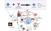
Figure 4
Schematic diagram of ultrasound combined with targeted microvesicle therapy for PCa. (red arrow: BST-2-EGFR-STAT3/ERK1/2 pathway inhibition; green arrow: HIFU thermal effect activates immunogenic cell death (ICD); purple arrow: HIFU cavitation effect generates cellular debris; yellow arrow: apoptosis due to multiple effects of HIFU; blue arrow: activation of immune pathways). (Reproduced from Zhou. et al60 under the Creative Commons CC-BY 4.0 license)"

| [1] | Bray F, Laversanne M, Sung H, Ferlay J, Siegel RL, Soerjomataram I, et al. Global cancer statistics 2022: GLOBOCAN estimates of incidence and mortality worldwide for 36 cancers in 185 countries. CA Cancer J Clin 2024; 74: 229-263. |
| [2] | Bouleftour W, Rowinski E, Louati S, Sotton S, Wozny AS, Moreno-Acosta P, et al. A review of the role of hypoxia in radioresistance in cancer therapy. Med Sci Monit 2021; 27: e934116 |
| [3] |
He J, Liu Z, Zhu X, Xia H, Gao H, Lu J. Ultrasonic microbubble cavitation enhanced tissue permeability and drug diffusion in solid tumor therapy. Pharmaceutics 2022; 14: 1642.
doi: 10.3390/pharmaceutics14081642 |
| [4] | Pillai K, Akhter J, Chua TC, Shehata M, Alzahrani N, Al-Alem I, et al. Heat sink effect on tumor ablation characteristics as observed in monopolar radiofrequency, bipolar radiofrequency, and microwave, using ex vivo calf liver model. Medicine (Baltimore) 2015; 94: e580 |
| [5] |
Eisenbrey JR, Merton DA, Marshall A, Liu JB, Fox TB, Sridharan A, et al. Comparison of photoacoustically derived hemoglobin and oxygenation measurements with contrast-enhanced ultrasound estimated vascularity and immunohistochemical staining in a breast cancer model. Ultrason Imaging 2015; 37: 42-52.
doi: 10.1177/0161734614527435 |
| [6] |
Gummadi SE, Eisenbrey JJ, Li J, Li Z, Forsberg F, Lyshchik A, et al. Advances in modern clinical ultrasound. Advanced Ultrasound in Diagnosis and Therapy 2018; 2: 51-63.
doi: 10.37015/AUDT.2018.180801 |
| [7] |
Frinking P, Segers T, Luan Y, Tranquart F. Three decades of ultrasound contrast agents: a review of the past, present and future improvements. Ultrasound Med Biol 2020; 46: 892-908.
doi: 10.1016/j.ultrasmedbio.2019.12.008 |
| [8] |
Pitt WG, Husseini GA, Staples BJ. Ultrasonic drug delivery—a general review. Expert Opin Drug Deliv 2004; 1: 37-56.
doi: 10.1517/17425247.1.1.37 |
| [9] |
Chen KT, Wei KC, Liu HL. Focused ultrasound combined with microbubbles in central nervous system applications. Pharmaceutics 2021; 13: 1084.
doi: 10.3390/pharmaceutics13071084 |
| [10] |
Sharma D, Czarnota GJ. Using ultrasound and microbubble to enhance the effects of conventional cancer therapies in clinical settings. Cancer Metastasis Rev 2025; 44: 39.
doi: 10.1007/s10555-025-10255-5 |
| [11] | Carpentier A, Canney M, Vignot A, Reina V, Beccaria K, Horodyckid C, et al. Clinical trial of blood-brain barrier disruption by pulsed ultrasound. Sci Transl Med 2016; 8: 343re2 |
| [12] |
Dimcevski G, Kotopoulis S, Bjånes T, Hoem D, Schjøtt J, Gjertsen BT, et al. A human clinical trial using ultrasound and microbubbles to enhance gemcitabine treatment of inoperable pancreatic cancer. J Control Release 2016; 243: 172-181.
doi: 10.1016/j.jconrel.2016.10.007 |
| [13] |
Sridharan A, Eisenbrey JR, Forsberg F, Lorenz N, Steffgen L, Ntoulia A. Ultrasound contrast agents: microbubbles made simple for the pediatric radiologist. Pediatr Radiol 2021; 51: 2117-2127.
doi: 10.1007/s00247-021-05080-1 |
| [14] |
Strom JB, Mulvagh SL, Porter TR, Wei K, Stout JL, Main ML. Contemporary safety of ultrasound enhancing agents in a nationwide analysis. J Am Heart Assoc 2025; 14: e039480.
doi: 10.1161/JAHA.124.039480 |
| [15] |
Czarnota GJ, Karshafian R, Burns PN, Wong S, Al Mahrouki A, Lee JW, et al. Tumor radiation response enhancement by acoustical stimulation of the vasculature. Proc Natl Acad Sci U S A 2012; 109: E2033-E2041.
doi: 10.1073/pnas.1108438109 |
| [16] |
El Kaffas A, Al-Mahrouki A, Hashim A, Law N, Giles A, Czarnota GJ. Role of acid sphingomyelinase and ceramide in mechano-acoustic enhancement of tumor radiation responses. J Natl Cancer Inst 2018; 110: 1009-1018.
doi: 10.1093/jnci/djy011 |
| [17] |
Eisenbrey JR, Forsberg F, Wessner CE, Delaney LJ, Bradigan K, Gummadi S, et al. US-triggered microbubble destruction for augmenting hepatocellular carcinoma response to transarterial radioembolization: a randomized pilot clinical trial. Radiology 2021; 298: 450-457.
doi: 10.1148/radiol.2020202321 |
| [18] |
Eisenbrey JR, Shraim R, Liu JB, Li J, Stanczak M, Oeffinger B, et al. Sensitization of hypoxic tumors to radiation therapy using ultrasound-sensitive oxygen microbubbles. Int J Radiat Oncol Biol Phys 2018; 101: 88-96.
doi: 10.1016/j.ijrobp.2018.01.042 |
| [19] |
Hill RM, Rocha S, Parsons JL. Overcoming the impact of hypoxia in driving radiotherapy resistance in head and neck squamous cell carcinoma. Cancers (Basel) 2022; 14: 4130.
doi: 10.3390/cancers14174130 |
| [20] |
Lacerda Q, Falatah H, Liu JB, Wessner CE, Oeffinger B, Rochani A, et al. Improved tumor control following radiosensitization with ultrasound-sensitive oxygen microbubbles and tumor mitochondrial respiration inhibitors in a preclinical model of head and neck cancer. Pharmaceutics 2023; 15: 1302.
doi: 10.3390/pharmaceutics15041302 |
| [21] |
Lacerda Q, Rochani A, Oeffinger B, Liu JB, Wessner CE, Tahmasebi A, et al. Tumoral oxygenation and biodistribution of Lonidamine oxygen microbubbles following localized ultrasound-triggered delivery. Int J Pharm 2022; 625: 122072.
doi: 10.1016/j.ijpharm.2022.122072 |
| [22] |
Falatah HA, Lacerda Q, Wessner CE, Lo S, Wheatley MA, Liu JB, et al. Influence of phase change droplet activation and microbubble cavitation on the microenvironment of hepatocellular carcinoma. Ultrasound Med Biol 2024; 50: 1387-1394.
doi: 10.1016/j.ultrasmedbio.2024.05.015 |
| [23] | He Y, Dong XH, Zhu Q, Xu YL, Chen ML, Liu Z. Ultrasound-triggered microbubble destruction enhances the radiosensitivity of glioblastoma by inhibiting PGRMC1-mediated autophagy in vitro and in vivo. Mil Med Res 2022; 9: 9 |
| [24] |
Wessner CE, Forsberg F, Lyshchik A, O'Kane P, Bradigan K, Liu JB, et al. Microbubble-based radiosensitization of hepatocellular carcinoma: evaluation of safety and efficacy in a phase II randomized trial. Radiology 2025; 316: e250083.
doi: 10.1148/radiol.250083 |
| [25] |
Wessner CE, Siu Xiao T, Chang A, Liu J, Chong W, Liu JB, et al. Radiosensitization of intrahepatic cholangiocarcinoma and metastatic disease in the liver using microbubble cavitation: results from a phase 2 clinical trial. Cancer Lett 2025; 633: 218026.
doi: 10.1016/j.canlet.2025.218026 |
| [26] |
Moore-Palhares D, Dasgupta A, Saifuddin M, Anzola Pena ML, Prasla S, Ho L, et al. Radiation enhancement using focussed ultrasound-stimulated microbubbles for breast cancer: a phase 1 clinical trial. PLoS Med 2024; 21: e1004408.
doi: 10.1371/journal.pmed.1004408 |
| [27] |
Dasgupta A, Saifuddin M, McNabb E, Ho L, Lu L, Vesprini D, et al. Novel MRI-guided focussed ultrasound stimulated microbubble radiation enhancement treatment for breast cancer. Sci Rep 2023; 13: 13566.
doi: 10.1038/s41598-023-40551-5 |
| [28] |
Moore-Palhares D, Saifuddin M, Dasgupta A, Anzola Pena ML, Prasla S, Ho L, et al. Radiation enhancement using focussed ultrasound-stimulated microbubbles for head and neck cancer: a phase 1 clinical trial. Radiother Oncol 2024; 198: 110380.
doi: 10.1016/j.radonc.2024.110380 |
| [29] |
Sharma D, McNabb E, Law N, Cumal A, Czarnota GJ. Ultrasound-stimulated microbubbles enhancement of fractionated radiation for tumor treatment. BMC Cancer 2023; 23: 693.
doi: 10.1186/s12885-023-10981-5 |
| [30] |
Klein J, Tran W, Lai P, Al-Mahrouki A, Giles A, Czarnota GJ. Effect of treatment sequencing on the tumor response to combined treatment with ultrasound-stimulated microbubbles and radiotherapy. J Ultrasound Med 2020; 39: 2415-2425.
doi: 10.1002/jum.15363 |
| [31] |
Michon S, Rodier F, Yu FTH. Targeted anti-cancer provascular therapy using ultrasound, microbubbles, and nitrite to increase radiotherapy efficacy. Bioconjug Chem 2022; 33: 1093-1105.
doi: 10.1021/acs.bioconjchem.1c00510 |
| [32] |
Petchiny TN, Sharma D, Giles A, Leong KX, Yang W, Sannachi L, et al. ASMase activation in ultrasound-stimulated radiation enhancement using MRI-guided focused ultrasound. Cells 2025; 14: 1618.
doi: 10.3390/cells14201618 |
| [33] |
Fletcher SP, Chisholm A, Lavelle M, Guthier R, Zhang Y, Power C, et al. A study combining microbubble-mediated focused ultrasound and radiation therapy in the healthy rat brain and a F98 glioma model. Sci Rep 2024; 14: 4831.
doi: 10.1038/s41598-024-55442-6 |
| [34] |
Delaney LJ, Ciraku L, Oeffinger BE, Wessner CE, Liu JB, Li J, et al. Breast cancer brain metastasis response to radiation after microbubble oxygen delivery in a murine model. J Ultrasound Med 2019; 38: 3221-3228.
doi: 10.1002/jum.15031 |
| [35] |
Minchinton AI, Tannock IF. Drug penetration in solid tumours. Nat Rev Cancer 2006; 6: 583-592.
doi: 10.1038/nrc1893 |
| [36] |
Lentacker I, De Cock I, Deckers R, De Smedt SC, Moonen CT. Understanding ultrasound induced sonoporation: definitions and underlying mechanisms. Adv Drug Deliv Rev 2014; 72: 49-64.
doi: 10.1016/j.addr.2013.11.008 |
| [37] |
Kooiman K, Vos HJ, Versluis M, de Jong N. Acoustic behavior of microbubbles and implications for drug delivery. Adv Drug Deliv Rev 2014; 72: 28-48.
doi: 10.1016/j.addr.2014.03.003 |
| [38] |
Idbaih A, Canney M, Belin L, Desseaux C, Vignot A, Bouchoux G, et al. Safety and feasibility of repeated and transient blood-brain barrier disruption by pulsed ultrasound in patients with recurrent glioblastoma. Clin Cancer Res 2019; 25: 3793-3801.
doi: 10.1158/1078-0432.CCR-18-3643 |
| [39] |
Sonabend AM, Gould A, Amidei C, Ward R, Schmidt KA, Zhang DY, et al. Repeated blood-brain barrier opening with an implantable ultrasound device for delivery of albumin-bound paclitaxel in patients with recurrent glioblastoma: a phase 1 trial. Lancet Oncol 2023; 24: 509-522.
doi: 10.1016/S1470-2045(23)00112-2 |
| [40] | Gould A, Luan Y, Hou Y, Korobova FV, Chen L, Arrieta VA, et al. Endothelial response to blood-brain barrier disruption in the human brain. JCI Insight 2024; 10: e187328 |
| [41] |
Carpentier A, Stupp R, Sonabend AM, Dufour H, Chinot O, Mathon B, et al. Repeated blood-brain barrier opening with a nine-emitter implantable ultrasound device in combination with carboplatin in recurrent glioblastoma: a phase I/II clinical trial. Nat Commun 2024; 15: 1650.
doi: 10.1038/s41467-024-45818-7 |
| [42] |
Habashy KJ, Synold TW, Feng Y, Gomez C, Amidei C, Ward R, et al. Pharmacokinetic analysis of carboplatin and fluorescein brain retention following ultrasound-based blood-brain barrier opening. Clin Cancer Res 2025; 31: 3562-3570.
doi: 10.1158/1078-0432.CCR-25-0080 |
| [43] |
Mainprize T, Lipsman N, Huang Y, Meng Y, Bethune A, Ironside S, et al. Blood-brain barrier opening in primary brain tumors with non-invasive MR-guided focused ultrasound: a clinical safety and feasibility study. Sci Rep 2019; 9: 321.
doi: 10.1038/s41598-018-36340-0 |
| [44] |
Woodworth GF, Anastasiadis P, Ozair A, Chabros J, Bettegowda C, Chen C, et al. Microbubble-enhanced transcranial focused ultrasound with temozolomide for patients with high-grade glioma (BT008NA): a multicentre, open-label, phase 1/2 trial. Lancet Oncol 2025; 26: 1651-1664.
doi: 10.1016/S1470-2045(25)00492-9 |
| [45] |
Wu CC, Szalontay L, Pouliopoulos AN, Bae S, Berg X, Wei HJ, et al. Blood-brain barrier opening with neuronavigation-guided focused ultrasound in pediatric patients with diffuse midline glioma. Sci Transl Med 2025; 17: eadq6645.
doi: 10.1126/scitranslmed.adq6645 |
| [46] |
Heldin CH, Rubin K, Pietras K, Ostman A. High interstitial fluid pressure—an obstacle in cancer therapy. Nat Rev Cancer 2004; 4: 806-813.
doi: 10.1038/nrc1456 |
| [47] |
Li YJ, Wen G, Wang Y, Wang DX, Yang L, Deng YJ, et al. Perfusion heterogeneity in breast tumors for assessment of angiogenesis. J Ultrasound Med 2013; 32: 1145-1155.
doi: 10.7863/ultra.32.7.1145 |
| [48] |
Zhou B, Lian Q, Jin C, Lu J, Xu L, Gong X, Zhou P. Human clinical trial using diagnostic ultrasound and microbubbles to enhance neoadjuvant chemotherapy in HER2-negative breast cancer. Frontiers in Oncology 2022; 12: 992774.
doi: 10.3389/fonc.2022.992774 |
| [49] | Dong Y, Zhang D, Feng W, Huang Y, Ge Z, Shi XQ. Effect of ultrasound combined with microbubbles on blood perfusion in invasive breast cancer—a prospective clinical trial. Technol Cancer Res Treat 2026; 25: 15330338261423051 |
| [50] | Kane S, Engelhart A, Guadagno J, Jones A, Usoro I, Brutcher E. Pancreatic ductal adenocarcinoma: characteristics of tumor microenvironment and barriers to treatment. J Adv Pract Oncol 2020; 11: 693-698. |
| [51] |
Schultz CW, Ruiz de Garibay G, Langer A, Liu JB, Dhir T, Leitch C, et al. Selecting the optimal parameters for sonoporation of pancreatic cancer in a pre-clinical model. Cancer Biol Ther 2021; 22: 204-215.
doi: 10.1080/15384047.2021.1881026 |
| [52] |
Han F, Wang Y, Dong X, Lin Q, Wang Y, Gao W, et al. Clinical sonochemotherapy of inoperable pancreatic cancer using diagnostic ultrasound and microbubbles: a multicentre, open-label, randomised, controlled trial. Eur Radiol 2024; 34: 1481-1492.
doi: 10.1007/s00330-023-10210-4 |
| [53] |
Haram M, Hansen R, Bouget D, Myhre OF, Davies CL, Hofsli E. Treatment of liver metastases with focused ultrasound and microbubbles in patients with colorectal cancer receiving chemotherapy. Ultrasound Med Biol 2023; 49: 2081-2088.
doi: 10.1016/j.ultrasmedbio.2023.05.013 |
| [54] |
Yao Y, Yang K, Cao Y, Zhou X, Xu J, Liu J, et al. Comparison of the synergistic effect of lipid nanobubbles and SonoVue microbubbles for high intensity focused ultrasound thermal ablation of tumors. PeerJ 2016; 4: e1716.
doi: 10.7717/peerj.1716 |
| [55] |
Chen Z, Zhao H, Qiao X, Yi C, Gao S, Gao W, et al. Effect of microbubble-enhanced ultrasound on radiofrequency ablation of rabbit liver. Ultrasound Med Biol 2018; 44: 1451-1459.
doi: 10.1016/j.ultrasmedbio.2018.03.008 |
| [56] |
Zhou H, Li F, Luo L, Xiong X, Zhou K, Zhu H, et al. Safety of Sonazoid in assisting high-intensity focused ultrasound ablation therapy for advanced liver malignant lesions: a single-arm clinical study. Ultrasound Med Biol 2024; 50: 134-141.
doi: 10.1016/j.ultrasmedbio.2023.09.014 |
| [57] |
Xiong X, Zhou H, Xu X, Fu Q, Wan Y, Cao Y, et al. Ultrasound molecular imaging enhances high-intensity focused ultrasound ablation on liver cancer with B7-H3-targeted microbubbles. Cancer Med 2024; 13: e70341.
doi: 10.1002/cam4.70341 |
| [58] |
Yang Y, Bai W, Chen Y, Zhang W, Wang M, Hu B. Low-frequency and low-intensity ultrasound-mediated microvessel disruption enhance the effects of radiofrequency ablation on prostate cancer xenografts in nude mice. Mol Med Rep 2015; 12: 7517-7525.
doi: 10.3892/mmr.2015.4375 |
| [59] |
Yang Y, Bai W, Chen Y, Lin Y, Hu B. Optimization of low-frequency low-intensity ultrasound-mediated microvessel disruption on prostate cancer xenografts in nude mice using an orthogonal experimental design. Oncol Lett 2015; 10: 2999-3007.
doi: 10.3892/ol.2015.3716 |
| [60] |
Zhou H, Tang J, Wan Y, Gong J, Wang Q, Huang K, et al. Platform-based BST-2-targeted microbubbles enhance HIFU therapy and effectively inhibit prostate cancer residual growth. Int J Hyperthermia 2025; 42: 2511035.
doi: 10.1080/02656736.2025.2511035 |
| [61] |
Xi F, Feng Y, Chen Q, Chen L, Liu J. Microbubbles ultrasonic cavitation regulates tumor interstitial fluid pressure and enhances sonodynamic therapy. Front Oncol 2022; 12: 852454.
doi: 10.3389/fonc.2022.852454 |
| [62] |
Yang H, Qu Y, Tian Y, Wang C, Sun Y, Dai Z, et al. Ultrasound-targeted microbubble destruction enhances the inhibitory effect of sonodynamic therapy against hepatocellular carcinoma. ACS Omega 2024; 9: 51253-51263.
doi: 10.1021/acsomega.4c07746 |
| [1] | Li Na, Li Fan. Focused Ultrasound Combined with Microbubbles for Inducing Blood-Brain Barrier Opening: Cavitation Monitoring and Control [J]. Advanced Ultrasound in Diagnosis and Therapy, 2026, 10(1): 51-58. |
| [2] | Mohamed Tantawi, MD, Ji-Bin Liu, MD, John R Eisenbrey, PhD. Recent Advances in Microbubble-Augmented Cancer Therapy [J]. Advanced Ultrasound in Diagnosis and Therapy, 2020, 4(3): 155-168. |
| [3] | Xianghong Luo, MD, Jianhui Zhang, MD, Sihui Shao, MD, Rong Wu, MD, Lianfang Du, MD, Jie Yuan, PhD, Zhaojun Li, MD. Improving Ultrasound Gene Transfection Efficiency in Vitro [J]. Advanced Ultrasound in Diagnosis and Therapy, 2019, 3(3): 81-86. |
| Viewed | ||||||
|
Full text |
|
|||||
|
Abstract |
|
|||||
Share: WeChat
Copyright ©2018 Advanced Ultrasound in Diagnosis and Therapy
|
 Advanced Ultrasound in Diagnosis and Therapy (AUDT)
is licensed under a Creative Commons Attribution 4.0 International License.
