
Advanced Ultrasound in Diagnosis and Therapy ›› 2026, Vol. 10 ›› Issue (4): 317-333.doi: 10.26599/AUDT.2026.260037
• REVIEW ARTICLE • Previous Articles Next Articles
Sun Yanga,*,1( ), Luo Wanga,b,1, Zhang Qunxiaa, Ren Jianlia, Wang Zhiganga, Ran Haitaoa,*()
Received:2026-09-14
Revised:2026-09-25
Accepted:2026-11-04
Online:2026-12-30
Published:2026-11-02
Contact:
Sun Yang, Ran Haitao,
E-mail:sunyang@cqmu.edu.cn;ranhaitao@cqmu.edu.cn
About author:First author contact:1Yang Sun and Wang Luo contributed equally to this study.
Sun Yang, Luo Wang, Zhang Qunxia, Ren Jianli, Wang Zhigang, Ran Haitao. Ultrasound-Based Precision Assessment and Intervention of Vulnerable Atherosclerotic Plaques: From Plaque Characterization to Therapeutic Modulation. Advanced Ultrasound in Diagnosis and Therapy, 2026, 10(4): 317-333.
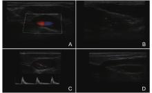
Figure 1
Representative Doppler ultrasound appearances of carotid plaque. (A) Plaque with moderate echogenicity and a smooth surface; (B) Color-flow signals within the plaque region; (C) Plaque at the carotid bifurcation with a spectral Doppler trace; (D) Long-axis view of a plaque with sparse color-flow signals. Reproduced from Wang et al. [20]."
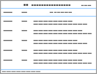
Table 1
Advanced ultrasound assessment of carotid plaque"
| Method | Primary quantity | Evidence | Key limitation | Role in plaque assessment |
| CEUS, contrast-enhanced ultrasound; IPN, intraplaque neovascularization; SMI, superb microvascular imaging; SWE, shear-wave elastography; ULM, ultrasound localization microscopy | ||||
| B-mode and duplex Doppler | Anatomy, burden, surface, stenosis, flow velocity | Established stenosis framework; structured morphology lexicon [ | Shadowing, plane dependence, scanner settings, and limited tissue specificity | Stenosis grading and structural description |
| 3D ultrasound and automated segmentation | Plaque volume and geometry | Histology/surgical-volume agreement and improved efficiency [ | Segmentation failure, registration drift, and uncertain outcome meaning of volume change | Quantification of burden; serial volume change as a research endpoint |
| CEUS | Intravascular enhancement and perfusion related to IPN | Histology correlation, symptom association, small prospective cohorts [ | Protocol and threshold heterogeneity; calcified plaques may be unevaluable; limited specificity for inflammation | Perfused neovessel assessment |
| SMI and related microvascular flow modes | Filtered low-velocity intraplaque flow | Agreement with CEUS and histologic neovessel density; pooled diagnostic studies [ | Vendor-specific clutter filtering and settings; signal differs from contrast kinetics | Noncontrast vascularity assessment within a defined scanner protocol |
| SWE and strain elastography | Stiffness or deformation | Histology studies and symptom-association meta-analysis [ | Pressure, cardiac phase, calcification, geometry, and reconstruction affect estimates | Regional mechanical characterization alongside plaque morphology |
| Ultrafast/vector-flow methods | Flow vectors and wall-shear-related indices | Small human surgical-validation and prospective technical cohorts [ | Boundary conditions, segmentation, and fitted composites | Characterization of local flow and loading |
| ULM | Super-resolved microvascular tracks, density, and velocity | Two small human feasibility/discrimination studies [ | Acquisition and processing dependence; outcome validation unavailable | Human feasibility mapping of microvascular architecture |
| AI/radiomics | Segmentation, quantification, feature integration, classification | Accurate volume measurement; high internal and emerging external discrimination [ | Patient leakage, vendor shift, and exclusion of calcified plaques | Measurement automation and research classification of plaque phenotypes |

Figure 2
Contrast-enhanced ultrasound (CEUS) and histologic neovascularization in human carotid plaque. (A) Color Doppler ultrasound at the carotid bifurcation; the red dashed contour marks the plaque; (B) CEUS of the same region, with focal intraplaque enhancement outlined by the yellow box; dashed lines connect this region to the corresponding histology in panel C; (C) Anti-CD31 immunohistochemistry from the corresponding plaque region; arrows identify CD31-positive neovessels, and the scale bar is 500 μm. Reproduced from Schmidt et al. [25]."

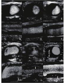
Figure 3
Standard carotid ultrasound and superb microvascular imaging (SMI) in community participants at high stroke risk. (A-F) show transverse and longitudinal views with limited intraplaque vascular signal (SMI grade 1); (G-L) show multiple short linear or strip-like signals in transverse and longitudinal views (SMI grade 2), marked by arrows in the source. Reproduced from Wang et al. [33]."
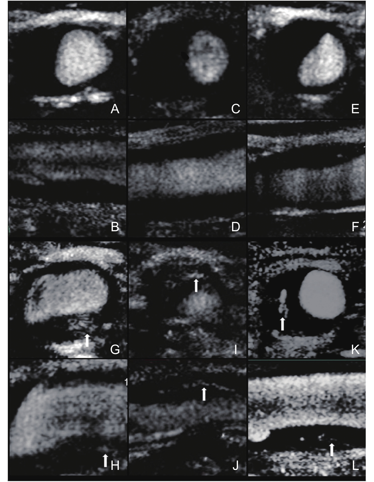
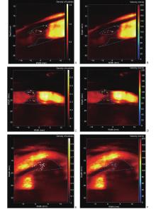
Figure 4
Ultrasound localization microscopy (ULM) density and velocity maps in three patients with histologically detected carotid plaque neovessels. (A, C, and E) show localized-bubble density; (B, D, and F) show the corresponding tracked-bubble velocity. Blue dashed contours are the source study's plaque segmentations. Adapted from Leroy et al. [34]."

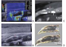
Figure 5
Multiparametric assessment of the same human carotid plaque. (A) Shear-wave elastography, (B) B-mode ultrasound, (C) computed tomography angiography, and (D) gross endarterectomy specimen in longitudinal view. In the source, the red arrow marks a hemorrhagic region corresponding to low stiffness, low echogenicity, and low computed-tomography density; the white arrow marks a fibrous calcified region corresponding to high stiffness, high echogenicity, and high density. Ci, internal carotid artery; Cc, common carotid artery. Reproduced from Goudot et al. [38]."
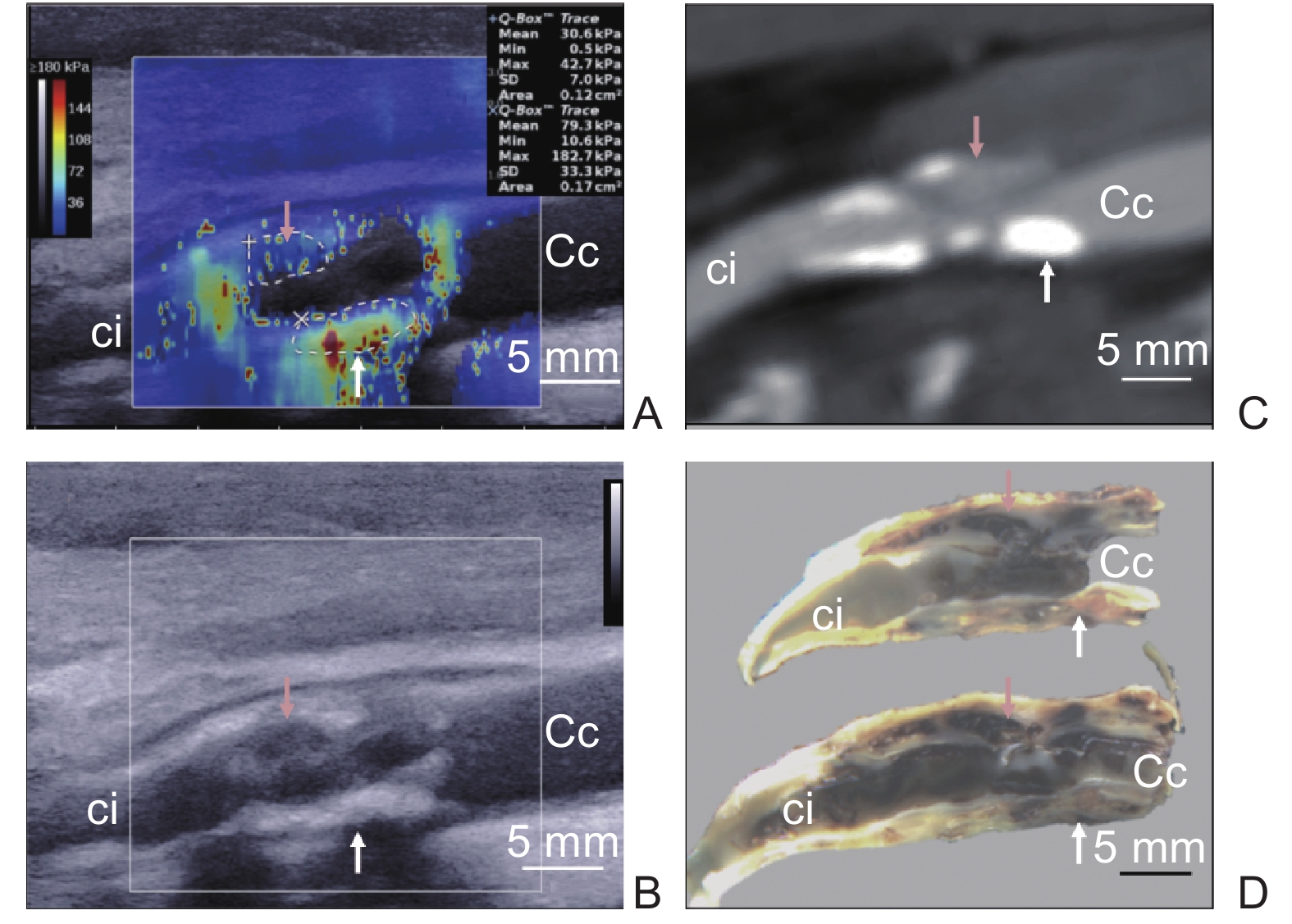
Table 2
Evidence linking plaque imaging, treatment, and response assessment"
| Research area | Target or treatment | Measurements | Evidence | Unresolved clinical question |
| CEUS, contrast-enhanced ultrasound; EDB, extra-domain B; siRNA, small interfering RNA; SMI, superb microvascular imaging; UTMD, ultrasound-targeted microbubble destruction; VCAM-1, vascular cell adhesion molecule-1; vWF, von Willebrand factor. Human ex vivo evidence refers to excised plaque studies. Imaging responses and peripheral functional outcomes are distinct from carotid stroke-prevention outcomes | ||||
| Endothelial inflammation | Luminal VCAM-1 and P-selectin; experimental targeted microbubbles | Targeted molecular signal, target expression, and ex vivo agent binding | Animal molecular imaging and treatment-response studies [ | Can a clinical-grade agent measure carotid target engagement in vivo and inform treatment? |
| Platelet adhesion | Accessible vWF and activated platelet epitopes; experimental targeted microbubbles | Platelet/vWF-associated imaging signal and experimental treatment effects | Human ex vivo plaque target expression [ | How do timing and antiplatelet exposure affect signal interpretation and its relation to ipsilateral events? |
| Plaque-directed delivery | UTMD-assisted gene, antibody, siRNA, or drug delivery; EDB-fibronectin and other experimental targets | Payload delivery, target modulation, CEUS or stiffness, and histology | Animal delivery and imaging studies [ | What human dose, biodistribution, and repeat-treatment safety permit carotid application? |
| Sonodynamic therapy | Sensitizers and ultrasound in vascular or macrophage-rich experimental plaque | Vascular, inflammatory, and matrix changes; early clinical imaging and functional outcomes | Animal studies [ | Can carotid target engagement translate into durable benefit with acceptable neurological safety? |
| Treatment-response monitoring | Lipid-lowering or antithrombotic therapy | CEUS/SMI vascularity, plaque volume, and other ultrasound measurements | Human studies with treatment-related changes and neutral findings [ | Do imaging changes predict a reduction in stroke? |
Figure 6
Experimental molecular ultrasound with cyclic Arg-Gly-Asp-modified microbubbles (MBR) in mice. (A) Background-subtracted color-coded signal after MBR or control microbubbles (MBC) in C57BL/6 mice and ApoE−/− mice receiving a normal or high-cholesterol diet, with separate receptor-blocked groups; (B) Quantified video intensity (n = 6 per group); (C-F) Associations between MBR video intensity and endothelial or plaque glycoprotein IIb/IIIa, plaque vulnerability index, and necrotic-core/fibrous-cap ratio. Reproduced from Guo et al. [51]."
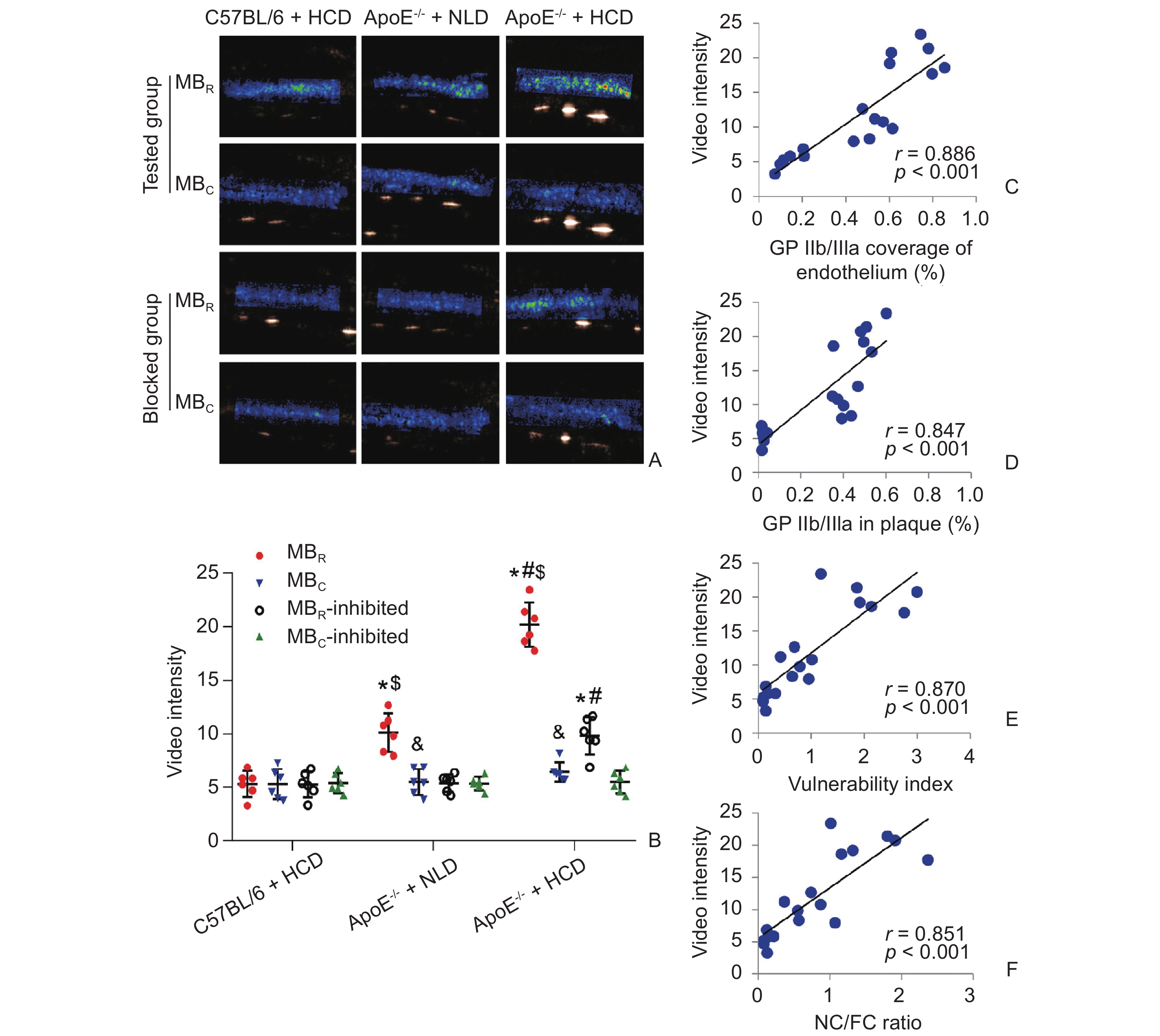
Figure 7
Plaque-directed delivery using platelet-membrane biomimetic rapamycin nanoparticles combined with ultrasound-targeted microbubble destruction (UTMD) in ApoE−/− mice. (A) Thirty-day treatment schedule; (B) En face Oil Red O-stained aortas; (C) Quantified aortic plaque area. Reproduced from Zhou et al. [65]."
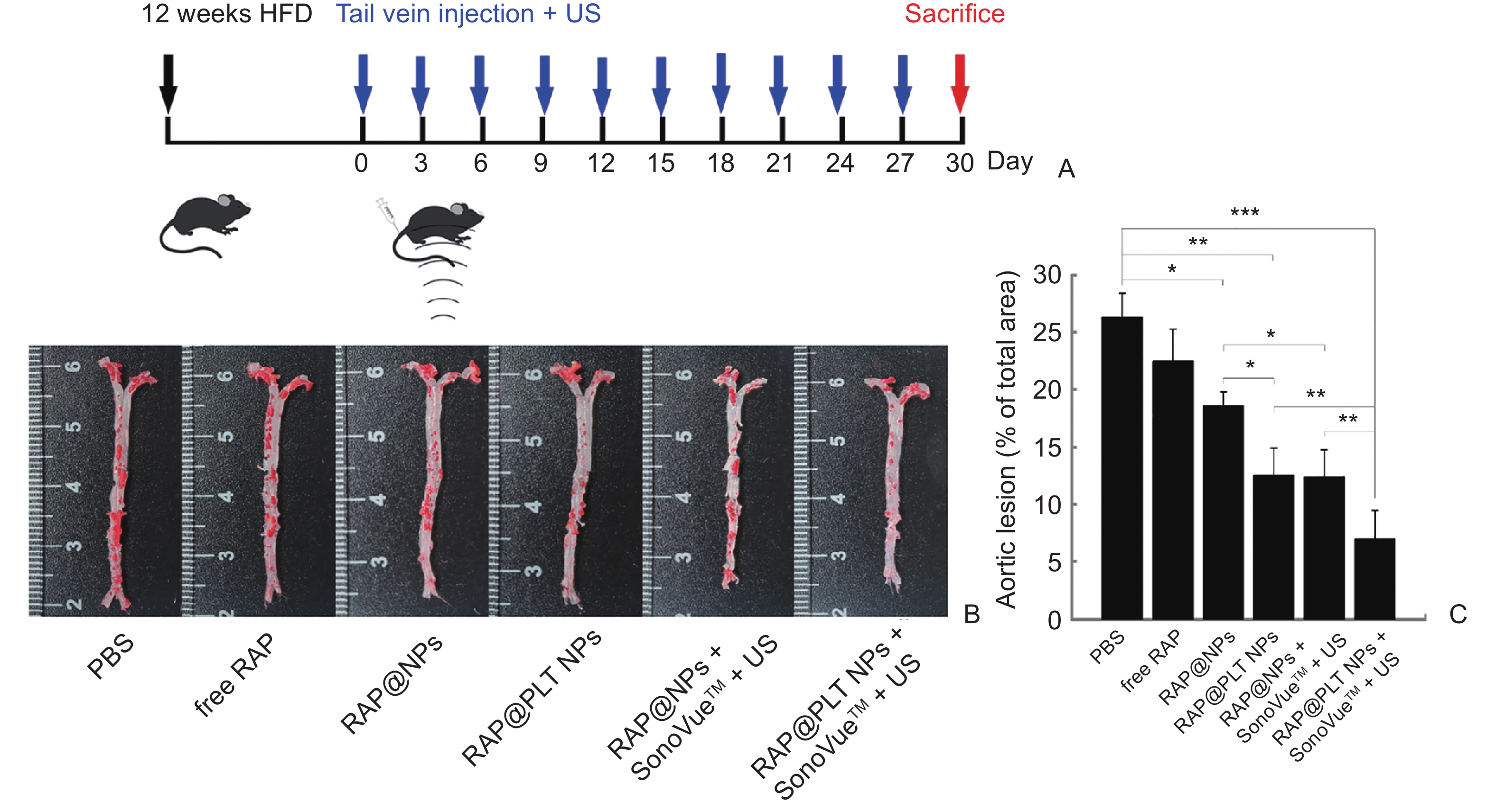
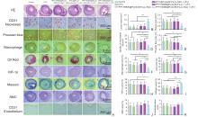
Figure 8
Low-intensity focused-ultrasound-responsive nanoplatform for sonodynamic modulation of advanced rabbit femoral plaque. (A) Representative histopathology 28 days after treatment, including hematoxylin and eosin, CD31, Prussian blue, macrophage, Oil Red O, HIF-1α, Masson, smooth-muscle-cell, and endothelial staining. Red arrows in the source mark abnormal adventitial neovessels and white arrows mark intraplaque hemorrhage; (B-K) Quantification (n = 7; mean ± SD; analysis of variance with Dunnett post hoc comparisons as defined in the source). Reproduced from Yao et al. [70]."

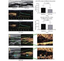
Figure 9
Serial carotid ultrasound during lipid-lowering therapy in a prospective single-arm study. (A) Representative B-mode images at baseline and 6 months show little visible change in plaque morphology; (B) CEUS images and time–intensity curves from the same representative patient; (C, D) Cohort-level reductions in plaque-to-lumen area under the curve and enhanced-intensity ratios at 6 months; (E) Magnified plaque regions in the representative CEUS images; red arrows mark intraplaque vessels and blue arrows mark calcification in the source. Reproduced from Chen et al. [82]."

| [1] |
Polak JF, Alexandrov AV. Accuracy of the society of radiologists in ultrasound (sru) carotid doppler velocity criteria for grading north american symptomatic carotid endarterectomy trial (nascet) stenosis: a meta-analysis. J Ultrasound Med 2023; 42: 1423-1435.
doi: 10.1002/jum.16150 |
| [2] |
Saba L, Brinjikji W, Spence JD, Wintermark M, Castillo M, de Borst GJ, et al. Roadmap consensus on carotid artery plaque imaging and impact on therapy strategies and guidelines: an international, multispecialty, expert review and position statement. AJNR Am J Neuroradiol 2021; 42: 1566-1575.
doi: 10.3174/ajnr.A7223 |
| [3] |
Saba L, Cau R, Murgia A, Nicolaides AN, Wintermark M, Castillo M, et al. Carotid plaque-rads: a novel stroke risk classification system. JACC Cardiovasc Imaging 2024; 17: 62-75.
doi: 10.1016/j.jcmg.2023.09.005 |
| [4] |
Ouyang Y, Lyu G, Huang S, Lin Y, Tong X. The predictive value of Plaque-RADS classification in two-dimensional carotid ultrasound for stroke risk stratification. Quant Imaging Med Surg 2026; 16: 156.
doi: 10.21037/qims-2025-1152 |
| [5] |
Naylor R, Rantner B, Ancetti S, de Borst GJ, De Carlo M, Halliday A, et al. Editor's Choice - European society for vascular surgery (esvs) 2023 clinical practice guidelines on the management of atherosclerotic carotid and vertebral artery disease. Eur J Vasc Endovasc Surg 2023; 65: 7-111.
doi: 10.1016/j.ejvs.2022.04.011 |
| [6] |
Saba L, Antignani PL, Gupta A, Cau R, Paraskevas KI, Poredos P, et al. International Union of Angiology (IUA) consensus paper on imaging strategies in atherosclerotic carotid artery imaging: From basic strategies to advanced approaches. Atherosclerosis 2022; 354: 23-40.
doi: 10.1016/j.atherosclerosis.2022.06.1014 |
| [7] |
Bouakaz A, Escoffre JM. From concept to early clinical trials: 30 years of microbubble-based ultrasound-mediated drug delivery research. Adv Drug Deliv Rev 2024; 206: 115199.
doi: 10.1016/j.addr.2024.115199 |
| [8] |
Yang S, Zhang R, Zhang H, Lu Y. Ultrasound-targeted microbubble destruction: a prospective strategy for treating cardiovascular disease. Front Cardiovasc Med 2025; 12: 1634059.
doi: 10.3389/fcvm.2025.1634059 |
| [9] |
Shao Y, Luo J, Tu S, Xie Y, Zhu J, Wu A, et al. Ultrasound-driven atherosclerosis nanomedicine: from mechanical, cavitation, and sonodynamic therapies to bedside translation. J Nanobiotechnology 2025; 23: 758.
doi: 10.1186/s12951-025-03857-2 |
| [10] |
Phair AS, Carreira J, Bowling FL, Ghosh J, Smith C, Rogers SK. Accelerated measurement of carotid plaque volume using artificial intelligence enhanced 3D ultrasound. Ann Vasc Surg 2024; 98: 317-324.
doi: 10.1016/j.avsg.2023.06.018 |
| [11] |
Pakizer D, Kozel J, Elmers J, Feber J, Michel P, Školoudík D, et al. Histologically verified carotid plaque characteristics by ultrasound: a diagnostic accuracy systematic review. Ultrasound Med Biol 2025; 51: 1646-1659.
doi: 10.1016/j.ultrasmedbio.2025.07.001 |
| [12] |
Coli S, Magnoni M, Sangiorgi G, Marrocco-Trischitta MM, Melisurgo G, Mauriello A, et al. Contrast-enhanced ultrasound imaging of intraplaque neovascularization in carotid arteries: correlation with histology and plaque echogenicity. J Am Coll Cardiol 2008; 52: 223-230.
doi: 10.1016/j.jacc.2008.02.082 |
| [13] |
Li C, He W, Guo D, Chen L, Jin X, Wang W, et al. Quantification of carotid plaque neovascularization using contrast-enhanced ultrasound with histopathologic validation. Ultrasound Med Biol 2014; 40: 1827-1833.
doi: 10.1016/j.ultrasmedbio.2014.02.010 |
| [14] |
Meng Q, Xie X, Li L, Jiang C, Zhao K, Bai Z, et al. Assessment of neovascularization of carotid artery atherosclerotic plaques using superb microvascular imaging: a comparison with contrast-enhanced ultrasound imaging and histology. Quant Imaging Med Surg 2021; 11: 1958-1969.
doi: 10.21037/qims-20-933 |
| [15] |
Zamani M, Skagen K, Scott H, Lindberg B, Russell D, Skjelland M. Carotid plaque neovascularization detected with superb microvascular imaging ultrasound without using contrast media. Stroke 2019; 50: 3121-3127.
doi: 10.1161/STROKEAHA.119.025496 |
| [16] |
Liu Y, Yang S, Wang Y, Hu Y, Liu X, Yang G, et al. Linking carotid plaque immune subtypes to multimodal ultrasound features. Acad Radiol 2026; 33: 4668-4680.
doi: 10.1016/j.acra.2026.07.046 |
| [17] |
Sultan SR, Albin Hajji F, Fatani M, Albngali A, Alfatni A, Alsalamah A, et al. Quantitative ultrasound swe of carotid plaque in symptomatic and asymptomatic patients: a systematic review and meta-analysis. Diagnostics (Basel) 2026; 16: 1085.
doi: 10.3390/diagnostics16071085 |
| [18] |
Goudot G, Sitruk J, Jimenez A, Julia P, Khider L, Alsac JM, et al. Carotid plaque vulnerability assessed by combined shear wave elastography and ultrafast doppler compared to histology. Transl Stroke Res 2022; 13: 100-111.
doi: 10.1007/s12975-021-00920-6 |
| [19] |
Ruisch J, Holewijn S, Vermeer SE, Reijnen MMPJ, de Korte CL, Saris AECM. Blood flow characteristics in patients with asymptomatic carotid artery stenosis using ultrasound-based velocity vector imaging. Ultrasound Med Biol 2026; 52: 2204-2212.
doi: 10.1016/j.ultrasmedbio.2026.06.009 |
| [20] |
Wang B, Li Y, Qiu L. Retrospective study on the association between carotid plaque stability assessed by ultrasound imaging features and stroke risk. Front Neurol 2026; 16: 1638989.
doi: 10.3389/fneur.2025.1638989 |
| [21] |
Sultan SR, Bashmail FT, Alzahrani NA, Alharbi SI, Anbar R, Alkharaiji M. Contrast-enhanced ultrasound for the evaluation of symptomatic and asymptomatic carotid plaques: A systematic review and meta-analysis. Echocardiography 2022; 39: 1032-1043.
doi: 10.1111/echo.15407 |
| [22] |
Hou C, Xuan JQ, Zhao L, Li MX, He W, Liu H. Comparison of the diagnostic performance of contrast-enhanced ultrasound and high-resolution magnetic resonance imaging in the evaluation of histologically defined vulnerable carotid plaque: a systematic review and meta-analysis. Quant Imaging Med Surg 2024; 14: 5814-5830.
doi: 10.21037/qims-24-540 |
| [23] |
Morelli N, Rota E, Spallazzi M, Biondi M, Gallerini S. Sequential contrast-enhanced ultrasound assessment of intraplaque enhancement in carotid plaques. J Ultrasound 2026; 29: 397-406.
doi: 10.1007/s40477-026-01146-8 |
| [24] | Qiu S, Shi J, Tang S, Zheng R, Hu Z, Jing X, et al. Application of high frame rate contrast-enhanced ultrasound in carotid plaque neovascularization: a preliminary inter-physician comparison study. Med Ultrason 2026. |
| [25] |
Schmidt C, Fischer T, Rückert RI, Oberwahrenbrock T, Harms L, Kronenberg G, et al. Identification of neovascularization by contrast-enhanced ultrasound to detect unstable carotid stenosis. PLoS One 2017; 12: e0175331.
doi: 10.1371/journal.pone.0175331 |
| [26] |
Camps-Renom P, Prats-Sánchez L, Casoni F, González-de-Echávarri JM, Marrero-González P, Castrillón I, et al. Plaque neovascularization detected with contrast-enhanced ultrasound predicts ischaemic stroke recurrence in patients with carotid atherosclerosis. Eur J Neurol 2020; 27: 809-816.
doi: 10.1111/ene.14157 |
| [27] |
Cui L, Xing Y, Wang L, Liu K, Chen H, Li C, et al. Carotid Intraplaque Neovascularization and future vascular events in patients with asymptomatic carotid stenosis. Front Pharmacol 2022; 13: 804810.
doi: 10.3389/fphar.2022.804810 |
| [28] |
Chung H, Kim BY, Kim HS, Kim HO, Lee JM, Woo JS, et al. Long-term clinical effects of carotid intraplaque neovascularization in patients with coronary artery disease. Korean J Radiol 2020; 21: 900-907.
doi: 10.3348/kjr.2019.0550 |
| [29] |
Marfella R, Marfella LV, Fumagalli C, Rinaldi L, Sasso FC, Cozzolino D, et al. Intra-plaque vascularization assessed in contrast-enhanced ultrasound predicts cardiovascular events in type 2 diabetes with no significant carotid atherosclerotic stenosis: a prospective study with therapeutic implications for SGLT2 inhibitor use. Eur Heart J Imaging Methods Pract 2025; 3: qyaf129.
doi: 10.1093/ehjimp/qyaf129 |
| [30] |
Zhou S, Hui P. Predictive value of contrast-enhanced carotid ultrasound features for stroke risk: a systematic review and meta-analysis. Front Neurol 2025; 16: 1487850.
doi: 10.3389/fneur.2025.1487850 |
| [31] |
Zhao L, Han Y, Che Y, Zhang X, Luo M, Li J. Diagnostic accuracy of superb microvascular imaging for detecting intraplaque neovascularization: a systematic review and meta-analysis. Cardiovasc Diagn Ther 2023; 13: 893-905.
doi: 10.21037/cdt-23-202 |
| [32] |
Wang J, Tang S, Zhang S, Lin H, Wang Y, Lu X, et al. Superb Microvascular Imaging-based nomogram for acute stroke risk prediction in carotid plaque: integrating ultrasonic neovascularization and clinical biomarkers. Med Ultrason 2026; 28: 194-204.
doi: 10.11152/mu-4556 |
| [33] |
Wang Y, Yao M, Zou M, Li S, Ge Z, Hong Y, et al. Assessment of carotid intraplaque neovascularization using superb microvascular imaging in high risk of stroke individuals: results from a community-based study. Front Neurol 2019; 10: 1146.
doi: 10.3389/fneur.2019.01146 |
| [34] |
Leroy H, Wang LZ, Jimenez A, Mohamedi N, Papadacci C, Julia P, et al. Assessment of microvascular flow in human atherosclerotic carotid plaques using ultrasound localization microscopy. EBioMedicine 2025; 111: 105528.
doi: 10.1016/j.ebiom.2024.105528 |
| [35] |
Wang T, Zhang Y, Chen J, Chen P, Kandi F, Yao J, et al. Ultrasound localization microscopy in identifying high-risk carotid plaque using low-frame-rate ultrasound imaging. Ultrasound Med Biol 2026; 52: 123-132.
doi: 10.1016/j.ultrasmedbio.2025.08.030 |
| [36] |
Di Leo N, Venturini L, de Soccio V, Forte V, Lucchetti P, Cerone G, et al. Multiparametric ultrasound evaluation with CEUS and shear wave elastography for carotid plaque risk stratification. J Ultrasound 2018; 21: 293-300.
doi: 10.1007/s40477-018-0320-7 |
| [37] |
Zhang Y, Cao J, Zhou J, Zhang C, Li Q, Chen S, et al. Plaque Elasticity and intraplaque neovascularisation on carotid artery ultrasound: a comparative histological study. Eur J Vasc Endovasc Surg 2021; 62: 358-366.
doi: 10.1016/j.ejvs.2021.05.026 |
| [38] |
Goudot G, Khider L, Pedreira O, Poree J, Julia P, Alsac JM, et al. Innovative multiparametric characterization of carotid plaque vulnerability by ultrasound. Front Physiol 2020; 11: 157.
doi: 10.3389/fphys.2020.00157 |
| [39] | Wang L, Zhang S, Gao S, Chen J. Carotid plaque mechanical heterogeneity and stroke risk: a comprehensive study integrating multi-site viscoelastic analysis and RADS classification. BMC Cardiovasc Disord 2026; 26: 908 |
| [40] |
Huang Z, Cheng XQ, Liu HY, Bi XJ, Liu YN, Lv WZ, et al. Relation of carotid plaque features detected with ultrasonography-based radiomics to clinical symptoms. Transl Stroke Res 2022; 13: 970-982.
doi: 10.1007/s12975-021-00963-9 |
| [41] |
Jin ZY, Zhou CY, Tan S, Fan XZ, Li JK, Li SY, et al. Features of B-mode ultrasound and contrast-enhanced ultrasound of carotid plaque based on deep learning enhance the prediction of vulnerable plaques associated with acute ischemic stroke. Eur Radiol 2026; 36: 5257-5268.
doi: 10.1007/s00330-026-12376-z |
| [42] |
Hu B, Zhang H, Jia C, Chen K, Tang X, He D, et al. Automatic multi-task segmentation and vulnerability assessment of carotid plaque on contrast-enhanced ultrasound images and videos via deep learning. IEEE J Biomed Health Inform 2026; 30: 4634-4645.
doi: 10.1109/JBHI.2025.3581686 |
| [43] |
Jiang L, Sun Y, Huang W, Wang S, Pan D, Zhang Y, et al. Deep learning-based early prediction of carotid plaque response to lipid-lowering therapy using longitudinal multimodal ultrasound imaging. Insights Imaging 2026; 17: 109.
doi: 10.1186/s13244-026-02286-5 |
| [44] |
Zhang L, Jia C, Gu S, Chen J, Wu R. Intraplaque neovascularization combined with plaque elasticity for predicting ipsilateral stroke in patients with asymptomatic mild carotid stenosis. Quant Imaging Med Surg 2024; 14: 4815-4824.
doi: 10.21037/qims-24-202 |
| [45] | Liu R, Xie J, Huang Z, Wei L. Contrast-enhanced ultrasound for reclassification of moderate-risk carotid plaques (Plaque-RADS 3). BMC Med Imaging 2026; 26: 512 |
| [46] |
Kaufmann BA, Sanders JM, Davis C, Xie A, Aldred P, Sarembock IJ, et al. Molecular imaging of inflammation in atherosclerosis with targeted ultrasound detection of vascular cell adhesion molecule-1. Circulation 2007; 116: 276-284.
doi: 10.1161/CIRCULATIONAHA.106.684738 |
| [47] |
Kaufmann BA, Carr CL, Belcik JT, Xie A, Yue Q, Chadderdon S, et al. Molecular imaging of the initial inflammatory response in atherosclerosis: implications for early detection of disease. Arterioscler Thromb Vasc Biol 2010; 30: 54-59.
doi: 10.1161/ATVBAHA.109.196386 |
| [48] |
Weinkauf CC, Concha-Moore K, Lindner JR, Marinelli ER, Hadinger KP, Bhattacharjee S, et al. Endothelial vascular cell adhesion molecule 1 is a marker for high-risk carotid plaques and target for ultrasound molecular imaging. J Vasc Surg 2018; 68: 105S-113S.
doi: 10.1016/j.jvs.2017.10.088 |
| [49] |
Moccetti F, Weinkauf CC, Davidson BP, Belcik JT, Marinelli ER, Unger E, et al. Ultrasound molecular imaging of atherosclerosis using small-peptide targeting ligands against endothelial markers of inflammation and oxidative stress. Ultrasound Med Biol 2018; 44: 1155-1163.
doi: 10.1016/j.ultrasmedbio.2018.01.001 |
| [50] |
Tian J, Weng Y, Sun R, Zhu Y, Zhang J, Liu H, et al. Contrast-enhanced ultrasound molecular imaging of activated platelets in the progression of atherosclerosis using microbubbles bearing the von Willebrand factor A1 domain. Exp Ther Med 2021; 22: 721.
doi: 10.3892/etm.2021.10153 |
| [51] |
Guo S, Zhang S, Chen K, Chen X, Hu F. Effects of diagnostic ultrasound with cRGD-microbubbles on simultaneous detection and treatment of atherosclerotic plaque in ApoE−/− mice. Front Cardiovasc Med 2022; 9: 946557.
doi: 10.3389/fcvm.2022.946557 |
| [52] |
Kim Y, Lobatto ME, Kawahara T, Lee Chung B, Mieszawska AJ, Sanchez-Gaytan BL, et al. Probing nanoparticle translocation across the permeable endothelium in experimental atherosclerosis. Proc Natl Acad Sci U S A 2014; 111: 1078-1083.
doi: 10.1073/pnas.1322725111 |
| [53] |
Kim I, Elliott JC, Lawanprasert A, Koehle AM III, Wood GM, Castro R, et al. Twinkling peptide nanoemulsions enable precision ultrasound detection of atherosclerotic plaques. Adv Funct Mater 2025; 35: 2415609.
doi: 10.1002/adfm.202415609 |
| [54] |
Li X, Wu Y, Lin C, Wang W, Song W, Yan F, et al. Extradomain B fibronectin-targeted small interfering RNA-loaded biosynthetic gas vesicles for ultrasound-imaging-guided vulnerable plaque treatment. Int J Biol Macromol 2025; 320: 145934.
doi: 10.1016/j.ijbiomac.2025.145934 |
| [55] |
Razansky D, Harlaar NJ, Hillebrands JL, Taruttis A, Herzog E, Zeebregts CJ, et al. Multispectral optoacoustic tomography of matrix metalloproteinase activity in vulnerable human carotid plaques. Mol Imaging Biol 2012; 14: 277-285.
doi: 10.1007/s11307-011-0502-6 |
| [56] |
Yan F, Sun Y, Mao Y, Wu M, Deng Z, Li S, et al. Ultrasound molecular imaging of atherosclerosis for early diagnosis and therapeutic evaluation through leucocyte-like multiple targeted microbubbles. Theranostics 2018; 8: 1879-1891.
doi: 10.7150/thno.22070 |
| [57] |
Punjabi M, Xu L, Ochoa-Espinosa A, Kosareva A, Wolff T, Murtaja A, et al. Ultrasound molecular imaging of atherosclerosis with nanobodies: translatable microbubble targeting murine and human VCAM (vascular cell adhesion molecule) 1. Arterioscler Thromb Vasc Biol 2019; 39: 2520-2530.
doi: 10.1161/ATVBAHA.119.313088 |
| [58] |
Memari E, Helfield B. Shear stress preconditioning and microbubble flow pattern modulate ultrasound-assisted plasma membrane permeabilization. Mater Today Bio 2024; 27: 101128.
doi: 10.1016/j.mtbio.2024.101128 |
| [59] |
Liu Y, Davidson BP, Yue Q, Belcik T, Xie A, Inaba Y, et al. Molecular imaging of inflammation and platelet adhesion in advanced atherosclerosis effects of antioxidant therapy with NADPH oxidase inhibition. Circ Cardiovasc Imaging 2013; 6: 74-82.
doi: 10.1161/CIRCIMAGING.112.975193 |
| [60] |
Shentu W, Ozawa K, Nguyen TA, Wu MD, Packwood W, Xie A, et al. Echocardiographic molecular imaging of the effect of anticytokine therapy for atherosclerosis. J Am Soc Echocardiogr 2021; 34: 433-442.e3.
doi: 10.1016/j.echo.2020.11.012 |
| [61] |
Willmann JK, Bonomo L, Testa AC, Rinaldi P, Rindi G, Valluru KS, et al. Ultrasound molecular imaging with BR55 in patients with breast and ovarian lesions: first-in-human results. J Clin Oncol 2017; 35: 2133-2140.
doi: 10.1200/JCO.2016.70.8594 |
| [62] |
Yang L, Chen L, Fang Y, Ma S. Downregulation of GSK-3β expression via ultrasound-targeted microbubble destruction enhances atherosclerotic plaque stability in new zealand rabbits. Ultrasound Med Biol 2021; 47: 710-722.
doi: 10.1016/j.ultrasmedbio.2020.11.002 |
| [63] |
Yang H, Sun Y, Wei J, Xu L, Tang Y, Yang L, et al. The effects of ultrasound-targeted microbubble destruction (UTMD) carrying IL-8 monoclonal antibody on the inflammatory responses and stability of atherosclerotic plaques. Biomed Pharmacother 2019; 118: 109161.
doi: 10.1016/j.biopha.2019.109161 |
| [64] |
Wu Y, Deng C, Xu J, Wang W, Chen Y, Qin X, et al. Enhanced local delivery of microRNA-145a-5P into mouse aorta via ultrasound-targeted microbubble destruction inhibits atherosclerotic plaque formation. Mol Pharm 2023; 20: 1086-1095.
doi: 10.1021/acs.molpharmaceut.2c00799 |
| [65] |
Zhou J, Niu C, Huang B, Chen S, Yu C, Cao S, et al. Platelet membrane biomimetic nanoparticles combined with UTMD to improve the stability of atherosclerotic plaques. Front Chem 2022; 10: 868063.
doi: 10.3389/fchem.2022.868063 |
| [66] |
Zhong C, Bai J, Yang X, Ji Y, Huang J, Tan X, et al. A study of saponin-encapsulated ultrasound microbubbles Rb3NPs@MBs for atherosclerosis targeted treatment. Biomater Sci 2025; 13: 4984-5000.
doi: 10.1039/D5BM00078E |
| [67] |
Li Z, Sun X, Guo S, Wang L, Wang T, Peng C, et al. Rapid stabilisation of atherosclerotic plaque with 5-aminolevulinic acid-mediated sonodynamic therapy. Thromb Haemost 2015; 114: 793-803.
doi: 10.1160/TH14-12-1030 |
| [68] |
Li B, Gong J, Sheng S, Lu M, Guo S, Yao J, et al. Sonodynamic therapy reduces iron retention of hemorrhagic plaque. Bioeng Transl Med 2021; 6: e10193.
doi: 10.1002/btm2.10193 |
| [69] |
Tian Y, Sheng S, Gao W, Yao J, Tian Y. Sonodynamic therapy suppresses matrix collagen degradation in vulnerable atherosclerotic plaque by modulating caspase 3 - PEDF/HIF-1α - MMP-2/MMP-9 signaling in macrophages. PLoS One 2022; 17: e0279191.
doi: 10.1371/journal.pone.0279191 |
| [70] |
Yao J, Yang Z, Huang L, Yang C, Wang J, Cao Y, et al. Low-intensity focused ultrasound-responsive ferrite-encapsulated nanoparticles for atherosclerotic plaque neovascularization theranostics. Adv Sci (Weinh) 2021; 8: e2100850.
doi: 10.1002/advs.202100850 |
| [71] | Hu J, Jiao M, Tang C, Liang P, Tang J, Wan L, et al. Flexible ultrasound patch for noninvasive, targeted anti-angiogenic therapy of unstable carotid plaques. Ann Biomed Eng 2026. |
| [72] |
Liu J, Chen S, Zhang W, Xu Z, Liu Y, Rong X, et al. A biomimetic nanoplatform for endogenous self-replenishing of oxygen to promote sonodynamic therapy for targeted ablation of activated macrophages in atherosclerotic plaques. Mater Today Bio 2025; 35: 102338.
doi: 10.1016/j.mtbio.2025.102338 |
| [73] |
Li Y, Yang Z, Ma X, Fu P, Zhang L, Gao P, et al. Non-lethal sonodynamic therapy-engineered foam cell exosomes reprograms endothelial immunometabolic crosstalk to stabilize atherosclerotic plaques. Int Immunopharmacol 2025; 167: 115675.
doi: 10.1016/j.intimp.2025.115675 |
| [74] | Jia Y, Li B, Gu Q, Zhang Y, Sang S, Ma F, et al. AIEgen-based biomimetic nanoplatform for targeted synergistic sonodynamic therapy and macrophage reprogramming of hyperhomocysteinemic atherosclerosis. Small 2026:e74733. |
| [75] |
Jiang Y, Fan J, Li Y, Wu G, Wang Y, Yang J, et al. Rapid reduction in plaque inflammation by sonodynamic therapy in patients with symptomatic femoropopliteal peripheral artery disease: a randomized controlled trial. Int J Cardiol 2021; 325: 132-139.
doi: 10.1016/j.ijcard.2020.09.035 |
| [76] |
Sun X, Guo S, Yao J, Wang H, Peng C, Li B, et al. Rapid inhibition of atherosclerotic plaque progression by sonodynamic therapy. Cardiovasc Res 2019; 115: 190-203.
doi: 10.1093/cvr/cvy139 |
| [77] | Wang Y, Sun YH, Liu Y, Fan JX, Wang YQ, Jiang YX, et al. Sonodynamic therapy promotes luminal gain in carotid and femoral atherosclerotic plaques: a single-center prospective clinical study. Zhonghua Xin Xue Guan Bing Za Zhi 2025; 53: 1216-1224. |
| [78] | First Affiliated Hospital of Harbin Medical University. Sonodynamic therapy in the treatment of carotid atherosclerosis (SMART-C). ClinicalTrials.gov identifier NCT03871725; 2019 [cited 2026 Sep 28]. |
| [79] |
Korn A, Simsek S, Fiet MD, Waas ISE, Kooiman K, Niessen HWM, et al. Targeted microbubble cavitation has no therapeutic effect on atherosclerotic plaques of the aorta in diabetic ApoE−/− mice. Ultrasound Med Biol 2026; 52: 1173-1180.
doi: 10.1016/j.ultrasmedbio.2026.02.023 |
| [80] |
Cui E, Kersche G, Grubic N, Hétu MF, Pang SC, Sillesen H, et al. Effect of pharmacologic anti-atherosclerotic therapy on carotid intraplaque neovascularization: A systematic review. J Clin Lipidol 2023; 17: 315-326.
doi: 10.1016/j.jacl.2023.04.009 |
| [81] |
Zhu YC, Jiang XZ, Bai QK, Deng SH, Zhang Y, Zhang ZP, et al. Evaluating the Efficacy of Atorvastatin on Patients with Carotid Plaque by an Innovative Ultrasonography. J Stroke Cerebrovasc Dis 2019; 28: 830-837.
doi: 10.1016/j.jstrokecerebrovasdis.2018.11.027 |
| [82] |
Chen J, Zhao F, Lei C, Qi T, Xue X, Meng Y, et al. Effect of evolocumab on the progression of intraplaque neovascularization of the carotid based on contrast-enhanced ultrasonography (EPIC study): A prospective single-arm, open-label study. Front Pharmacol 2023; 13: 999224.
doi: 10.3389/fphar.2022.999224 |
| [83] |
Huang X, Hou X, Wang M, Wang Z, Liu G, Wang F, et al. Intravascular ultrasound-guided local theranostics enables precise treatment of atherosclerotic plaques. ACS Nano 2026; 20: 14681-14695.
doi: 10.1021/acsnano.6c01815 |
| [84] |
Jessen ML, Yeung K, Collet-Billon A, Eiberg J, Sillesen H, Eldrup N. Rivaroxaban does not alter carotid plaque echolucency, volume, or neovascularisation. Eur J Vasc Endovasc Surg 2026; 71: 354-363.
doi: 10.1016/j.ejvs.2025.10.056 |
| [85] | Sidhu PS, Cantisani V, Dietrich CF, Gilja OH, Saftoiu A, Bartels E, et al. The EFSUMB guidelines and recommendations for the clinical practice of contrast-enhanced ultrasound (CEUS) in non-hepatic applications: update 2017 (long version). Ultraschall Med 2018; 39: e2-e44. |
| [86] | U.S. Food and Drug Administration. Principles of Premarket Pathways for Combination Products: Guidance for Industry and FDA Staff 2022 [cited 2026 Sep 6]. |
| [87] | U.S. Food and Drug Administration. Early Development Considerations for Innovative Combination Products: Guidance for Industry and FDA Staff 2006 [cited 2026 Sep 6]. |
| [1] | Zhao Chenyang, Sun Desheng, Tan Li, Chen Xiangmei. Update of Photodynamic Therapy and Sonodynamic Therapy in Treating Rheumatoid Arthritis [J]. Advanced Ultrasound in Diagnosis and Therapy, 2025, 9(2): 138-145. |
| [2] | Wang Yixuan, Jin Lin, Chen Jianxiong, Yang Huixian, Shen Cuiqin, Xu Wenzhe, Shen Yuzhou, Huang Jun, Sun Liwan, Du Lianfang, Wang Bei, Li Fan, Li Zhaojun. Is the Adventitial Vasa Vasorum in Vulnerable Carotid Plaques Increased or Decreased? [J]. Advanced Ultrasound in Diagnosis and Therapy, 2025, 9(1): 56-64. |
| [3] | Lingyun Jia, MD, PhD, Yuan Li, PhD, Yang Hua, MD, Yumei Liu, MD, Nan Zhang, MD, Mingjie Gao, MD, Ke Zhang, MD, Jingzhi Li, MD, Benchi Chen, BS, Jidong Mi, MS, Nan Zhao, PhD. Evaluation of Atherosclerosis Development by Vascular Duplex Ultrasonography in ApoE-deficient Dogs Fed with a High-fat Diet [J]. Advanced Ultrasound in Diagnosis and Therapy, 2024, 8(2): 49-56. |
| [4] | Qiaoer Gong, BS, Nianyu Xue, MS. Application and Progress of Ultrasound Technology in Atherosclerosis [J]. Advanced Ultrasound in Diagnosis and Therapy, 2023, 7(1): 8-15. |
| [5] | Rifei Li, MM, Yuanmei Zhang, MM, Chengkai Zhang, MM, Xuenian Huang, MM, Shangwei Ding, MD. Contrast Echocardiography Evaluation of Microcirculation of Myocardial Infarction Caused by Takotsubo Syndrome: Case Report and Literature Review [J]. Advanced Ultrasound in Diagnosis and Therapy, 2021, 5(3): 258-261. |
| [6] | Yumei Liu, MD, Beibei Liu, MD, MS, Boyu Li, MD, PhD, Yang Hua, MD. Coronary Heart Disease Concomitant with Atherosclerotic Cerebrovascular Disease [J]. Advanced Ultrasound in Diagnosis and Therapy, 2019, 3(3): 76-80. |
| [7] | Vishal Thumar, MD, Ji-Bin Liu, MD, John Eisenbrey, PhD. Applications in Molecular Ultrasound Imaging: Present and Future [J]. Advanced Ultrasound in Diagnosis and Therapy, 2019, 3(3): 62-75. |
| Viewed | ||||||
|
Full text |
|
|||||
|
Abstract |
|
|||||
Share: WeChat
Copyright ©2018 Advanced Ultrasound in Diagnosis and Therapy
|
 Advanced Ultrasound in Diagnosis and Therapy (AUDT)
is licensed under a Creative Commons Attribution 4.0 International License.
