

| [1] |
Wenderott K , Krups J , Zaruchas F , Weigl M . Effects of artificial intelligence implementation on efficiency in medical imaging-a systematic literature review and meta-analysis. NPJ Digital Medicine 2024; 7: 265.
doi: 10.1038/s41746-024-01248-9 |
| [2] | G. S , Gopalakrishnan U , Parthinarupothi RK , Madathil T . Deep learning supported echocardiogram analysis: A comprehensive review. Artif Intell Med 2024; 151: 26. |
| [3] |
Narang A , Bae R , Hong H , Thomas Y , Surette S , Cadieu C , et al . Utility of a deep-learning algorithm to guide novices to acquire echocardiograms for limited diagnostic use. JAMA cardiology 2021; 6: 624-632.
doi: 10.1001/jamacardio.2021.0185 |
| [4] |
Leclerc S , Smistad E , Pedrosa J , Østvik A , Cervenansky F , Espinosa F , et al . Deep learning for segmentation using an open large-scale dataset in 2D echocardiography. IEEE transactions on medical imaging 2019; 38: 2198-2210.
doi: 10.1109/TMI.2019.2900516 |
| [5] |
Ouyang D , He B , Ghorbani A , Yuan N , Ebinger J , Langlotz CP , et al . Video-based AI for beat-to-beat assessment of cardiac function. Nature 2020; 580: 252-256.
doi: 10.1038/s41586-020-2145-8 |
| [6] | IEEE approved draft guide for an architectural framework for explainable artificial intelligence. IEEE P2894/D9, August 2023 2024:1-51. |
| [7] |
Gao X , Li W , Loomes M , Wang L . A fused deep learning architecture for viewpoint classification of echocardiography. Information Fusion 2017; 36: 103-113.
doi: 10.1016/j.inffus.2016.11.007 |
| [8] |
Madani A , Ong JR , Tibrewal A , Mofrad MRK . Deep echocardiography: data-efficient supervised and semi-supervised deep learning towards automated diagnosis of cardiac disease. NPJ Digital Medicine 2018; 1: 59.
doi: 10.1038/s41746-018-0065-x |
| [9] |
Madani A , Arnaout R , Mofrad M , Arnaout R . Fast and accurate view classification of echocardiograms using deep learning. NPJ Digital Medicine 2018; 1: 6.
doi: 10.1038/s41746-017-0013-1 |
| [10] |
Christensen M , Vukadinovic M , Yuan N , Ouyang D . Vision–language foundation model for echocardiogram interpretation. Nature Medicine 2024; 30: 1481-1488.
doi: 10.1038/s41591-024-02959-y |
| [11] |
Zamzmi G , Rajaraman S , Hsu L-Y , Sachdev V , Antani S . Real-time echocardiography image analysis and quantification of cardiac indices. Medical Image Analysis 2022; 80: 102438.
doi: 10.1016/j.media.2022.102438 |
| [12] |
Tokodi M , Magyar B , Soós A , Takeuchi M , Tolvaj M , Lakatos BK , et al . Deep learning-based prediction of right ventricular ejection fraction using 2D echocardiograms. JACC: Cardiovascular Imaging 2023; 16: 1005-1018.
doi: 10.1016/j.jcmg.2023.02.017 |
| [13] |
Cikes M , Sanchez‐Martinez S , Claggett B , Duchateau N , Piella G , Butakoff C , et al . Machine learning‐based phenogrouping in heart failure to identify responders to cardiac resynchronization therapy. European Journal of Heart Failure 2019; 21: 74-85.
doi: 10.1002/ejhf.1333 |
| [14] |
Holste G , Oikonomou EK , Mortazavi BJ , Coppi A , Faridi KF , Miller EJ , et al . Severe aortic stenosis detection by deep learning applied to echocardiography. Eur Heart J 2023; 44: 4592-4604.
doi: 10.1093/eurheartj/ehad456 |
| [15] |
Wang J , Liu X , Wang F , Zheng L , Gao F , Zhang H , et al . Automated interpretation of congenital heart disease from multi-view echocardiograms. Medical Image Analysis 2021; 69: 101942.
doi: 10.1016/j.media.2020.101942 |
| [16] |
Ulloa Cerna AE , Jing L , Good CW , vanMaanen DP , Raghunath S , Suever JD , et al . Deep-learning-assisted analysis of echocardiographic videos improves predictions of all-cause mortality. Nat Biomed Eng 2021; 5: 546-554.
doi: 10.1038/s41551-020-00667-9 |
| [17] |
Valsaraj A , Kalmady SV , Sharma V , Frost M , Sun W , Sepehrvand N , et al . Development and validation of echocardiography-based machine-learning models to predict mortality. EBioMedicine 2023; 90: 104479.
doi: 10.1016/j.ebiom.2023.104479 |
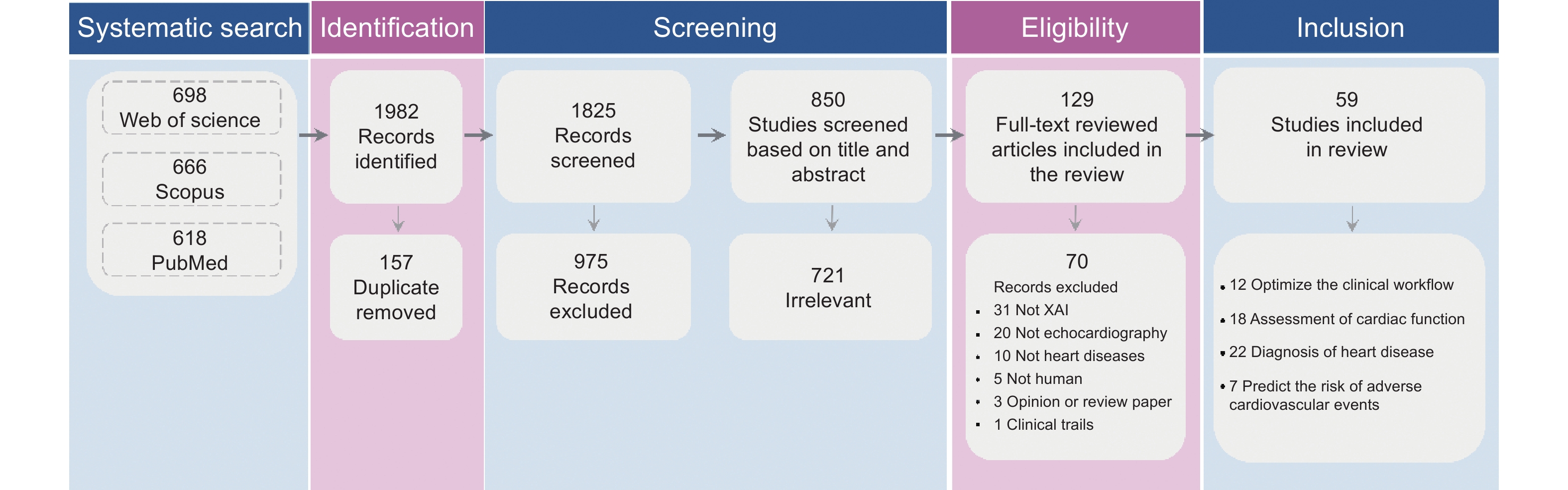
| [18] | Page MJ , McKenzie JE , Bossuyt PM , Boutron I , Hoffmann TC , Mulrow CD , et al . The PRISMA 2020 statement: an updated guideline for reporting systematic reviews. BMJ 2021; 372: n71 |
| [19] |
Barredo Arrieta A , Díaz-Rodríguez N , Del Ser J , Bennetot A , Tabik S , Barbado A , et al . Explainable artificial intelligence (XAI): concepts, taxonomies, opportunities and challenges toward responsible AI. Information Fusion 2020; 58: 82-115.
doi: 10.1016/j.inffus.2019.12.012 |
| [20] |
Du M , Liu N , Hu X . Techniques for interpretable machine learning. Commun Acm 2019; 63: 68-77.
doi: 10.1145/3359786 |
| [21] |
Sengupta PP , Shrestha S , Berthon B , Messas E , Donal E , Tison GH , et al . Proposed requirements for cardiovascular imaging-related machine learning evaluation (PRIME): a checklist: reviewed by the american college of cardiology healthcare innovation council. JACC: Cardiovascular Imaging 2020; 13: 2017-2035.
doi: 10.1016/j.jcmg.2020.07.015 |
| [22] | Charton J , Ren H , Khambhati J , DeFrancesco J , Cheng J , Waheed AA , et al . View classification of color doppler echocardiography via automatic alignment between doppler and b-mode imaging. In: Aylward S, Noble JA, Hu Y, Lee S-L, Baum Z, Min Z, editors. Simplifying Medical Ultrasound. Cham: Springer International Publishing; 2022. p. 64-71. |
| [23] | Liao Z , Jafari MH , Girgis H , Gin K , Rohling R , Abolmaesumi P , Tsang T . Echocardiography view classification using quality transfer star generative adversarial networks. In: Shen D, Liu T, Peters TM, Staib LH, Essert C, Zhou S, et al., editors. Medical image computing and computer assisted intervention – MICCAI 2019. Cham: Springer International Publishing; 2019. p. 687-695. |
| [24] |
Zhang J , Gajjala S , Agrawal P , Tison GH , Hallock LA , Beussink-Nelson L , et al . Fully automated echocardiogram interpretation in clinical practice. Circulation 2018; 138: 1623-1635.
doi: 10.1161/CIRCULATIONAHA.118.034338 |
| [25] | Huang M , Lin W , Chen Y , Hsiao T , Liu P , Tsai W . Explainable deep neural network for echocardiography view classification. European Heart Journal - Cardiovascular Imaging 2022; 23. |
| [26] | Charton J , Ren H , Kim S , Gonzalez CM , Khambhati J , Cheng J , et al . Multi-task learning for hierarchically-structured images: study on echocardiogram view classification. In: Kainz B, Noble A, Schnabel J, Khanal B, Müller JP, Day T, editors. Simplifying Medical Ultrasound. Cham: Springer Nature Switzerland; 2023. p. 185-194. |
| [27] |
Abdi AH , Luong C , Tsang T , Allan G , Nouranian S , Jue J , et al . Automatic quality assessment of echocardiograms using convolutional neural networks: feasibility on the apical four-chamber view. IEEE transactions on medical imaging 2017; 36: 1221-1230.
doi: 10.1109/TMI.2017.2690836 |
| [28] |
Huang M-S , Wang C-S , Chiang J-H , Liu P-Y , Tsai W-C . Automated recognition of regional wall motion abnormalities through deep neural network interpretation of transthoracic echocardiography. Circulation 2020; 142: 1510-1520.
doi: 10.1161/CIRCULATIONAHA.120.047530 |
| [29] |
Hsu C-C , Wang Y-W , Lin L-C , Chang R-F . Spatiotemporal feature disentanglement for quality surveillance of left ventricular echocardiographic video using ST-R(2 + 1)D-ConvNeXt. Biomedical Signal Processing and Control 2025; 105: 107671.
doi: 10.1016/j.bspc.2025.107671 |
| [30] |
Tromp J , Seekings PJ , Hung C-L , Iversen MB , Frost MJ , Ouwerkerk W , et al . Automated interpretation of systolic and diastolic function on the echocardiogram: a multicohort study. The Lancet Digital Health 2022; 4: e46-e54.
doi: 10.1016/S2589-7500(21)00235-1 |
| [31] |
Olaisen S , Smistad E , Espeland T , Hu J , Pasdeloup D , Østvik A , et al . Automatic measurements of left ventricular volumes and ejection fraction by artificial intelligence: clinical validation in real time and large databases. European Heart Journal - Cardiovascular Imaging 2024; 25: 383-395.
doi: 10.1093/ehjci/jead280 |
| [32] | Duffy G , Jain I , He B , Ouyang D . Interpretable deep learning prediction of 3d assessment of cardiac function. Biocomputing 2022: WORLD SCIENTIFIC; 2021. p. 231-241. |
| [33] |
Chen X , Yang F , Zhang P , Lin X , Wang W , Pu H , et al . Artificial intelligence–assisted left ventricular diastolic function assessment and grading: multiview versus single view. J Am Soc Echocardiogr 2023; 36: 1064-1078.
doi: 10.1016/j.echo.2023.07.001 |
| [34] |
Barzegar N , Khatibi T , Hosseinsabet A . Proposing novel methods for simultaneous cardiac cycle phase identification and estimating maximal and minimal left atrial volume (LAV) from apical four-chamber view in 2-D echocardiography. Informatics in Medicine Unlocked 2021; 23: 100517.
doi: 10.1016/j.imu.2021.100517 |
| [35] |
Sanjeevi G , Gopalakrishnan U , Pathinarupothi RK , Madathil T . Automatic diagnostic tool for detection of regional wall motion abnormality from echocardiogram. J Med Syst 2023; 47: 13.
doi: 10.1007/s10916-023-01911-w |
| [36] |
Degerli A , Kiranyaz S , Hamid T , Mazhar R , Gabbouj M . Early myocardial infarction detection over multi-view echocardiography. Biomedical Signal Processing and Control 2024; 87: 105448.
doi: 10.1016/j.bspc.2023.105448 |
| [37] |
Konstam MA , Kiernan MS , Bernstein D , Bozkurt B , Jacob M , Kapur NK , et al . Evaluation and management of right-sided heart failure: a scientific statement from the american heart association. Circulation 2018; 137: e578-e622.
doi: 10.1161/CIRCULATIONAHA.117.032207 |
| [38] |
Hassoun Paul M . Pulmonary arterial hypertension. New England Journal of Medicine 2021; 385: 2361-2376.
doi: 10.1056/NEJMra2000348 |
| [39] |
Surkova E , Muraru D , Genovese D , Aruta P , Palermo C , Badano LP . Relative prognostic importance of left and right ventricular ejection fraction in patients with cardiac diseases. Journal of the American Society of Echocardiography 2019; 32: 1407-1415.e1403.
doi: 10.1016/j.echo.2019.06.009 |
| [40] |
Hagberg E , Hagerman D , Johansson R , Hosseini N , Liu J , Björnsson E , et al . Semi-supervised learning with natural language processing for right ventricle classification in echocardiography—a scalable approach. Comput Biol Med 2022; 143: 105282.
doi: 10.1016/j.compbiomed.2022.105282 |
| [41] |
Ragnarsdottir H , Ozkan E , Michel H , Chin-Cheong K , Manduchi L , Wellmann S , Vogt JE . Deep learning based prediction of pulmonary hypertension in newborns using echocardiograms. Int J Comput Vis 2024; 132: 2567-2584.
doi: 10.1007/s11263-024-01996-x |
| [42] |
Sun D , Hu Y , Li Y , Yu X , Chen X , Shen P , et al . Chamber attention network (CAN): towards interpretable diagnosis of pulmonary artery hypertension using echocardiography. J Adv Res 2024; 63: 103-115.
doi: 10.1016/j.jare.2023.10.013 |
| [43] |
Hirata Y , Tsuji T , Kotoku J , Sata M , Kusunose K . Echocardiographic artificial intelligence for pulmonary hypertension classification. Heart 2024; 110: 586-593.
doi: 10.1136/heartjnl-2023-323320 |
| [44] |
Liu L , Duan S , Li Y , Liu R , Wu Y , Zhang L . Development status and prospect of remote diagnosis and treatment of echocardiography worldwide. Advanced Ultrasound in Diagnosis and Therapy 2020; 4: 303-307.
doi: 10.37015/AUDT.2020.200047 |
| [45] |
Moor M , Banerjee O , Abad ZSH , Krumholz HM , Leskovec J , Topol EJ , Rajpurkar P . Foundation models for generalist medical artificial intelligence. Nature 2023; 616: 259-265.
doi: 10.1038/s41586-023-05881-4 |
| [46] |
Webster P . Six ways large language models are changing healthcare. Nature Medicine 2023; 29: 2969-2971.
doi: 10.1038/s41591-023-02700-1 |
| [47] |
Chen RJ , Ding T , Lu MY , Williamson DFK , Jaume G , Song AH , et al . Towards a general-purpose foundation model for computational pathology. Nature Medicine 2024; 30: 850-862.
doi: 10.1038/s41591-024-02857-3 |
| [48] |
Sanchez-Martinez S , Duchateau N , Erdei T , Kunszt G , Aakhus S , Degiovanni A , et al . Machine learning analysis of left ventricular function to characterize heart failure with preserved ejection fraction. Circ Cardiovasc Imaging 2018; 11: e007138.
doi: 10.1161/CIRCIMAGING.117.007138 |
| [49] |
Atsushi Kyodo , Koshiro Kanaoka , Ayaka Keshi , Maki Nogi , Kazutaka Nogi 1 , Satomi Ishihara , et al . Heart failure with preserved ejection fraction phenogroup classification using machine learning. ESC heart failure 2023; 10: 2019-2030.
doi: 10.1002/ehf2.14368 |
| [50] |
Shad R , Quach N , Fong R , Kasinpila P , Bowles C , Castro M , et al . Predicting post-operative right ventricular failure using video-based deep learning. Nat Commun 2021; 12: 5192.
doi: 10.1038/s41467-021-25503-9 |
| [51] |
Chen W , Xie Y , Zhang Z , Zhu Y , Zhang Y , Zhu S , et al . Artificial Intelligence-assisted medical imaging in interventional management of valvular heart disease. Advanced Ultrasound in Diagnosis and Therapy 2023; 7: 217-227.
doi: 10.37015/AUDT.2023.230030 |
| [52] |
Gu AN , Vaseli H , Tsang MY , Wu V , Ahmadi Amiri SN , Kondori N , et al . ProtoASNet: Comprehensive evaluation and enhanced performance with uncertainty estimation for aortic stenosis classification in echocardiography. Medical Image Analysis 2025; 103: 103600.
doi: 10.1016/j.media.2025.103600 |
| [53] |
Tang L , Wang X , Yang J , Wang Y , Qu M , Li H . DLFFNet: A new dynamical local feature fusion network for automatic aortic valve calcification recognition using echocardiography. Comput Methods Programs Biomed 2024; 243: 107882.
doi: 10.1016/j.cmpb.2023.107882 |
| [54] |
Bernard J , Yanamala N , Shah R , Seetharam K , Altes A , Dupuis M , et al . Integrating echocardiography parameters with explainable artificial intelligence for data-driven clustering of primary mitral regurgitation phenotypes. JACC: Cardiovascular Imaging 2023; 16: 1253-1267.
doi: 10.1016/j.jcmg.2023.02.016 |
| [55] | Vafaeezadeh M , Behnam H , Hosseinsabet A , Gifani P . CarpNet: Transformer for mitral valve disease classification in echocardiographic videos. International Journal of Imaging Systems and Technology 2023; 33: 1505-1514 |
| [56] | Hwang I-C , Choi D , Choi Y-J , Ju L , Kim M , Hong J-E , et al . Differential diagnosis of common etiologies of left ventricular hypertrophy using a hybrid CNN-LSTM model. Scientific Reports 2022; 12: 20998 |
| [57] | Chao C-J , Jeong J , Arsanjani R , Kim K , Tsai Y-L , Yu W-C , et al . Echocardiography-based deep learning model to differentiate constrictive pericarditis and restrictive cardiomyopathy. JACC: Cardiovascular Imaging 2024; 17: 349-360 |
| [58] | Li R , Zhang Y , Zhang C , Huang X , Ding S . Contrast echocardiography evaluation of microcirculation of myocardial infarction caused by takotsubo syndrome: case report and literature review. Advanced Ultrasound in Diagnosis and Therapy 2021; 5: 258-261 |
| [59] |
Zaman F , Ponnapureddy R , Wang YG , Chang A , Cadaret LM , Abdelhamid A , et al . Spatio-temporal hybrid neural networks reduce erroneous human “judgement calls” in the diagnosis of Takotsubo syndrome. EClinicalMedicine 2021; 40: 101115.
doi: 10.1016/j.eclinm.2021.101115 |
| [60] |
Zaman F , Isom N , Chang A , Wang YG , Abdelhamid A , Khan A , et al . Deep learning from atrioventricular plane displacement in patients with Takotsubo syndrome: lighting up the black-box. European Heart Journal - Digital Health 2024; 5: 134-143.
doi: 10.1093/ehjdh/ztad077 |
| [61] | Lee H , Eun Y , Hwang JY , Eun LY . Explainable deep learning algorithm for distinguishing incomplete Kawasaki disease by coronary artery lesions on echocardiographic imaging. Comput Methods Programs Biomed 2022; 223: 106970 |
| [62] |
Wang K , Tian J , Zheng C , Yang H , Ren J , Liu Y , et al . Interpretable prediction of 3-year all-cause mortality in patients with heart failure caused by coronary heart disease based on machine learning and SHAP. Comput Biol Med 2021; 137: 104813.
doi: 10.1016/j.compbiomed.2021.104813 |
| [63] |
Park J , Hwang I-C , Yoon YE , Park J-B , Park J-H , Cho G-Y . Predicting long-term mortality in patients with acute heart failure by using machine learning. J Card Fail 2022; 28: 1078-1087.
doi: 10.1016/j.cardfail.2022.02.012 |
| [64] |
Molenaar MA , Bouma BJ , Asselbergs FW , Verouden NJ , Selder JL , Chamuleau SAJ , Schuuring MJ . Explainable machine learning using echocardiography to improve risk prediction in patients with chronic coronary syndrome. Eur Heart J Digit Health 2024; 5: 170-182.
doi: 10.1093/ehjdh/ztae001 |
| [65] | Hirata Y , Kusunose K . AI in echocardiography: state-of-the-art automated measurement techniques and clinical applications. JMA J 2024; 8: 141-150 |
| [66] | Ahn SS , Ta K , Thorn SL , Onofrey JA , Melvinsdottir IH , Lee S , et al . Co-attention spatial transformer network for unsupervised motion tracking and cardiac strain analysis in 3D echocardiography. Medical Image Analysis 2023; 84: 102711 |
| [67] |
Cheng LH , Bosch PBJ , Hofman RFH , Brakenhoff TB , Bruggemans EF , van der Geest RJ , Holman ER . Revealing unforeseen diagnostic image features with deep learning by detecting cardiovascular diseases from apical 4‐chamber ultrasounds. Journal of the American Heart Association 2022; 11: e024168.
doi: 10.1161/JAHA.121.024168 |
| [68] |
Jin W , Li X , Fatehi M , Hamarneh G . Guidelines and evaluation of clinical explainable AI in medical image analysis. Medical Image Analysis 2023; 84: 102684.
doi: 10.1016/j.media.2022.102684 |
| [69] |
Behzad S , Tabatabaei SMH , Lu MY , Eibschutz LS , Gholamrezanezhad A . Pitfalls in interpretive applications of artificial intelligence in radiology. American Journal of Roentgenology 2024; 223: e2431493.
doi: 10.2214/AJR.24.31493 |
| [70] |
Duffy G , Cheng PP , Yuan N , He B , Kwan AC , Shun-Shin MJ , et al . High-throughput precision phenotyping of left ventricular hypertrophy with cardiovascular deep learning. JAMA cardiology 2022; 7: 386-395.
doi: 10.1001/jamacardio.2021.6059 |
| [71] | Kiranyaz S , Degerli A , Hamid T , Mazhar R , Fadil Ahmed RE , Abouhasera R , et al . Left ventricular wall motion estimation by active polynomials for acute myocardial infarction detection. Ieee Access 2020; 8: 210301-210317 |
| [72] |
Alessandrini M , Chakraborty B , Heyde B , Bernard O , De Craene M , Sermesant M , D’Hooge J . Realistic vendor-specific synthetic ultrasound data for quality assurance of 2-D speckle tracking echocardiography: simulation pipeline and open access database. IEEE Transactions on Ultrasonics, Ferroelectrics, and Frequency Control 2018; 65: 411-422.
doi: 10.1109/TUFFC.2017.2786300 |
| [73] | Huang Z , Long G , Wessler B , Hughes MC . A new semi-supervised learning benchmark for classifying view and diagnosing aortic stenosis from echocardiograms. In: Ken J, Serena Y, Mark S, Michael S, Rajesh R, editors. proceedings of the 6th machine learning for healthcare conference. Proceedings of Machine Learning Research: PMLR; 2021. p. 614-647. |
| [74] |
Qiu T , Wang X , Chen C , Atiquzzaman M , Liu L . TMED: a spider-web-like transmission mechanism for emergency data in vehicular ad hoc networks. IEEE Transactions on Vehicular Technology 2018; 67: 8682-8694.
doi: 10.1109/TVT.2018.2841348 |
| [75] | Kaikai L , Yiyu S , Zhuang J , Meiping H , Hongwen F , Boyang L , et al. Enhance regional wall segmentation by style transfer for regional wall motion assessment. 2023. |
| [76] | Yang J , Ding X , Zheng Z , Xu X , Li X . GraphEcho: graph-driven unsupervised domain adaptation for echocardiogram video segmentation. 2023. p. 11844-11853. |
| [77] | Yang J , Lin Y , Pu B , Guo J , Xu X , Li X . CardiacNet: learning to reconstruct abnormalities for cardiac disease assessment from echocardiogram videos. In: Leonardis A, Ricci E, Roth S, Russakovsky O, Sattler T, Varol G, editors. Computer Vision – ECCV 2024. Cham: Springer Nature Switzerland; 2025. p. 293-311. |
| [78] |
Carbonati T , Eslami P , Chaudhari A , Herbst E , Fiorina L , Porquet P , et al . Mimic-iv-ecg & mimic-iv-echo: detection of regional wall motion abnormalities from electrocardiogram using deep learning. Journal of the American College of Cardiology 2024; 83: 1172-1172.
doi: 10.1016/S0735-1097(24)03162-0 |
| [79] | Magyar B , Tokodi M , Soós A , Tolvaj M , Lakatos BK , Fábián A , et al. RVENet: A large echocardiographic dataset for the deep learning-based assessment of right ventricular function. In: Karlinsky L, Michaeli T, Nishino K, editors. Computer Vision – ECCV 2022 Workshops. Cham: Springer Nature Switzerland; 2023. p. 569-583. |
| [80] | Zhao D , Ferdian E , Maso Talou GD , Quill GM , Gilbert K , Wang VY , et al. MITEA: A dataset for machine learning segmentation of the left ventricle in 3D echocardiography using subject-specific labels from cardiac magnetic resonance imaging. Frontiers in Cardiovascular Medicine 2023;9. |
| [81] | Gearhart A , Goto S , Deo RC , Powell AJ . An automated view classification model for pediatric echocardiography using artificial intelligence. Journal of the American Society of Echocardiography 2022; 35: 1238-1246 |
| [82] |
Jwan A Naser , Eunjung Lee , Sorin V Pislaru , Gal Tsaban , Jeffrey G Malins , John I Jackson , et al . Artificial intelligence-based classification of echocardiographic views. European Heart Journal - Digital Health 2024; 5: 260-269.
doi: 10.1093/ehjdh/ztae015 |
| [83] | Thomas S , Tiago C , Andreassen BS , Aase S-A , Šprem J , Steen E , et al. Graph convolutional neural networks for automated echocardiography view recognition: a holistic approach. In: Kainz B, Noble A, Schnabel J, Khanal B, Müller JP, Day T, editors. Simplifying Medical Ultrasound. Cham: Springer Nature Switzerland; 2023. p. 44-54. |
| [84] | Howard JP , Tan J , Shun-Shin MJ , Mahdi D , Nowbar AN , Arnold AD , et al. Improving ultrasound video classification: an evaluation of novel deep learning methods in echocardiography. Journal of medical artificial intelligence 2019;3. |
| [85] |
Labs RB , Vrettos A , Loo J , Zolgharni M . Automated assessment of transthoracic echocardiogram image quality using deep neural networks. Intelligent Medicine 2023; 3: 191-199.
doi: 10.1016/j.imed.2022.08.001 |
| [86] | Shiokawa N , Izumo M , Shimamura T , Kurosaka Y , Sato Y , Okamura T , et al . Accuracy and efficacy of artificial intelligence-derived automatic measurements of transthoracic echocardiography in routine clinical practice. J Clin Med 2024; 13: 1861 |
| [87] |
Akerman Ashley P , Porumb M , Scott Christopher G , Beqiri A , Chartsias A , Ryu Alexander J , et al . Automated echocardiographic detection of heart failure with preserved ejection fraction using artificial intelligence. JACC: Advances 2023; 2: 100452.
doi: 10.1016/j.jacadv.2023.100452 |
| [88] |
Dai W , Li X , Ding X , Cheng KT . Cyclical self-supervision for semi-supervised ejection fraction prediction from echocardiogram videos. IEEE transactions on medical imaging 2023; 42: 1446-1461.
doi: 10.1109/TMI.2022.3229136 |
| [89] | Mokhtari M , Tsang T , Abolmaesumi P , Liao R . EchoGNN: explainable ejection fraction estimation with graph neural networks. In: Wang L, Dou Q, Fletcher PT, Speidel S, Li S, editors. Medical Image Computing and Computer Assisted Intervention – MICCAI 2022. Cham: Springer Nature Switzerland; 2022. p. 360-369. |
| [90] |
Zhang Z , Yu C , Zhang H , Gao Z . Embedding tasks into the latent space: cross-space consistency for multi-dimensional analysis in echocardiography. IEEE transactions on medical imaging 2024; 43: 2215-2228.
doi: 10.1109/TMI.2024.3362964 |
| [91] | Gomez C , Letizia A , Tufano V , Molinari F , Salvi M . A cascade approach for the early detection and localization of myocardial infarction in 2D-echocardiography. Medical Engineering & Physics 2025:104400. |
| [92] |
Zhao C , Chen W , Qin J , Yang P , Xiang Z , Frangi AF , et al . IFT-net: interactive fusion transformer network for quantitative analysis of pediatric echocardiography. Medical Image Analysis 2022; 82: 102648.
doi: 10.1016/j.media.2022.102648 |
| [93] |
Anand V , Weston AD , Scott CG , Kane GC , Pellikka PA , Carter RE . Machine learning for diagnosis of pulmonary hypertension by echocardiography. Mayo Clin Proc 2024; 99: 260-270.
doi: 10.1016/j.mayocp.2023.05.006 |
| [94] |
Morita SX , Kusunose K , Haga A , Sata M , Hasegawa K , Raita Y , et al . Deep learning analysis of echocardiographic images to predict positive genotype in patients with hypertrophic cardiomyopathy. Frontiers in Cardiovascular Medicine 2021; 8: 669860.
doi: 10.3389/fcvm.2021.669860 |
| [95] |
Liu B , Chang H , Yang D , Yang F , Wang Q , Deng Y , et al . A deep learning framework assisted echocardiography with diagnosis, lesion localization, phenogrouping heterogeneous disease, and anomaly detection. Scientific Reports 2023; 13: 3.
doi: 10.1038/s41598-022-27211-w |
| [96] | H T , G H , M S , M P , G H , A B-R , et al . Left ventricular myocardial dysfunction evaluation in thalassemia patients using echocardiographic radiomic features and machine learning algorithms. Journal of digital imaging 2023; 36: 2494-2506 |
| [97] | Peng B , Li X , Li X , Wang Z , Deng H , Luo X , et al. A deep learning-driven pipeline for differentiating hypertrophic cardiomyopathy from cardiac amyloidosis using 2D multi-view echocardiography. ArXiv 2024;abs/2404.16522. |
| [98] | Vafaeezadeh M , Behnam H , Hosseinsabet A , Gifani P . Automatic morphological classification of mitral valve diseases in echocardiographic images based on explainable deep learning methods. Int J CARS 2022; 17: 413-425 |
| [99] | Nurmaini S , Partan RU , Bernolian N , Sapitri AI , Tutuko B , Rachmatullah MN , et al . Deep learning for improving the effectiveness of routine prenatal screening for major congenital heart diseases. Journal of Clinical Medicine 2022; 11: 6454 |
| [100] | Jina L , Youngtaek H , Da Hae J , Yeonggul J , Sihyeon J , Taekgeun J , et al. Self supervised convolutional kernel based handcrafted feature harmonization: Enhanced left ventricle hypertension disease phenotyping on echocardiography. ArXiv 2023;abs/2310.08897. |
| [101] |
Lee H , Lee K , Lee MH , Kim S , Eun Y , Eun LY , Hwang JY . Expert-level differentiation of incomplete Kawasaki disease and pneumonia from echocardiography via multiple large receptive attention mechanisms. Computers in Biology and Medicine 2025; 195: 110478.
doi: 10.1016/j.compbiomed.2025.110478 |
| [102] |
Rhee T-M , Ko Y-K , Kim H-K , Lee S-B , Kim B-S , Choi H-M , et al . Machine learning-based discrimination of cardiovascular outcomes in patients with hypertrophic cardiomyopathy. JACC: Asia 2024; 4: 375-386.
doi: 10.1016/j.jacasi.2023.12.001 |
| [103] | Dutta A , Batabyal T , Basu M , Acton ST . An efficient convolutional neural network for coronary heart disease prediction. Expert Syst Appl 2020; 159: 113408 |
| [104] |
Petmezas G , Papageorgiou VE , Vassilikos V , Pagourelias E , Tachmatzidis D , Tsaklidis G , et al . Enhanced heart failure mortality prediction through model-independent hybrid feature selection and explainable machine learning. Journal of Biomedical Informatics 2025; 163: 104800.
doi: 10.1016/j.jbi.2025.104800 |
| [105] |
Zhu Y , Ma J , Zhang Z , Zhang Y , Zhu S , Liu M , et al . Automatic view classification of contrast and non-contrast echocardiography. Frontiers in Cardiovascular Medicine 2022; 9: 989091.
doi: 10.3389/fcvm.2022.989091 |
| No related articles found! |
|
||