[an error occurred while processing this directive]

Advanced Ultrasound in Diagnosis and Therapy ›› 2026, Vol. 10 ›› Issue (1): 59-68.doi: 10.26599/AUDT.2026.250053

• • 上一篇    下一篇


  • 收稿日期:2025-07-16 修回日期:2025-10-20 接受日期:2025-11-27 出版日期:2026-03-31 发布日期:2026-03-30

From Non-Mass Stage to Complex Type: Ultrasonographic Assessment of Granulomatous Lobular Mastitis Across Six Clinical Stages

Yin Yuliana,1, Cheng Yifana,1, Zhou Liangmeib, Zhong Yuanyuana, Wang Binga, Wu Jingjinga, Ren Yajuanb, Gao Dongwenb, Chen Hongfenga, Ye Meinaa,*(), Yin Haoqiangb,*()   

  1. aDepartment of Traditional Chinese Medicine Breast Surgery, Longhua Hospital, Shanghai University of Traditional Chinese Medicine, Shanghai, China
    bDepartment of Ultrasound Medicine, Longhua Hospital affiliated to Shanghai University of Traditional Chinese Medicine, Shanghai, China
  • Received:2025-07-16 Revised:2025-10-20 Accepted:2025-11-27 Online:2026-03-31 Published:2026-03-30
  • Contact: Department of Traditional Chinese Medicine Breast Surgery, Longhua Hospital, Shanghai University of Traditional Chinese Medicine, Shanghai,China (Meina Ye), e-mail: yemeina2002@126.com(MN Y); Department of Ultrasound Medicine, Longhua Hospital affiliated to Shanghai University of Traditional Chinese Medicine, Shanghai, China (Haoqiang Yin), e-mail: rvsvpjt20@outlook.com (HQ Y)
  • About author:1Yulian Yin and Yifan Cheng contributed equally to this study.

Abstract:

Objective

This study aims to summarize and validate the ultrasonographic features of granulomatous lobular mastitis (GLM) across its various clinical stages.

Methods

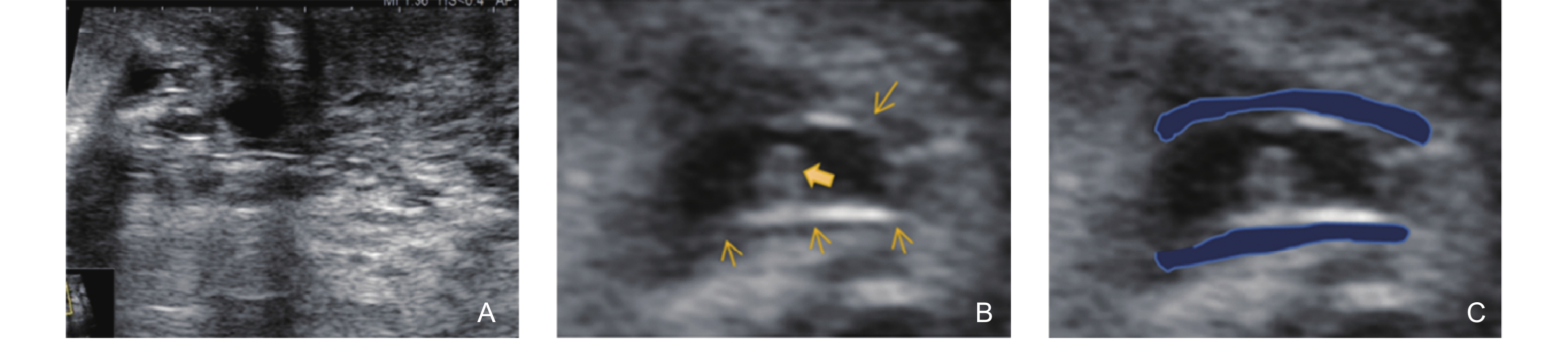
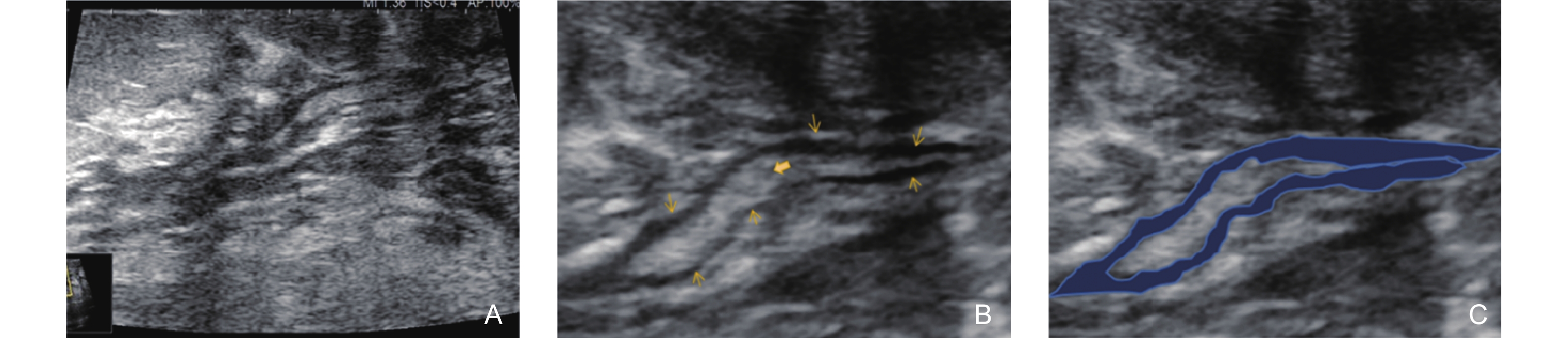
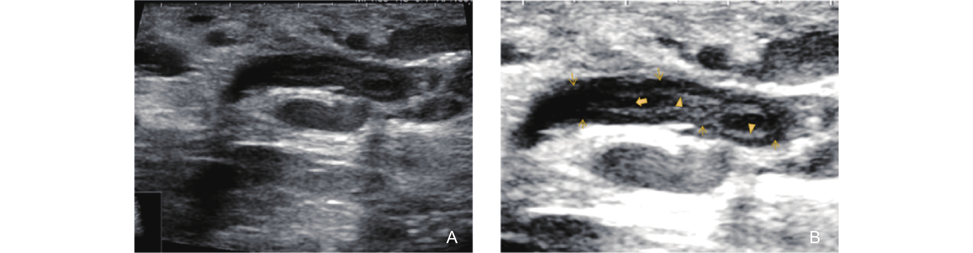
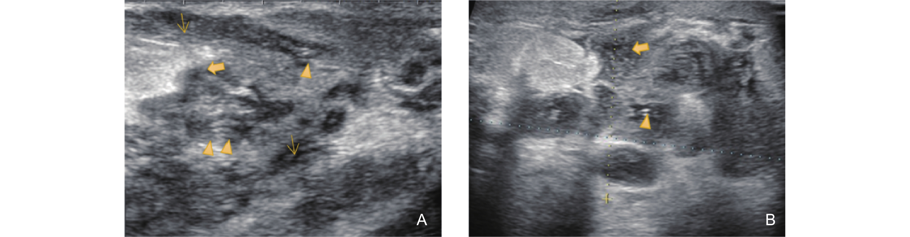
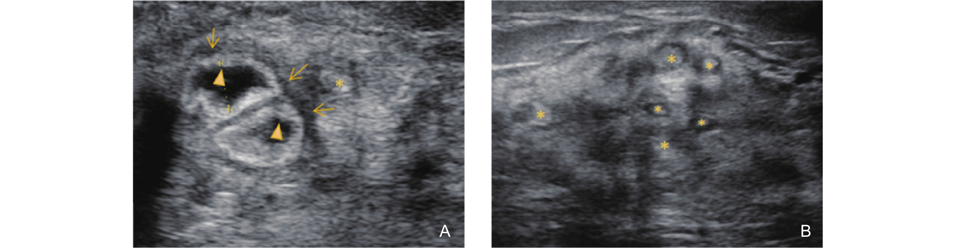
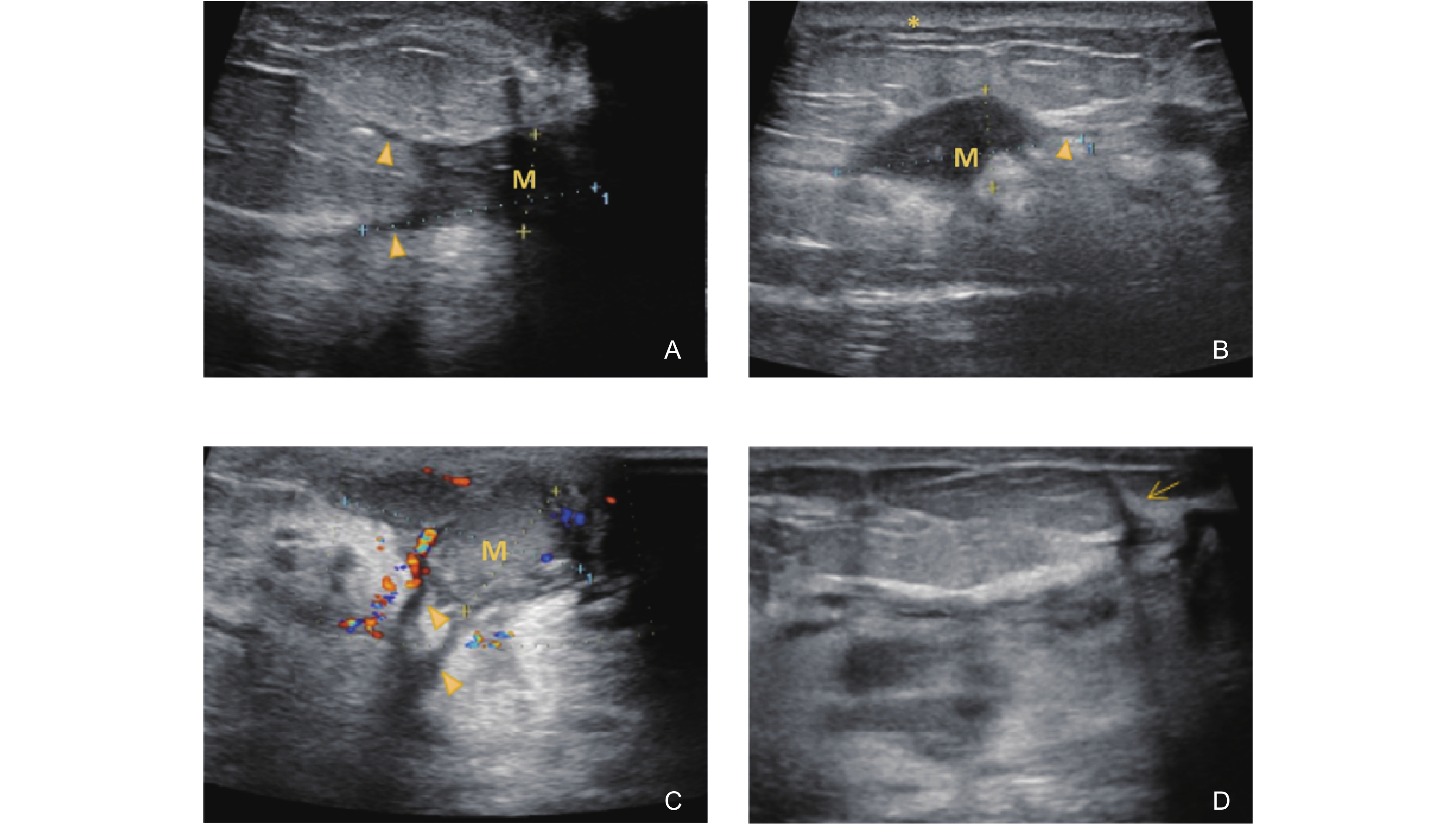
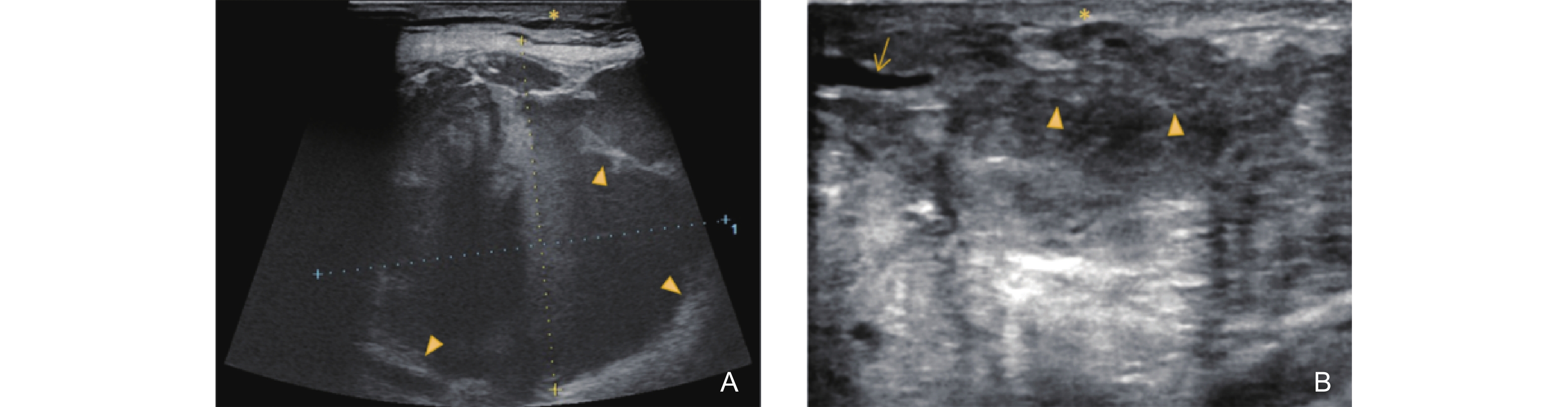
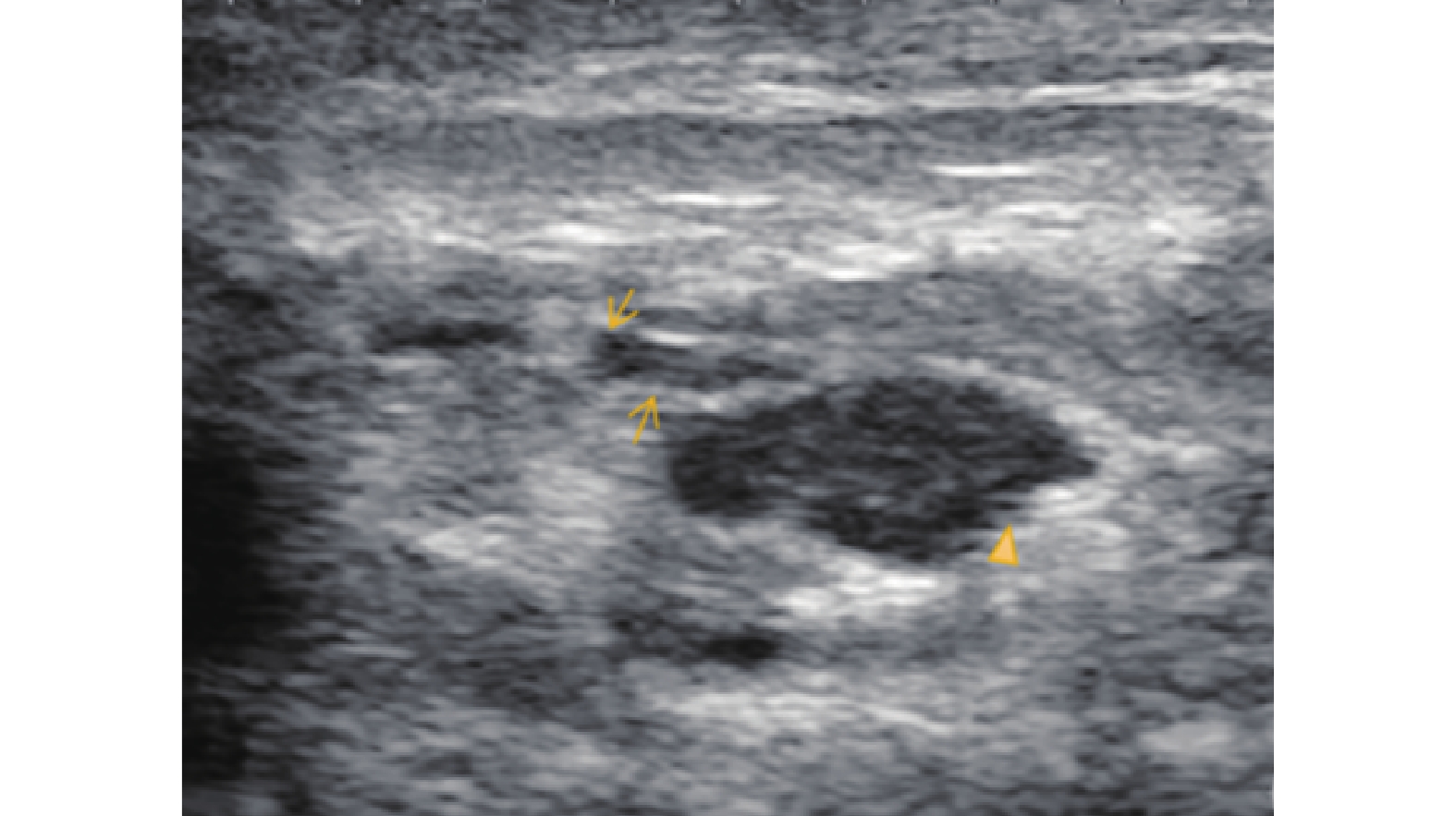
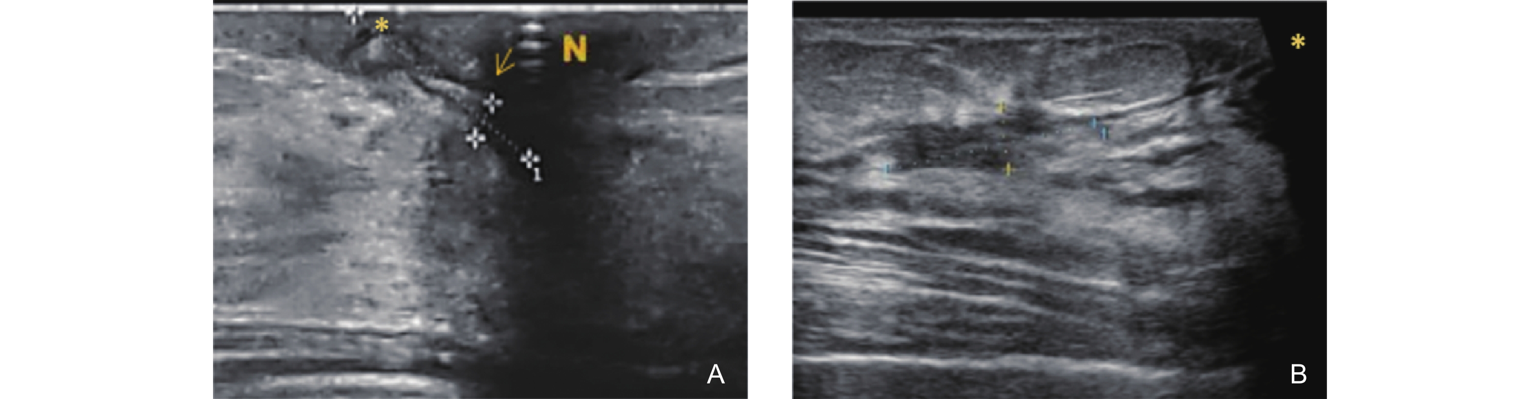
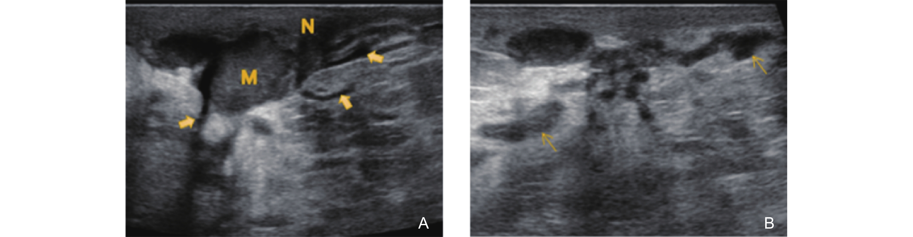
We have summarized and defined a set of disease-specific ultrasonographic features, including five subtypes of the “track sign,” as well as the “donut sign,” “false burr sign,” abscess formation, ulceration, and fistulous tracts. According to disease progression, GLM was categorized into six stages: Non-Mass Stage, Mass Stage, Abscess Stage, Late Ulceration Stage, Fistula Stage, and Complex Type (the simultaneous presence of ultrasonographic features corresponding to ≥ 2 different GLM stages within the same breast). In this study, a cohort of 273 patients was enrolled to evaluate GLM diagnosis and staging from both clinical and ultrasonographic perspectives. The diagnostic accuracy of ultrasonography was assessed, and the ultrasonographic manifestations of GLM were comprehensively delineated across its various clinical stages—with particular emphasis on the diagnostically challenging Non-Mass Stage.

Results

Ultrasound-based diagnosis of GLM achieved a sensitivity of 98.85%, specificity of 83.33%, and a positive predictive value (PPV) of 99.23%. Stage-specific ultrasonographic features demonstrated statistically significant associations with clinical staging: Track signs were most frequently observed in the Non-Mass Stage (92.86%). The donut sign was prevalent in both the Non-Mass (69.05%) and Abscess (66.67%) stages. The false burr sign was highly characteristic of the Mass Stage (78.69%). The Abscess Stage exhibited large mixed echogenic areas with irregular walls and internal fluid motion (100%). Late Ulceration Stage was marked by hypoechoic areas in the areolar region communicating with dilated ducts (70.37%). Fistula Stage showed duct-like hypoechoic tracts extending to the skin with thickened walls (92.68%). Complex Type cases showed overlapping features of multiple stages. These findings validate the diagnostic utility of stage-specific ultrasonographic signs and support the clinical relevance of the proposed classification system.

Conclusion

GLM exhibits distinct stage-specific ultrasonographic features. When combined with a standardized examination protocol and clearly defined imaging criteria, these features can significantly enhance the early detection and accurate staging of the disease. Additionally, representative images for each stage are provided to support the standardization of ultrasonographic evaluation for GLM.

Key words: Granulomatous lobular mastitis, Breast ultrasound, Diagnostic imaging, Ultrasonographic features, Non-mass lesion

"

Clinical assessmentFrequencyPercentageUltrasound assessment
SensitivitySpecificityPPVNPV
GLM, granulomatous lobular mastitis; PPV, positive predictive value; NPV, negative predictive value
GLM26195.60%98.85%83.33%99.23%76.92%
Not GLM124.40%

"

Ultrasonographic featuresNon-mass stage
(n = 42)
Mass stage
(n = 61)
Abscess stage
(n = 33)
Late ulceration stage
(n = 27)
Fistula stage
(n = 41)
Complex type#
(n = 57)
χ2P value
# Statistical analysis was not included for the Complex Type as this group may exhibit characteristics from the preceding stages, leading to potential confounding and dilution of effects. * Different types of track signs may occur concurrently within the same case; therefore, the sum of the frequencies of each track sign subtype exceeds the total frequency of the track sign (All Types).
a Fisher’s exact test; b The imaging features marked in this study are strictly stage-dependent (appearing only in specific pathological stages or later stages), and theoretically, they cannot appear prior to the specified stages (with observed values being 0 in this study). Given their 100% specificity (no false positives observed), we conducted the analysis only on subgroups where these features were actually present.
Track Ssign *
(All types)
39
(92.86%)
15
(24.59%)
4
(12.12%)
5
(18.52%)
3
(7.32%)
31
(54.39%)
92.207< 0.001
Track sign with high echogenicity on the inner wall*24
(57.14%)
7
(11.48%)
2
(6.06%)
3
(11.11%)
2
(4.88%)
16
(28.07%)
52.721< 0.001
Track sign with low echogenicity on the inner wall*17
(40.48%)
4
(6.56%)
1
(3.03%)
1
(3.7%)
1
(2.44%)
7
(12.28%)
31.689a< 0.001a
Track sign with isoechogenicity on the inner wall*11
(26.19%)
2
(3.28%)
1
(3.03%)
1
(3.7%)
1
(2.44%)
5
(8.77%)
17.256a< 0.001a
Track sign with periductal abscess formation*7
(16.67%)
2
(3.28%)
1
(3.03%)
1
(3.7%)
1
(2.44%)
3
(5.26%)
7.870a0.057a
Track sign coexisting with ductal inflammation, periductal inflammation, and nodular structures*10
(23.81%)
1
(1.64%)
1
(3.03%)
1
(3.7%)
1
(2.44%)
6
(10.53%)
16.773a<0.001a
Donut sign29
(69.05%)
4
(6.56%)
22
(66.67%)
2
(7.41%)
2
(4.88%)
7
(12.28%)
88.196< 0.001
False burr sign0
(0%)
48
(78.69%)
2
(6.06%)
8
(29.63%)
7
(17.07%)
17
(29.82%)
64.005b< 0.001b
Large areas of mixed echogenicity with irregularly thickened and rough abscess walls; a flowing sensation within the abscess cavity was observed upon probe compression.0
(0%)
0
(0%)
33
(100%)
8
(29.63%)
4
(9.76%)
11
(19.30%)
63.599b<0.001b
A relatively well-defined and smooth-edged hypoechoic area was observed in the areolar region, communicating with the ductal system.0
(0%)
0
(0%)
0
(0%)
19
(70.37%)
19
(46.34%)
23
(40.35%)
3.813b0.043b
A duct-like hypoechoic structure extending toward the skin was observed, with thickening of the inner wall of the duct.0
(0%)
0
(0%)
0
(0%)
3
(7.32%)
38
(92.68%)
26
(45.61%)
21.815b< 0.001b

"

"

"

"

"

"

"

"

"

"

[1] Bi J, Li Z, Lin X, Li F, Xu H, Yu X, et al. Etiology of granulomatous lobular mastitis based on metagenomic next-generation sequencing. Int J Infect Dis 2021; 113: 243-250.
[2] Yuan QQ, Xiao SY, Farouk O, Du YT, Sheybani F, Tan QT, et al. Management of granulomatous lobular mastitis: an international multidisciplinary consensus (2021 edition). Mil Med Res 2022; 9: 20.
[3] Yin Y, Le H, Cheng Y, Zhong Y, Cheng Y, Wang B, et al. A cohort study of prolactin and non-puerperal mastitis using real world data. Sci Rep 2025; 15: 8619.
doi: 10.1038/s41598-025-92504-9
[4] Meng T, Cheng YQ, Qiu WQ, Ye MN, Wang B, Wu JJ, et al. Clinical study on the treatment of 120 cases of acne mastoid carbuncle with GUs’ surgery comprehensive external therapy. CJTCMP 2021; 36: 3728-3731.
[5] Chen L, Zhang XY, Wang YW, Zhao QF, Ding HY. Granulomatous lobular mastitis: a clinicopathological analysis of 300 cases. Zhonghua Bing Li Xue Za Zhi 2019; 48: 231-236.
[6] Alikhassi A, Azizi F, Ensani F. Imaging features of granulomatous mastitis in 36 patients with new sonographic signs. J Ultrasound 2020; 23: 61-68.
[7] Dursun M, Yilmaz S, Yahyayev A, Salmaslioglu A, Yavuz E, Igci A, et al. Multimodality imaging features of idiopathic granulomatous mastitis: outcome of 12 years of experience. Radiol Med 2012; 117: 529-538.
[8] Hovanessian Larsen LJ, Peyvandi B, Klipfel N, Grant E, Iyengar G. Granulomatous lobular mastitis: imaging, diagnosis, and treatment. AJR Am J Roentgenol 2009; 193: 574-581.
[9] Wang M, Liu Y, Zhang D, et al. Analysis of the clinical character-istics and ultrasonographic features in 141 cases of cystic neutrophilic granulomatous mastitis. Acad Radiol 2025; 32: 2489-2496.
[10] Pluguez-Turull CW, Nanyes JE, Quintero CJ, Alizai H, Mais DD, Kist KA, et al. Idiopathic granulomatous mastitis: manifestations at multimodality imaging and pitfalls. Radiographics 2018; 38: 330-356.
[11] Guo JY, Yan XB, Cai ML, Lv GR. Diagnostic value of ultrasonography in different types of granulomatous lobular mastitis: a multicenter retrospective analysis of 120 cases. Fujian Med J 2022; 44: 51-54.
[12] Sripathi S, Ayachit A, Bala A, Kadavigere R, Kumar S. Idiopathic granulomatous mastitis: a diagnostic dilemma for the breast radiologist. Insights Imaging 2016; 7: 523-529.
[13] Villavicencio JJ, O'Brien SR, Hu T, Zuckerman S. Cystic neutrophilic granulomatous mastitis: Imaging features with histopathologic correlation. J Breast Imaging 2025; 7: 204-213.
[15] Kayadibi Y, Saracoglu MS, Kurt SA, et al. Differentiation of malignancy and idiopathic granulomatous mastitis presenting as nonmass lesions on MRI: Radiological, clinical, radiomics, and clinical-radiomics models. Acad Radiol 2024; 31: 3511-3523.
No related articles found!
Viewed
Full text


Abstract

Cited

  Shared   
  Discussed   
No Suggested Reading articles found!
[an error occurred while processing this directive]